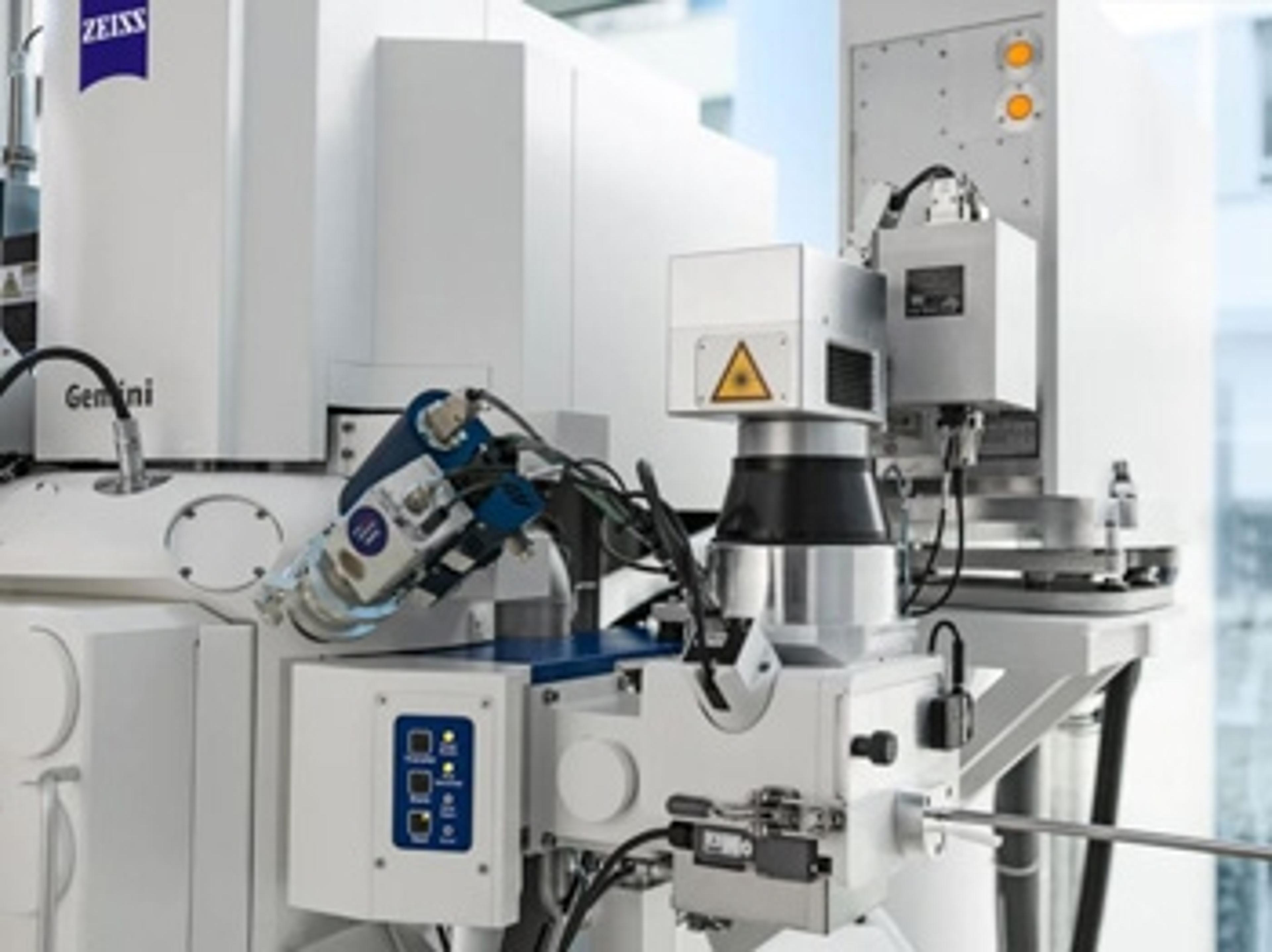
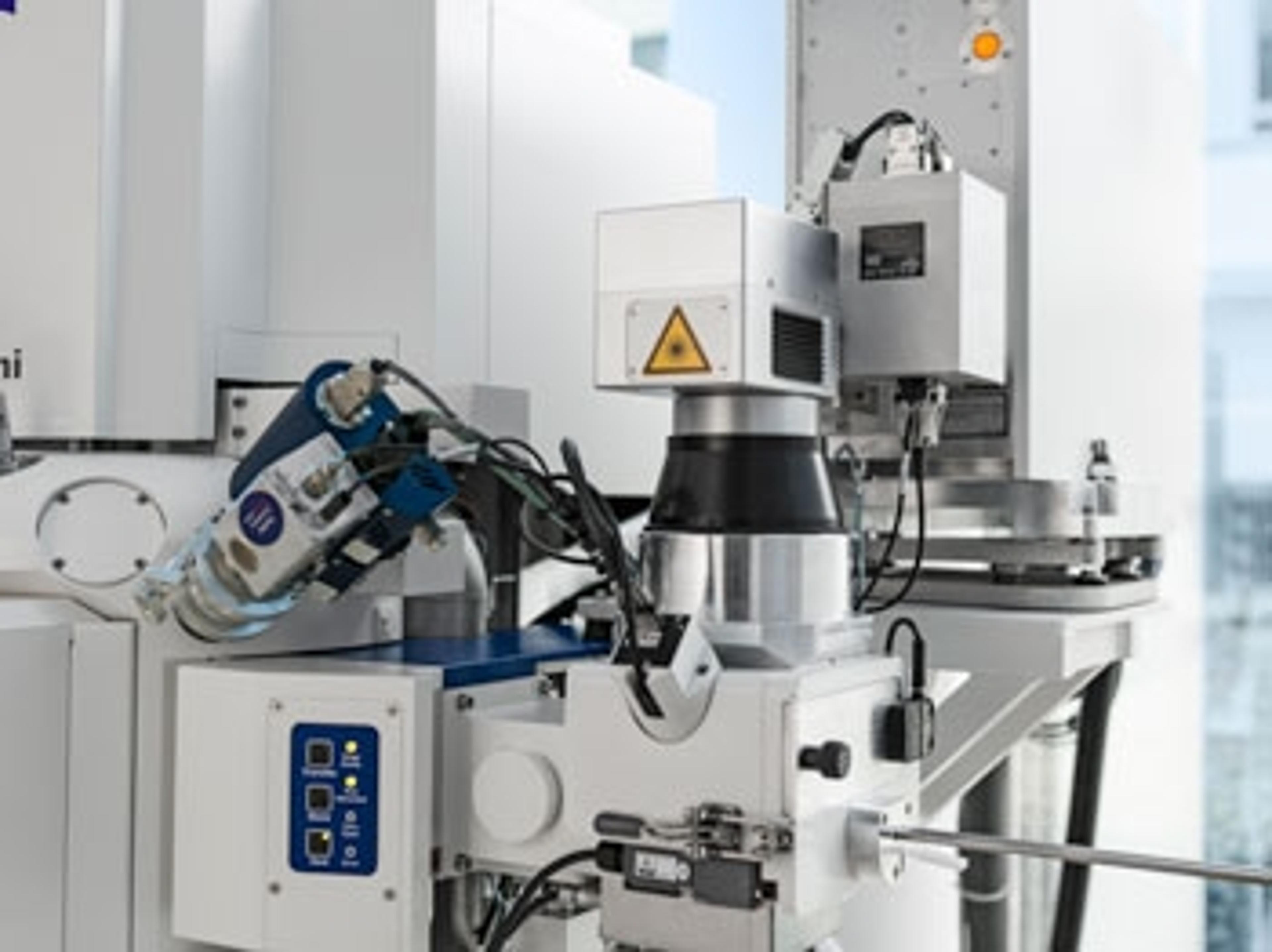
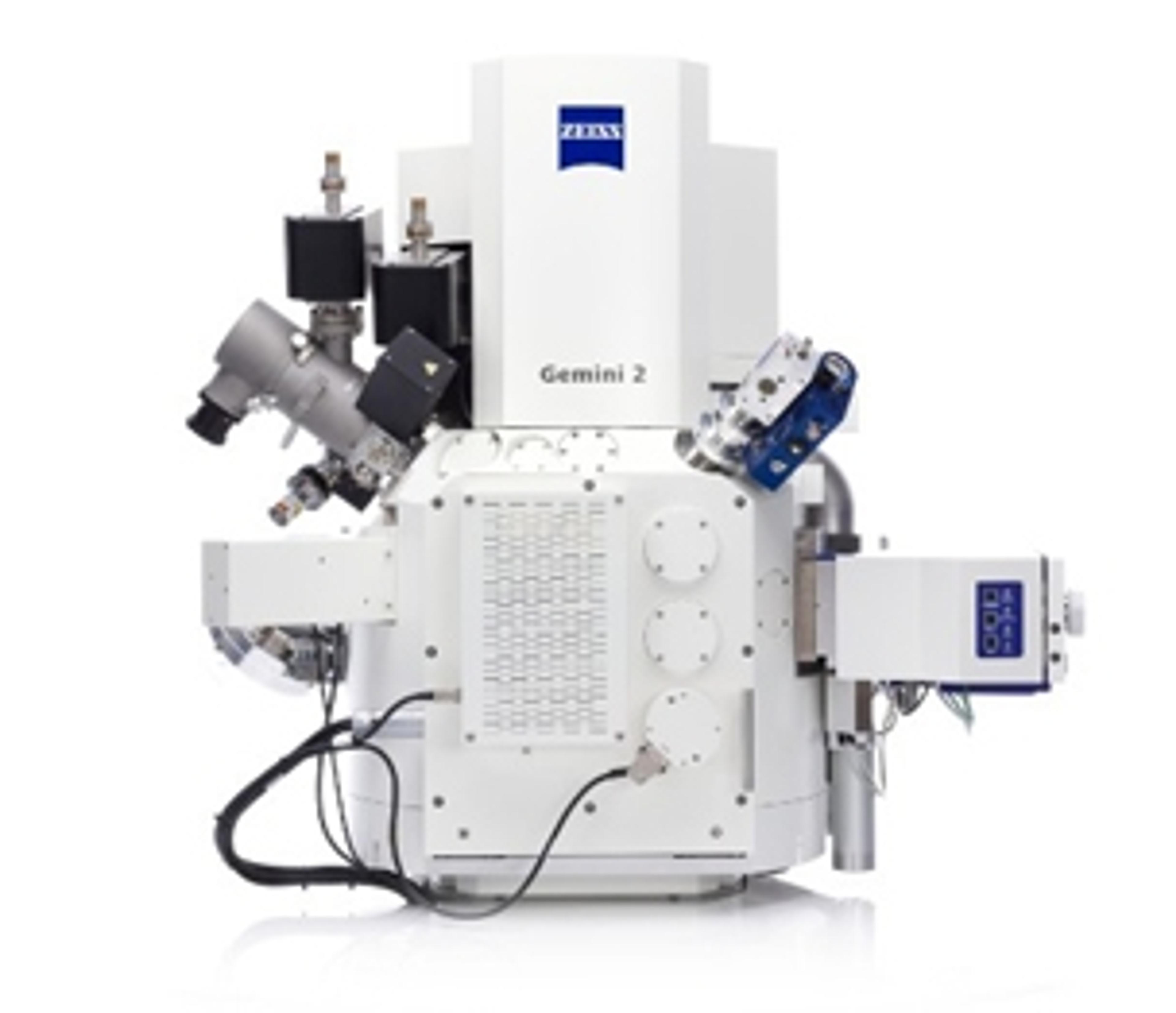
ZEISS Atlas 5
Large area imaging for SEM, FE-SEM & FIB-SEM ATLAS combines a 16 bit scan generator and dual super-sampling signal acquisition hardware with image processing and control software for your ZEISS electron microscope. Acquire images up to 32 k x 32 k pixels, with dwell times from 100 ns to > 100 s, adjustable in 100 ns increments. Save your images with eight or sixteen bits of intensity. With the ATLAS “Mosaic Tool” you create la…


Receive your quote directly from the manufacturer.
Large area imaging for SEM, FE-SEM & FIB-SEM
ATLAS combines a 16 bit scan generator and dual super-sampling signal acquisition hardware with image processing and control software for your ZEISS electron microscope. Acquire images up to 32 k x 32 k pixels, with dwell times from 100 ns to > 100 s, adjustable in 100 ns increments. Save your images with eight or sixteen bits of intensity. With the ATLAS “Mosaic Tool” you create large image montages, automatically moving from image tile to tile, and mosaic site to site, resulting in an “Extreme Field of View” image, at SEM nanometer scale resolution.
ATLAS provides
• reduced number of tiles to acquire, reducing stage motion delay and areal fraction of each image “lost” to overlap
• reduced number of overlap “seams”, leading to less beam damage and degradation of the sample
• reduced computational complexity

Investigating structure-property relationships in a carbon-fiber composite
Characterizing composite materials is a challenging task. Understanding the nucleation processes is critical toward engineering against failure, but traditional bulk testing methods are insufficient to describe this process. ZEISS presents how correlative microscopy is a viable conduit into the digital material testing approach. A carbon fiber reinforced composite hockey stick was used as the subject of the characterization study, though this same technique can apply to any variety of materials, from glass composites to metal matrix composites, as well as to monolithic materials. Correlative microscopy enabled a robust imaging-to-simulation workflow, producing a model that is available for further digital modification and analysis. Through implementation of this procedure in a regular basis, material development efficiencies may be enhanced, leading to high-performance products in a reduced amount of time.
Characterization of Solid Oxide Electrolysis Cells by Advanced FIB-SEM Tomography
Microstructural changes after cycling of a solid oxide electrolysis cell (SOEC) were studied by means of FIB-SEM tomography. The advanced tomography package ZEISS Atlas 5 3D Tomography allows high resolution 3D electron imaging and 3D EDS elemental imaging using two different sets of SEM conditions optimized for the respective task. The additional chemical information facilitated the correct segmentation of the different phases present in the sample. This was crucial to better understand the diverse mechanisms leading to deterioration of the cell.
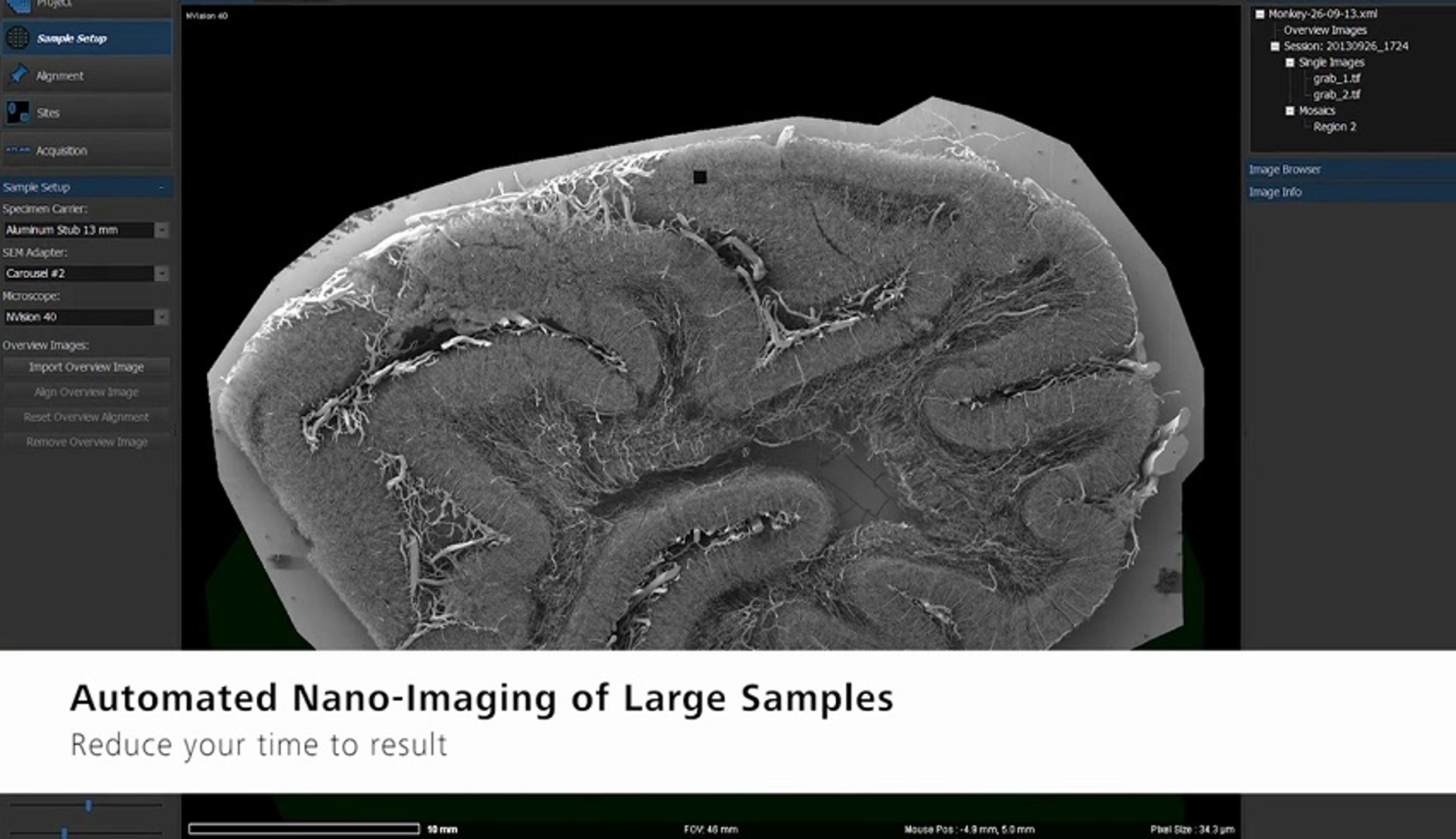
Large Area Imaging with High Throughput
This application note describes the automated acquisition of multi-image mosaics at multiple sample sites for electron microscopy, detailing the various sample collection capabilities.
Characterization of Solid Oxide Electrolysis Cells by Advanced Focused Ion Beam-SEM Tomography
This white paper investigates the microstructural changes after cycling of a solid oxide electrolysis cell (SOEC), studied by means of focused ion beam (FIB)-SEM tomography. The advanced tomography package, ZEISS Atlas 5 3D Tomography, enables high-resolution 3D electron imaging and 3D energy dispersive X-ray spectroscopy (EDS) elemental imaging, using two different sets of SEM conditions optimized for the respective task.
Multi-Scale Correlative Study of Corrosion Evolution in a Magnesium Alloy
This application note describes the results of a multi-scale correlative tomography study on the corrosion of a Magnesium alloy. Zeiss’ Atlas 5 is used to efficiently link and navigate between in situ sub-micron X-ray microscopy, nanoscale X-ray microscopy and FIB-SEM tomography. The study provides a description of the complex crack and corrosion byproduct geometries which can lead to a more complete understanding of the underlying mechanisms for corrosion.
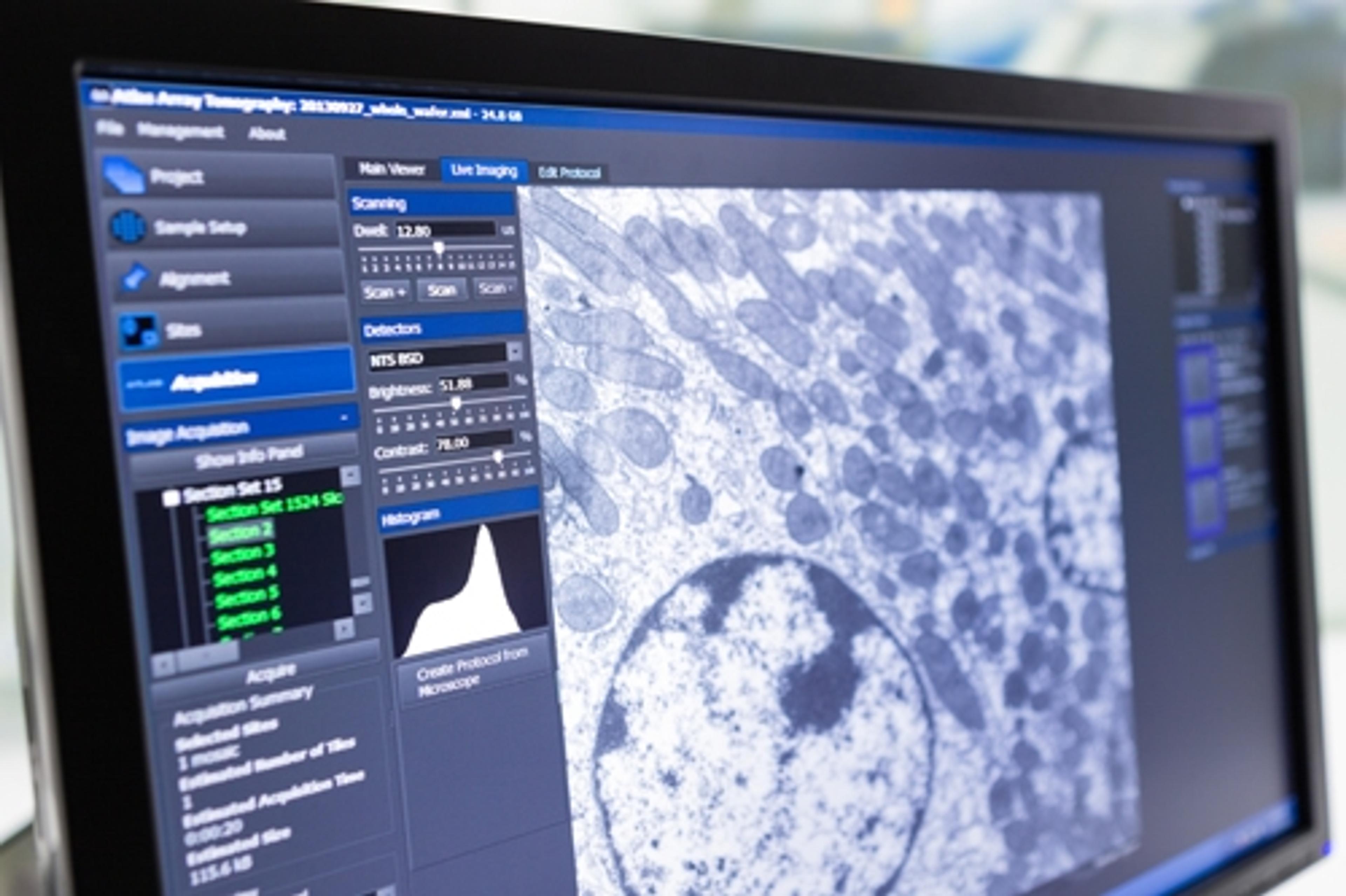
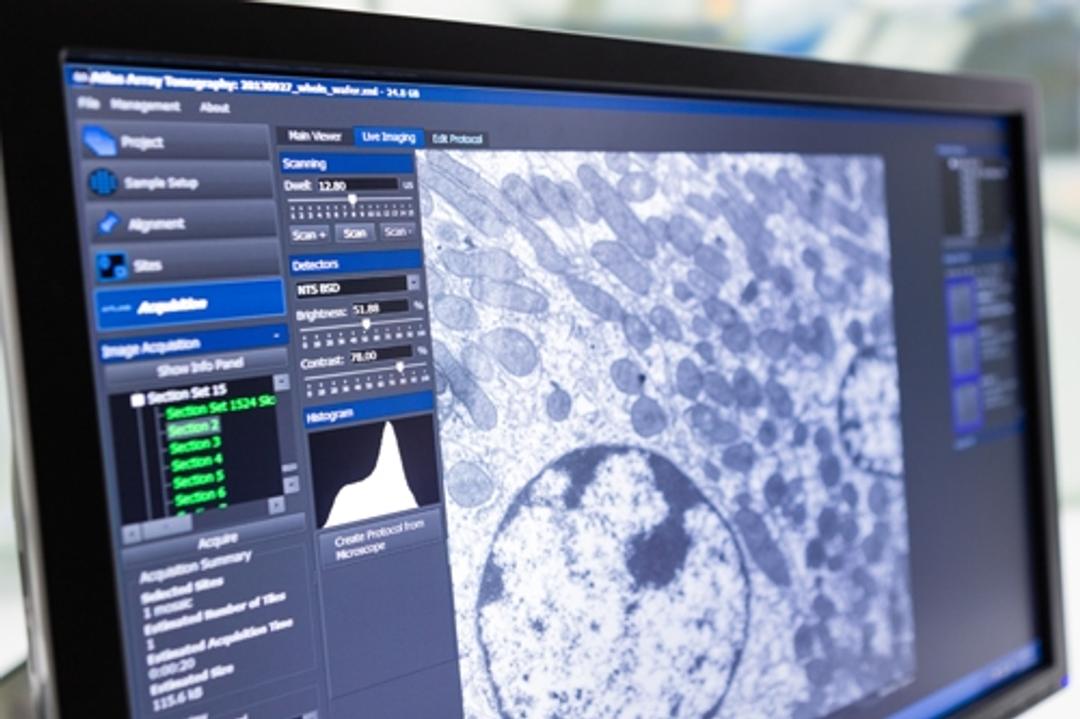
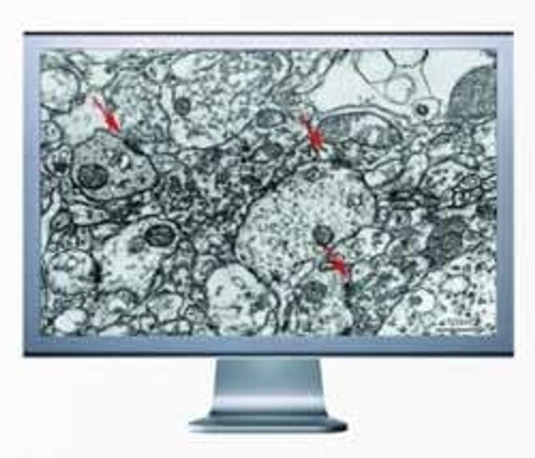
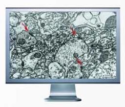
ZEISS Atlas 5 Array Tomography Image Your Serial Sections Fast and Efficiently
This application note describes how to use the ZEISS Atlas 5 Array Tomography to image serial sections fast and efficiently at nano scale resolution. Atlas 5 Array Tomography has been specifically designed for automated imaging of serial sections to enable 3D visualizations of large volumes.
ZEISS Legacy Continues: Advancing Microscopy at M&M 2017
Peter Lander discusses the legacy and development of ZEISS microscopy and the importance of conferences such as M&M. Its collaboration with scientists has enabled ZEISS to provide instrumentation that satisfies their needs, with the aim to develop simplified software that enables complementary and overlaying analyses over a range of its instrumentation.
ZEISS Atlas 5 Array Tomography
This video demonstrates the many applications of the ZEISS Atlas 5, allowing you to master your multi-scale challenges.
ZEISS Atlas 5 Array Tomography and Correlative Microscopy
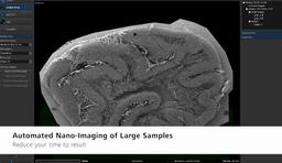
Reduce your time to result with automated nano-imaging of large samples: image your serial sections fast and efficiently. Atlas 5 Array Tomography is your unique, easy-to-use hardware and software package for your electron microscope (EM).
Optimized 3D Electron Microscope Workflow
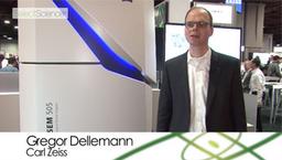
This video presents an integrated solution for your 3D electron microscopy workflow, from sample preparation to the final images. Discover the new ZEISS MultiSEM 505, the world’s fastest electron scanning microscope, and automated solutions for serial sectioning and imaging. Learn how these technologies will help to advance the field of neuroscience.
ZEISS enhances efficiency in multi-scale and multi-modal workflows
Researchers benefit from faster FIB-SEM sample preparation, more accurate 3D tomography, and greater integration in data reporting
ZEISS enhances efficiency in multi-scale and multi-modal workflows
Researchers benefit from faster FIB-SEM sample preparation, more accurate 3D tomography and greater integration in data reporting

ZEISS Introduces Enhanced Capabilities for Ion Beam Microscopes
Upgrade opens new opportunities in materials science and covers advances in analytics, tomography, sample preparation, and data integrity
Major improvements in workflow offerings for material sciences core facilities
Promise of enhanced analytics, tomography, sample preparation and data integrity

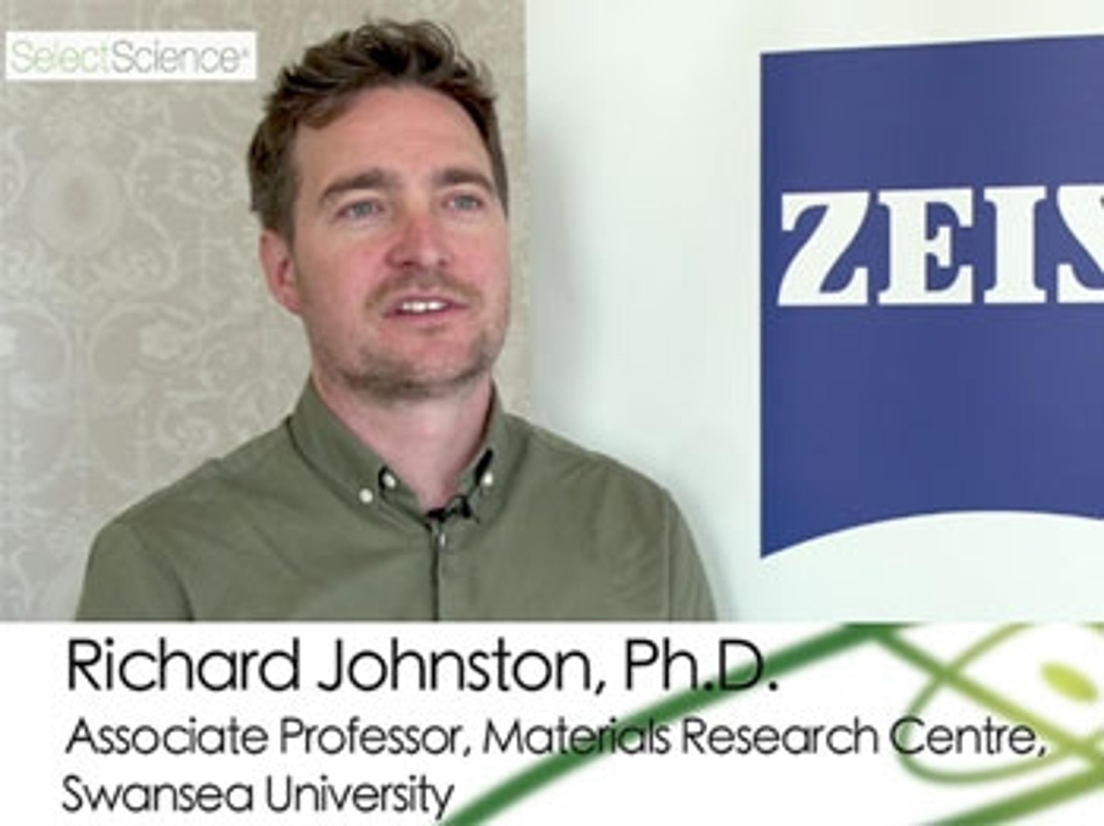
Use of Correlative Imaging to Bridge the Materials Gap Between Scientific Disciplines
Find out how Swansea University is developing correlative imaging workflows for multiple applications
Correlating Structural and Mechanical Properties Over Time: The Fourth Dimension of Materials Science
Find out how X-ray and focused ion beam microscopy are helping researchers to characterize the structure of materials
ZEISS Crossbeam 550 Sets New Standards in 3D Analytics and Sample Preparation
Enhanced resolution and faster FIB material processing for maximum efficiency

ZEISS Opens New Microscopy Customer Center
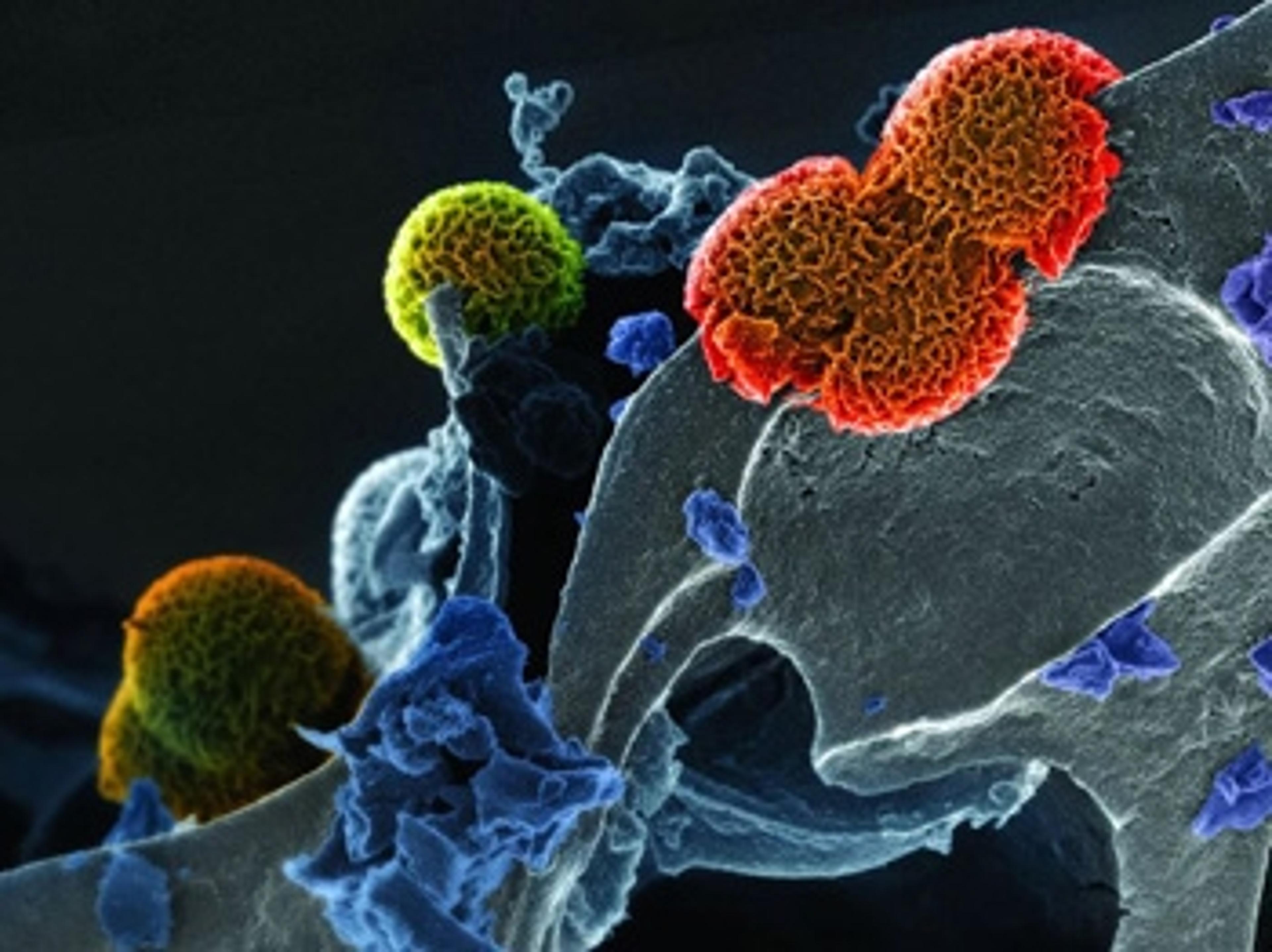
Using Molecular Spies to Understand Changes in Cell State in Health and Disease
SelectScience® spoke to Thomas Deerinck, National Center for Microscopy and Imaging Research at University of California, San Diego, about how correlative microscopy is helping scientists understand disease states