HALO® Image Analysis Platform
HALO provides easy-to-use, spatially-resolved cell and area-based image analysis for digital pathology and whole tissue images.


The supplier does not provide quotations for this product through SelectScience. You can search for similar products in our Product Directory.
User-friendly image analysis tool!
Quantification of Markers (IHC and RNAscope)
HALO Image analysis is a great tool, user-friendly and truly complementary to the day to day image analysis. Nuclei segmentation within HALO image analysis requires improvement in order to use it to its full potential and obtaining HALO AI just to access a better nuclei segmentation is currently too costly.
Review Date: 15 Jun 2022 | Indica Labs
Quick to learn and great results!
Analysis of immunofluorescence images from melanoma samples
The platform is very easy to use and quick to learn when you are new. I am very happy with the work that I have been able to do using Halo and the data that I have been able to have from my images. The people working in Indica labs are wonderful and always offer help. I would like to give a special thanks to Ghislaine Lioux. She has helped me numerous times and showed me lots of techniques when using Halo. I wouldn't have been able to achieve the amazing work without her help.
Review Date: 13 Jun 2022 | Indica Labs
My research depends on it.
Cell ecology analysis in colorectal tumours
HALO is just simply fabulous! Easy to use, and absolutely amazing at phenotyping cells. I’m interested in immune, stroma, and epithelial populations and HALO is perfect for them all. Well worth the price. I just love it!
Review Date: 9 Jun 2022 | Indica Labs
Great Product! Even better service/support! Exciting to watch our data roll in!
Antigentic intensity and Spatial studies in FFPE pancreatic tissue
Halo has been a game-changer for our lab. In addition to the (verifiable) accuracy of the data that we can now gather with speed and ease - there are now new layers of nuance to our questions that would have previously been impossible: whole section tissue and cellular segmentation, multiplex phenotyping, spatial relationships plotted within the software that allows for quick hypothesis testing and then the ability to build layers of data to answer multiple questions from our rare tissue. Even more impressive is the after-sales care: Every time we have a question or hit a barrier, the team responds quickly and creatively. Recently they have even offered to write a small algorithm that will make our studies even easier to run. I cannot fault them. Whatever our Halo, or Halo-data, needs are - whether it's associated with the applications specialists or the tech gurus - we are never left hanging. Truly (and surprisingly) still good after 4 years of a relationship. They will have your back! It isn't an inconsiderable amount to pay out - but the fact that we went from one HALO seat - to 2 HALO seats, one with AI, and now have just upgraded to another AI and with both machines are always busy we are considering HALO Link - says its worth every dime!
Review Date: 8 Jun 2022 | Indica Labs
A must-have!
Immuno-oncology
A great and user-friendly image analysis software for tissue segmentation, quantification and spatial analysis.
Review Date: 7 Jun 2022 | Indica Labs
Great results and intuitive workflow!
Hematopathology
The resources (tutorial videos e.g.) on the website were really helpful to learn how to get the best out of every module. Once you got familiar with the use, the platform is intuitive and the workflow works really well. If we encounter any kind of problem, we can always get in touch and get excellent and prompt help to solve the issue.
Review Date: 7 Jun 2022 | Indica Labs
Great instrument!
Area of tissues, immunohistochemistry analysis
I can get a high quality result, and it is easy to use!
Review Date: 6 Jun 2022 | Indica Labs
HALO has become a fundamental tool in our research activity.
Analysis of cell populations and spatial interactions within tissues
Our Lab (Tumor Immunology Laboratory, University of Palermo) investigates immune responses in cancer. We adopted several tools for quantitative assessment of immunolabeling with good results. HALO offers high-quality cell segmentation and allows accurate analysis of both brighfield and fluorescence-based microphotographs or slide scans. The possibility to easily extract cell features integrating spatial information enables detailed spatial interaction analyses.
Review Date: 3 Jun 2022 | Indica Labs
A software making all digital pathology analysis easier, and a great support team
Onco-immunology: characterization of the immune microenvironment in colorectal precancerous lesiosn
HALO and HALO AI have both been a big improvement for our image analysis. Being able to work on the whole slide and to develop artificial intelligence based tissue and nuclei classifier have been very useful for my research and made me gain a lot of time. In addition to the very complete software, the support team is very reactive and enjoyable to work with.
Review Date: 3 Jun 2022 | Indica Labs
Great results - easy to use
Analyze metabolites in tissue sampples
I am very satisfied with the effectiveness of the products and service for my applications. The software is super easy to use and I achieve highest quality and reproducible results. The after sales care is of high value and the application specialists are top.
Review Date: 3 Jun 2022 | Indica Labs
With unmatched ease-of-use, powerful analytic capabilities, and ultra-fast processing speeds, Laboratories around the world depend on HALO® to achieve high-throughput, accurate analysis of their digital pathology.
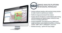
IMAGE ANALYSIS SIMPLIFIED
Spend less time learning software and more time analyzing data. HALO’s analysis tuning is fast and easy for experts and novices alike, without sacrificing data quality. No need to “build” analysis algorithms from scratch. HALO’s flexible, purpose-built modules provide quick, quantitative results in oncology, neuroscience, metabolism, toxicological pathology, and more.
ACCELERATE YOUR ANALYSIS
Digital slides are large and bog down conventional analysis systems. HALO’s parallel processing technology and optimized algorithms yield up to four times the analysis rate of competitive solutions using the same standard hardware. Organizations with greater throughput demands can couple HALO with our performance boosting analysis clusters.
EASILY EXPLORE CELLULAR DATA
HALO reports morphological and multiplexed expression data on a cell-by-cell basis across entire tissue sections and maintains an interactive link between cell data and cell image. Click on any cell in the image and immediately see analysis outputs for that specific cell. Sorting and filtering capabilities allow the user to mine millions of cells while visually assessing corresponding cell populations. For example, sort cells according to biomarker intensity and immediately locate cells with highest intensity in the image. Just imagine the endless possibilities.
GROW WITH HALO
HALO offers a modular platform that can expand with your needs. Start with a few modules, and add more as your needs change. Use HALO on a single workstation or ramp up to a server-based license for your entire group. HALO is flexible enough for any budget.
Brochures

HALO - quantitative tissue informatics
With ease of use, powerful analytic capabilities, and ultra-fast processing speeds, laboratories around the world depend on HALO® to achieve high-throughput, accurate analysis of their digital pathology slides in a broad range of research fields, including oncology, neuroscience, metabolism, transplant science, toxicological pathology and more.
Introduction of a robust workflow for the whole-slide acquisition and co-registration of multiplex immunofluorescence tissue images
In this application note, explore how Indica Labs designed and tested an 8-plex immunophenotyping panel using the InSituPlex® approach to detect and classify T cells, macrophages, and tumor cell in non-small cell lung cancer and colorectal cancer FFPE tissue.
Depicting the cellular architecture of the tumor microenvironment by integrating hyperplex immunofluorescence and automated image analysis
The tumor microenvironment (TME) is emerging as an important factor that shapes the dynamic of tumor growth, heterogeneity, and response to therapies. In this application eBook, Indica Labs focuses on the phenotyping of cells across different tumor types on a tissue microarray (TMA) with an immuno-oncology panel encompassing 20 biomarkers. They interrogated their TME with the use of the COMET™ automated staining and imaging system, and HALO® and HALO AI image analysis platforms.
RNAscope image analysis using HALO and HALO AI
In this application note, discover how the ISH module and FISH module available with the HALO® image analysis platform can be employed with HALO AI™ to quantitatively assess chromogenic and fluorescence RNAscope assays, respectively.
Characterization of immune cell phenotypes through quantification of the 12-plex spatial RNAscope HiPlex v2 assay using the HALO image analysis platform
In this application note, Advanced Cell Diagnostics demonstrates how the HALO® image analysis platform can be employed with HiPlex v2 to quantitatively assess differential expression of 12 RNA targets and quantify six distinct cell phenotypes based on gene expression. Plus, spatial relationships between cell phenotypes within the tumor microenvironment (TME) are analyzed.
RNAscope publications with the HALO Image Analysis Platform
RNAscope® image analysis with the HALO® image analysis platform continues to advance research areas as diverse as immunology and infectious disease, metabolism, neuroscience, as well as oncology and immuno-oncology. Download the white paper below to see recent publications according to research area and publications using ISH, FISH, and codetection assays using HALO image analysis.
Quantification and characterization of glomeruli across diverse stains using HALO AI™
In this application note, Indica Labs highlights the potential for HALO® and HALO AI to quantify biomarkers, gene expression, and morphological changes in kidney disease, during development and in toxicological studies.
Qualitative and quantitative evaluation of the tissue microenvironment by high-resolution 17-plex immunofluorescence reveals distinct populations
In this application note, Indica Labs highlights how the use of Orion imaging combined with HALO image analysis provides a powerful and intuitive workflow for visualization and quantification of distinct microenvironment populations.
High-impact publications with the HALO image analysis platform
Digital pathology with the HALO® image analysis platform continues to advance research areas as diverse as immunology, infectious disease, immuno-oncology, and neuroscience. Download the white paper below to see the caliber of publications that researchers are obtaining, using Indica's cloud-based software.
Quantitative RNAscope image analysis guide
The RNAscope® Duplex and Multiplex assays enable simultaneous detection of more than one target RNA thus facilitating the evaluation of multiple cell phenotypes and spatial relationships within a complex tissue microenvironment. Download the guide below to find out how HALO® imaging software can create an easier solution with RNAscope® assays.
Multiplex immunofluorescence staining of cells in tumor FFPE samples
In this scientific poster, Indica Labs presents a workflow using advanced multiplexing techniques to observe biologically and functionally distinct T cell and myeloid-derived suppressor cells (MDSC) populations. Using these techniques they were able to quantify their density and distribution within several tumor types.
HALO Image Analysis
Indica Labs' HALO® is an image analysis platform for quantitative tissue analysis in digital pathology. The platform promises ease-of-use and scalability, powerful analytic capabilities, and the fastest processing speeds available for digital pathology, pharmaceutical, healthcare and research organizations worldwide are using HALO® for high-throughput, quantitative tissue analysis in oncology, neuroscience, metabolism, toxicology and more.
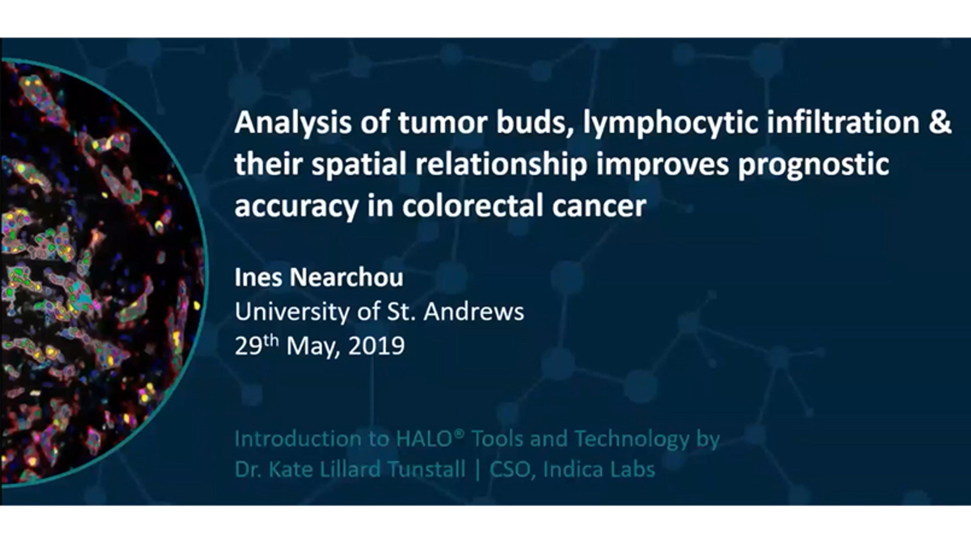
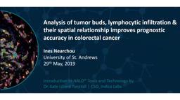
Improved prognostic accuracy in colorectal cancer
In this video, Indica Labs presents the HALO® Image Analysis Platform and Ines Nearchou from the University of St.Andrews describes how HALO can be used to analyse the tumour microenvironment in colorectal cancer.


Indica Labs receives top honor in Albuquerque Journal ‘Flying 40’ list of top tech firms in New Mexico
The company received the award due to its staggering growth rate over the past few years
Leica Biosystems announces partnership with Indica Labs to deliver integrated digital pathology workflow solutions for mutual customers
The collaboration is focused on delivering compatible digital pathology workflow solutions
Indica Labs announces collaboration with iCAIRD for the development of an AI-based algorithm for the automated reporting of lymph node status in colon cancer
This innovative research project aims to develop a tool that in the future may improve the efficiency of pathology teams reporting colorectal cancer cases and the detection of metastatic cancer in lymph nodes

Key laboratory automation products reviewed by our scientists
We’ve highlighted reviews of laboratory automation products we think could help make your lab's journey to becoming more automated hassle-free

Indica Labs earns work/life flexibility award
For the second year running, Indica Labs has been named a top workplace by the Albuquerque Journal

Indica Labs and Ibex partner to deliver AI-powered clinical workflows for digital pathology
The seamless integration of AI into digital pathology workflows aims to improve quality and efficiency of cancer diagnosis
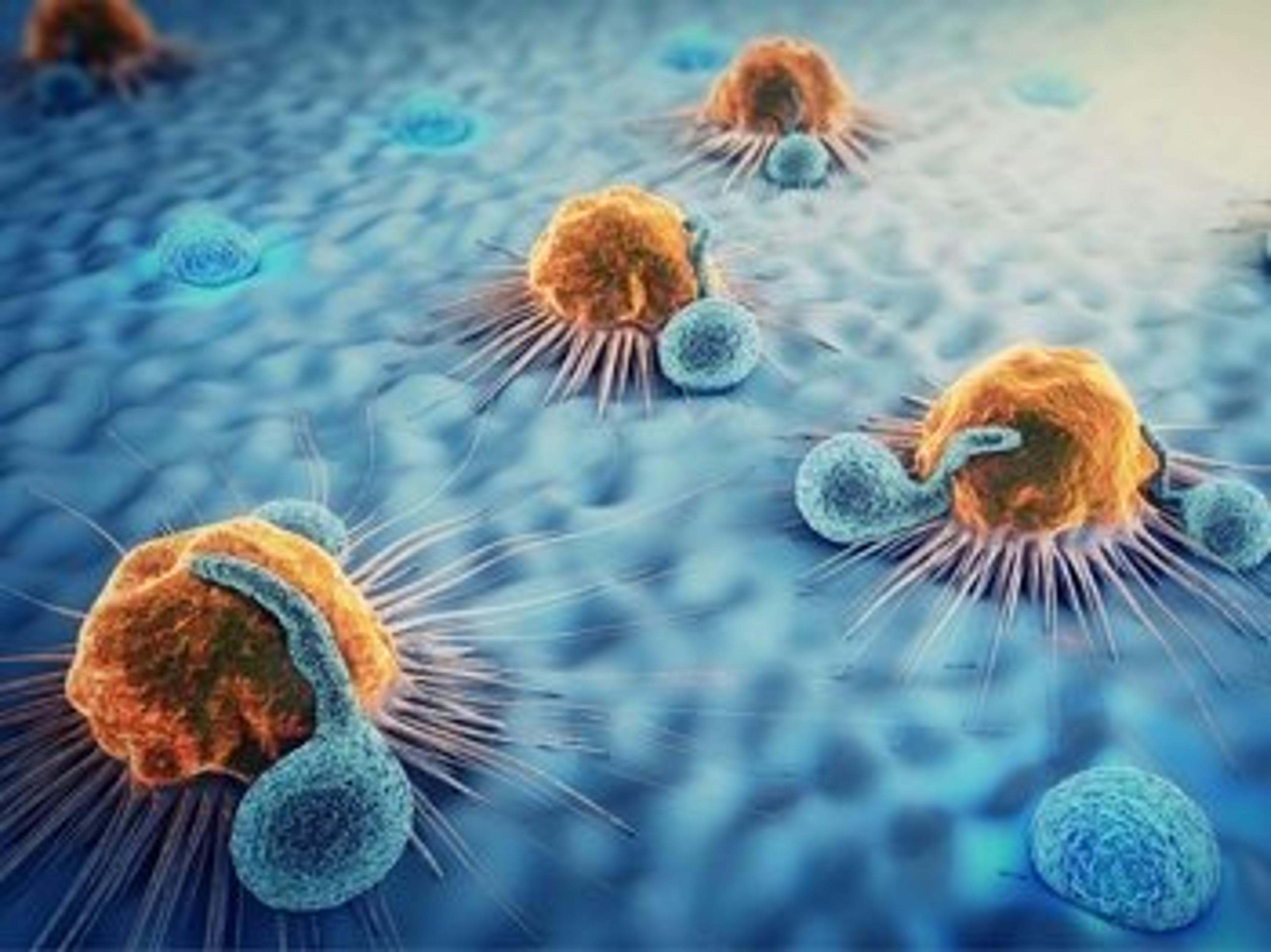
Immuno-oncology advances: From tumor microenvironments and CAR-T cell workflows to macrophage generation
Discover the latest technologies and techniques that are revolutionizing immuno-oncology research
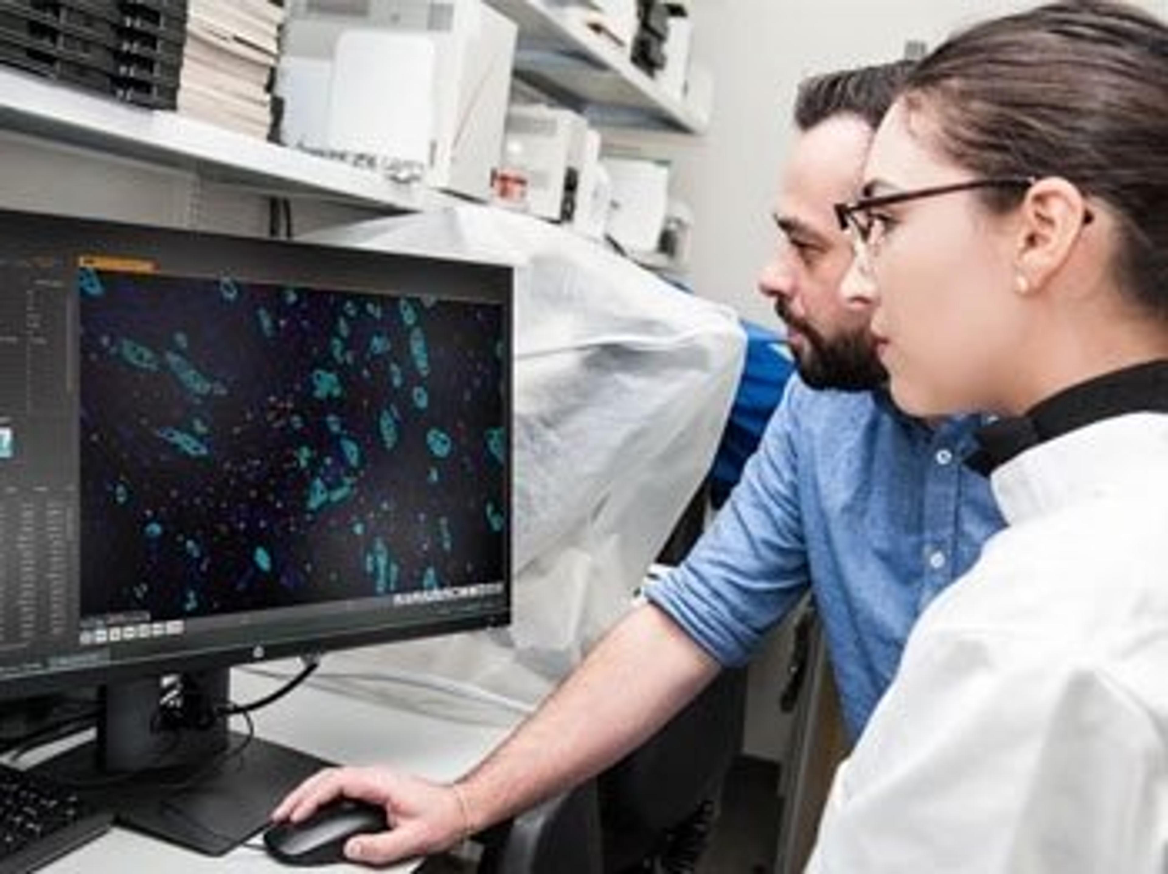
New prognostic test for colorectal cancer details spatial interaction between tumor and immune cells
Multiplexed immunofluorescence and next-generation image analysis offer spatial insights into the tumor microenvironment