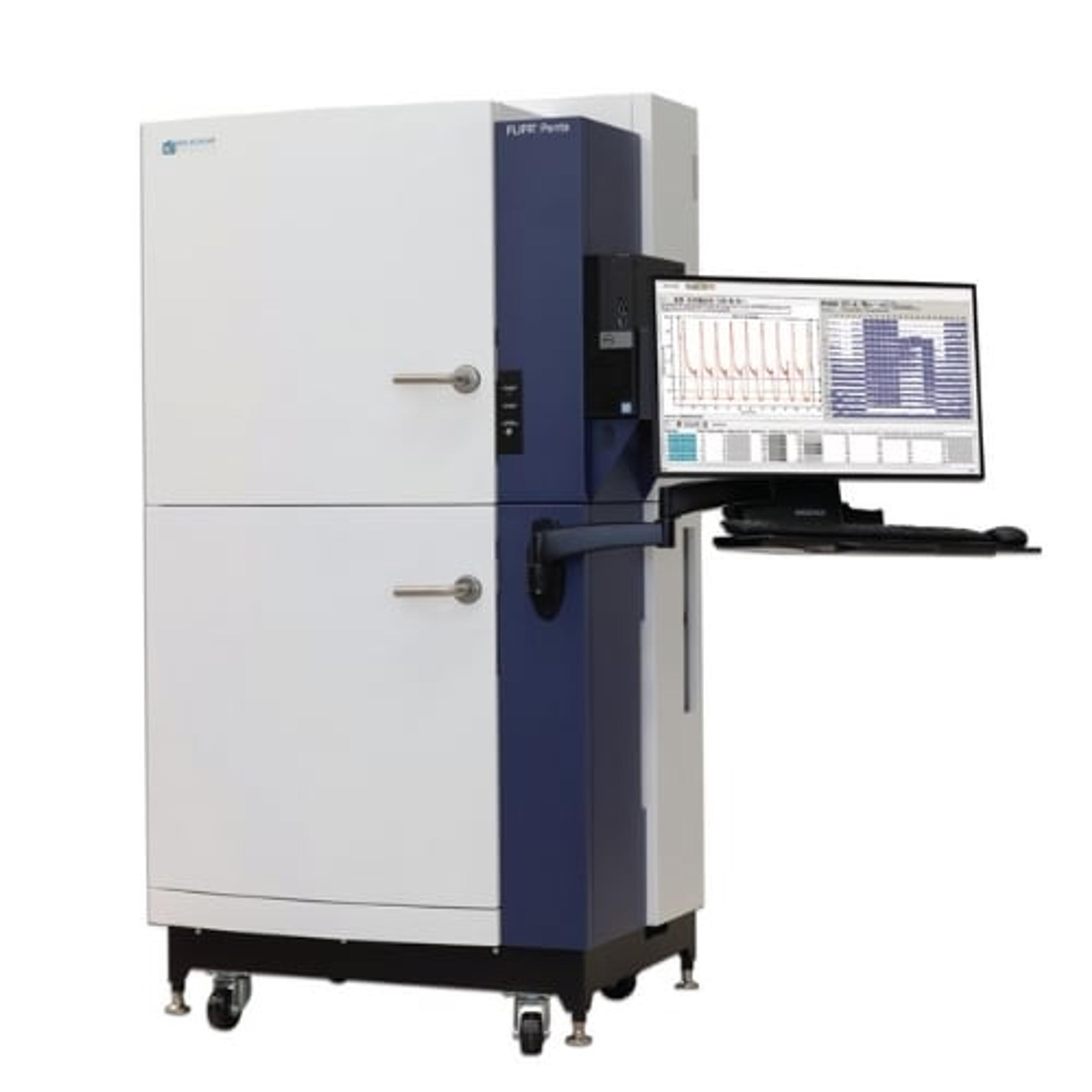
CloneSelect® Imager FL
The CloneSelect Imager FL is a next-generation fluorescent imaging solution for the assurance of monoclonality and automated confluence across diverse cell types.


Receive your quote directly from the manufacturer.
Demonstrating that cell lines are monoclonal – or that a gene was edited as expected – can be a time-consuming and highly-subjective process when relying on conventional technologies. The CloneSelect® Imager FL is a high-throughput automated solutions for imaging and analyzing mammalian cells. Tracking the formation of a colony from a single cell is effortless as barcoded plates are tracked over time. Automated acquisition and analysis provide accurate, objective, and consistent results.
The CloneSelect Imager FL features high contrast multichannel fluorescent and white light imaging that allows for accurate single-cell detection and proof of monoclonality at day 0. Streamline your workflow with comparative confluence assays to identify and verify gene edits.
Demonstrated IND success
The Monoclonality Report feature streamlines the creation of supporting documentation for regulatory agencies. Reports are automatically generated based on the parameters you select. The Monoclonality Report is an audit-ready document that supports filing for an Investigational New Drug (IND) Application with the FDA. (21 CFR Part 312)
Multichannel imaging and automated confluence
Algorithms are optimized for accurate cell detection and address varying cell types and conditions. Publication-ready high-resolution imaging provides automatic confluence analysis and monoclonality assurance.
Rapid single-cell confirmation
The imager delivers industry-leading acquisition times, imaging a 96-well plate in as little as two minutes, and can verify monoclonality from day zero with fluorescence.
Features
- White light and fluorescent imaging
Image every well in every plate with rapid acquisition times with label-free white light. Multichannel fluorescence imaging provides additional confidence of monoclonality and comparative confluence assays (red vs green). - Quickly image a variety of plate formats and cell types
Image a 96-well plate in under two minutes. Compatible with adherent or settled suspension cell types such as CHO, HEK, hybridomas, iPSCs, and many other cell types. - High-resolution images
High-speed fluorescence and high-resolution white light imaging with accurate detection of single cells including debris. Image visualization and data tracking over multiple days. - Data tools to accelerate research timelines
The software automatically calculates confluence measurements and generates growth curves, heatmaps, and image montages. Measurements for every well are automatically tracked over time. Streamline multiple steps (imaging, sample tracking, data analysis and report generation) in the cell line development workflow for IND filing. - Intelligent analysis with easy-to-use software
The software automatically calculates confluence for each imaging time point. Growth curve, image montage, total growth, and mean rate are generated automatically and are exportable. Guided software user interface allows for simple 1-hour training. - Custom automation options*
The Automation and Customization Team offers a variety of custom services from robotic plate loading to fully automated workstations with liquid handling and incubation.
*Price, time to deliver and specifications will vary based on mutually agreed technical requirements. Solution requirements may cause adjustment to standard performance.

Single-cell dispensing and screening of cell lines for monoclonality verification using the impedance-based single-cell dispenser and high-throughput fluorescence-based imager
There is an unmet need in the industry for a device that allows the fast and efficient isolation of single cells while preserving their integrity and providing insurance for their clonality. Performing gene editing, single-cell dispensing, and screening to develop a stable monoclonal cell line is a long and labor-intensive process. The optimization of these steps through different approaches, such as high-throughput screening and/or automation, can increase the efficiency and yield of the monoclonal cell-line development process. In this combined application note, Molecular Devices shows the impedance-based single-cell dispenser and automated fluorescence-based high-throughput imaging screening to obtain monoclonal cell lines through robust single-cell selection.

CRISPR expands horizons of genetic engineering
Uncover the technology allowing scientists to push boundaries in genetic engineering