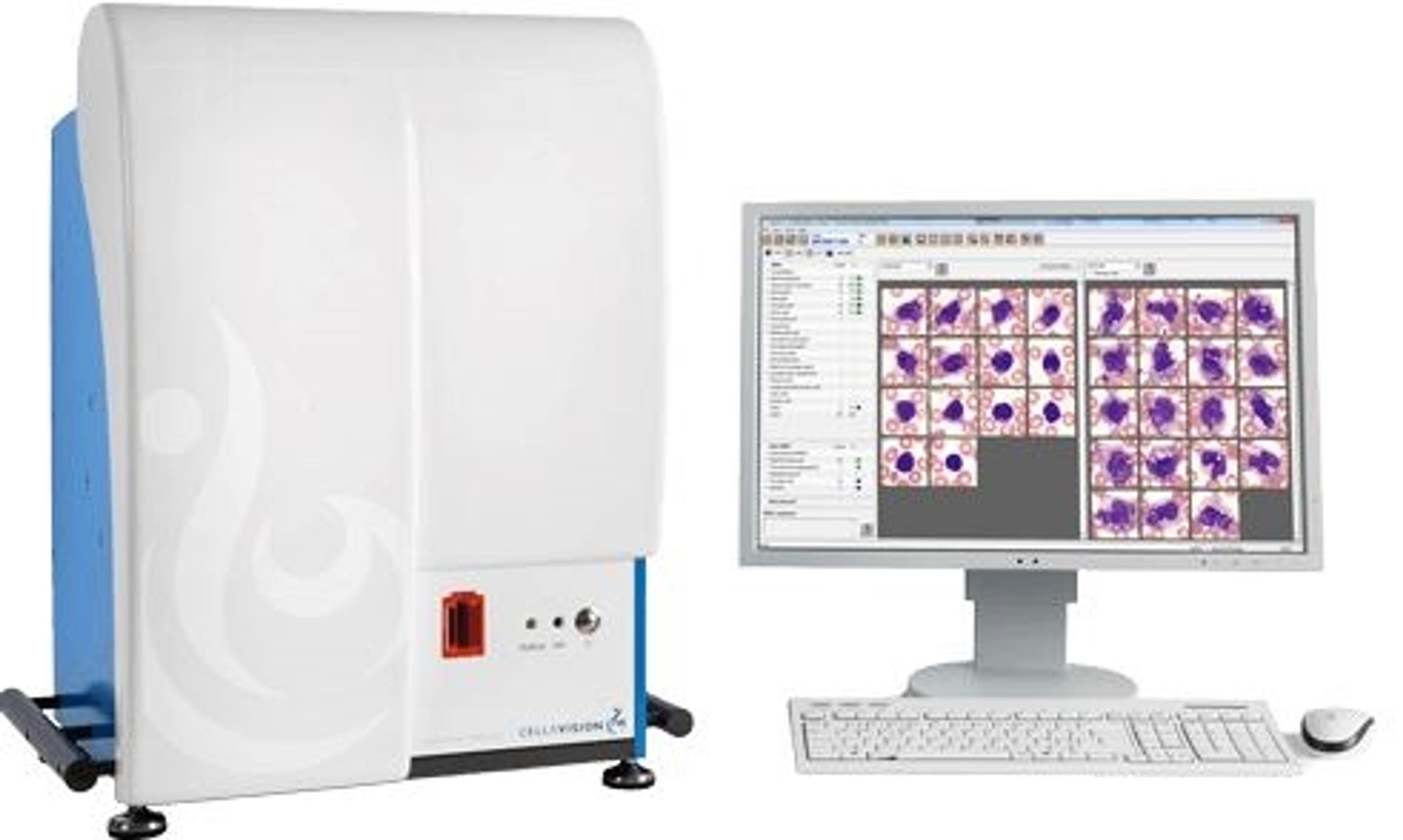
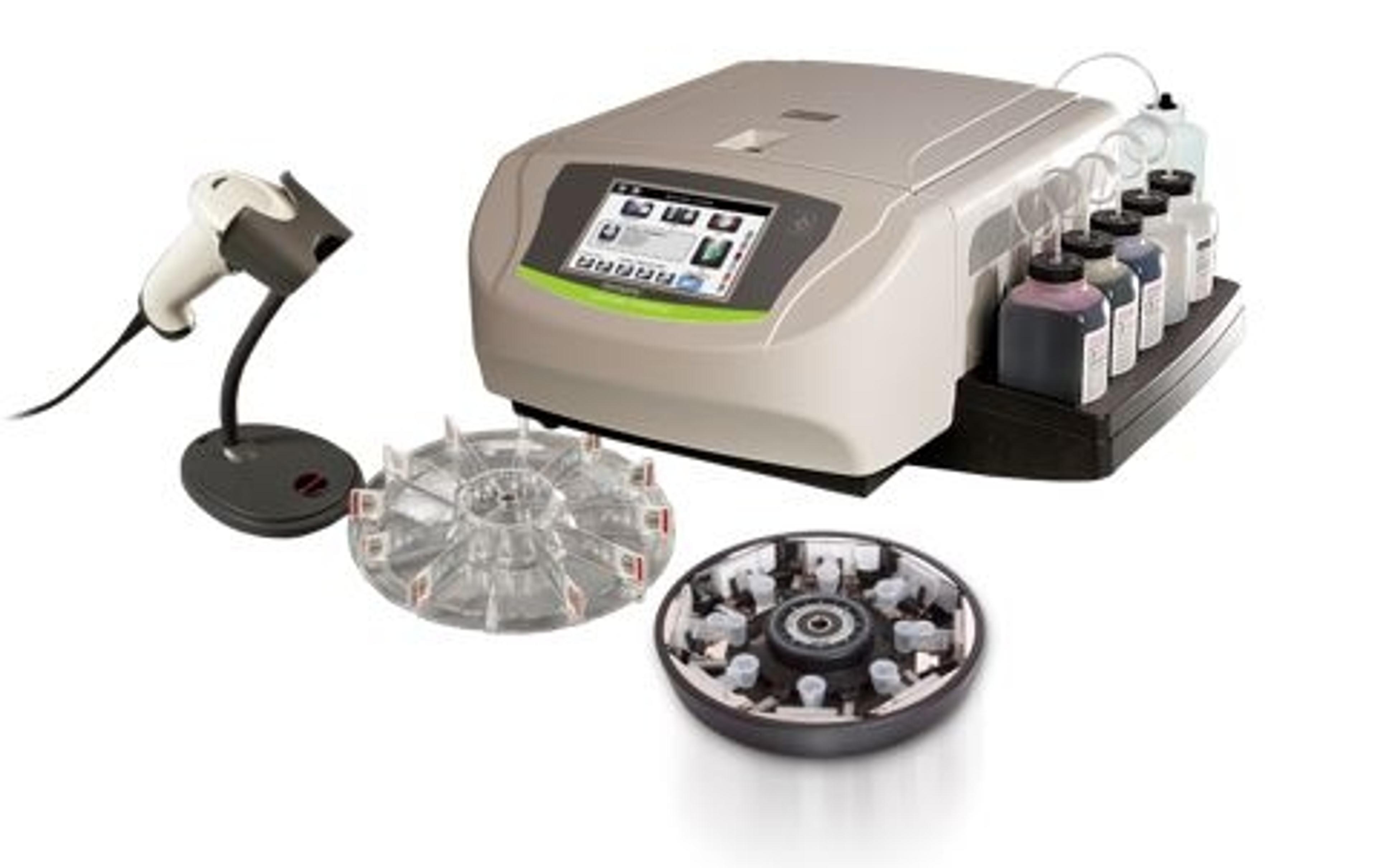
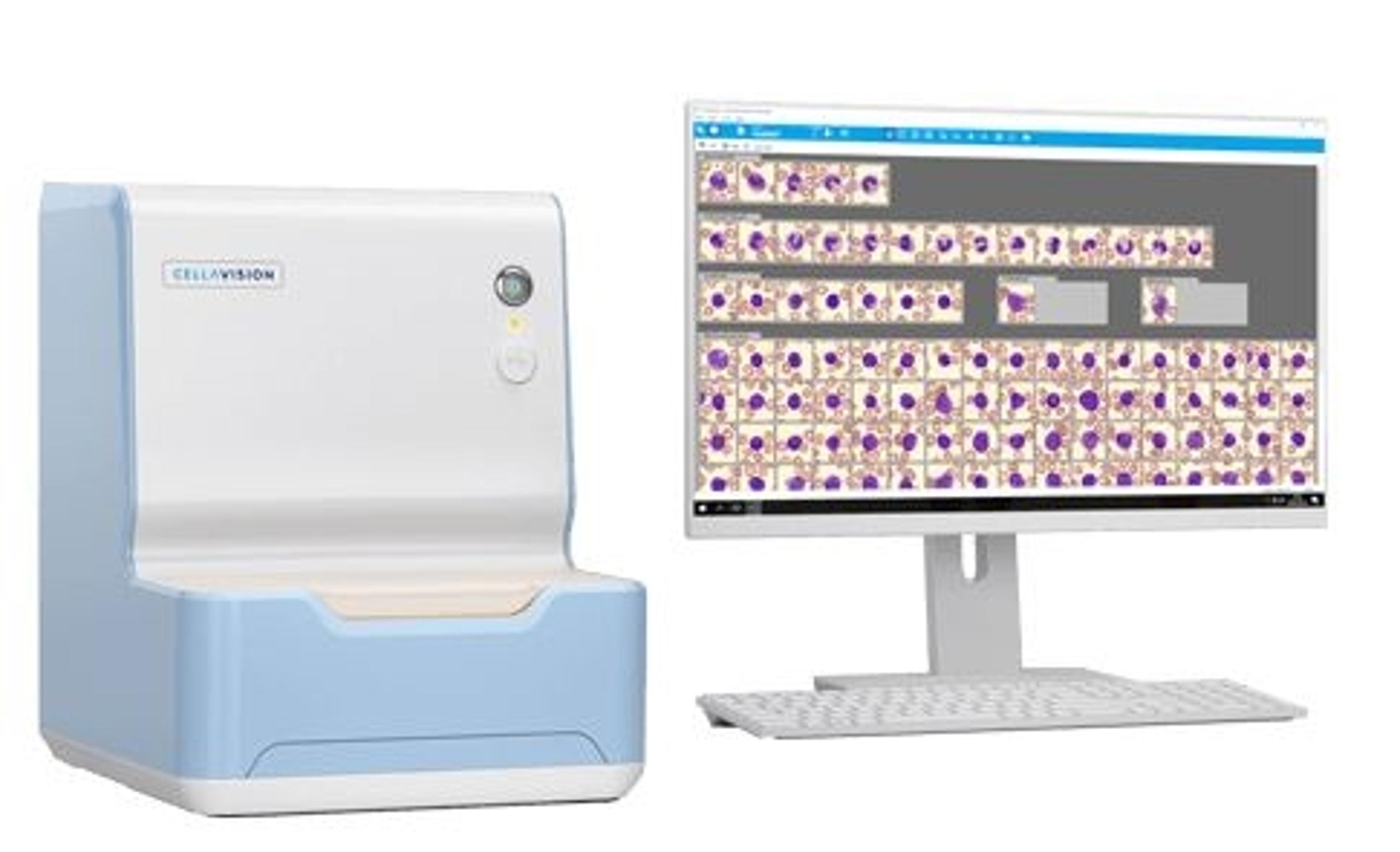
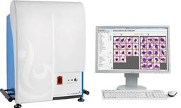
CellaVision® DM1200
Designed for the Mid Volume Lab - the CellaVision® DM1200 fills one of the remaining automation gaps in routine hematology testing. Designed to automate and simplify the process of performing blood and body fluid differentials, the system leverages high-speed robotics and digital imaging to automatically locate and capture high-quality images of cells. Download The Hematology Workflow White Paper When implemented together wit…

Receive your quote directly from the manufacturer.
Features
- Automatically captures digital images of cells from blood smears and body fluid preparations
- Loading Capacity of 12 slides
- Approximately 20 slides throughput / h*
- Creates digital scan of pre-defined area of any interesting specimen
*Processing time may vary depending on smear quality, WBC concentration and number of non-WBCs
How it works
- The system scans and reads the slide barcode and queries the Laboratory Information System for patient demographics and order data.
- The slide is automatically placed under the microscope and the monolayer is identified, cells are located and high-quality cell images are captured.
- When processing is complete, the Medical Technologist is presented with pre-classified white blood cells and pre-characterized red blood cells for review and verification.
The following software and applications are compatible with the CellaVision® DM1200
- CellaVision® Advanced RBC Application
- CellaVision® Body Fluid Application
- CellaVision® Remote Review Software
- CellaVision® Server Software
- CellaVision® Proficiency Software
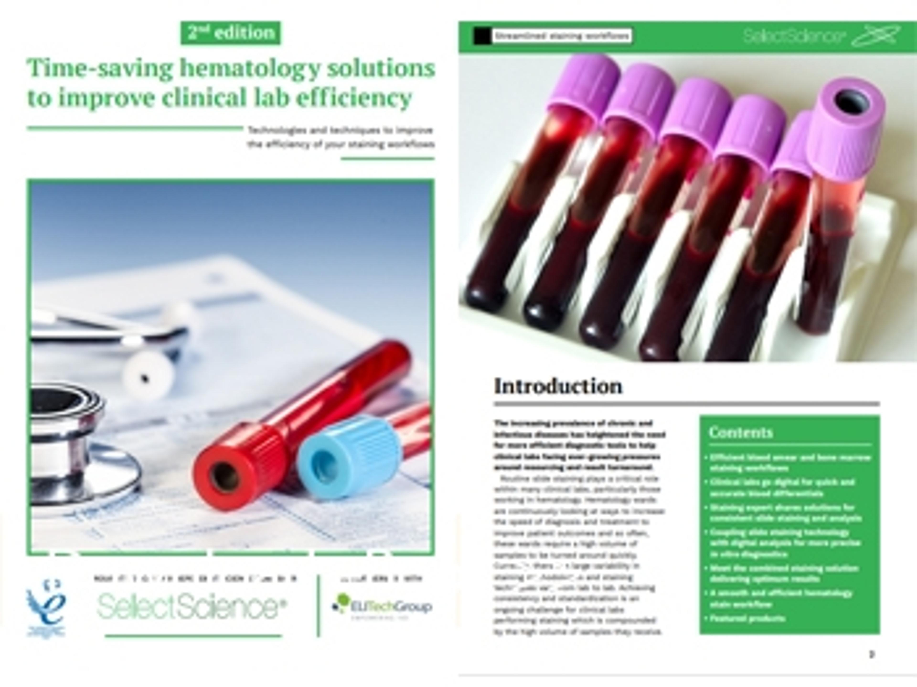
Time-saving hematology solutions to improve clinical lab efficiency
Routine slide staining plays a critical role within many clinical labs, particularly those working in hematology. Currently, there is a large variability in staining methodologies and staining techniques vary from lab to lab. Achieving consistency and standardization is an ongoing challenge for clinical labs performing staining and also looking at ways to accelerate the speed of diagnosis.
In this second edition application eBook, we present case studies and interviews with diagnostics experts and clinical scientists, providing insight and advice on streamlined and standardized staining workflows to help deliver a more precise and timely diagnosis.
Download your copy for free to learn more about:
- Efficient blood smear and bone marrow staining workflows
- Why clinical labs are choosing to go digital for quick and accurate blood differentials
- Coupling slide staining technology with digital analysis for more precise in vitro diagnostics
- Automated solutions for consistent slide staining and analysis

Standardized workflows to improve consistency within the clinical lab
The global in vitro diagnostics (IVD) market is expected to grow at a compound annual growth rate (CAGR) of 5% from 2020 to 2027. As the IVD market continues to expand, so does the prevalence of debilitating chronic and infectious diseases.
An increasingly aging population has heightened the need for highly accurate and standardized diagnostic tools to help better guide clinical decisions, optimize healthcare outcomes, and accelerate clinical laboratory processes.
Featuring exclusive interviews with a team of diagnostic experts, this eBook presents a range of state-of-the-art technologies and techniques designed to streamline and simplify slide staining and analysis. It explores why many clinical labs are now choosing to ‘go digital' in a bid to replace and upgrade current manual, labor-intensive processes and demonstrates how to effectively standardize workflows to improve consistency within the clinical lab setting.