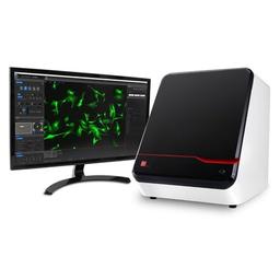
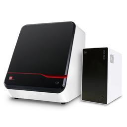
CELENA® X High Content Imaging System
The CELENA ® X High Content Imaging System is an integrated imaging system designed for rapid, high content image acquisition and analysis.

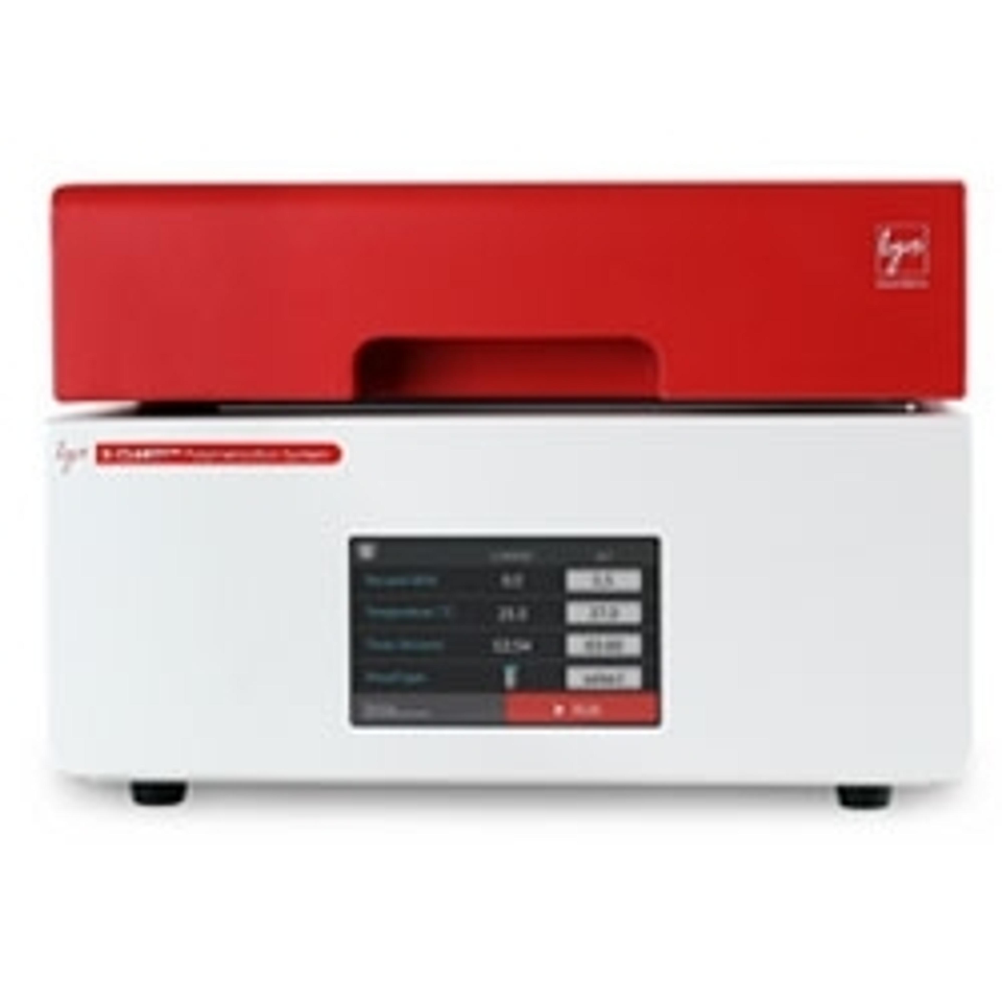
CELENA X High Content Imaging System
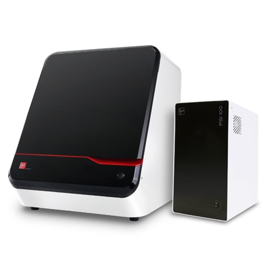
CELENA X High Content Imaging System and Controller

Receive your quote directly from the manufacturer.
Great to use, inexpensive - watch out big microscopy companies...
Cell Culture
The Celena-X is so easy to use, it has huge flexibility and it is oh so affordable making the 'big name brands' look like a rip off!
Review Date: 18 Jul 2021 | Logos Biosystems
Perfect combination of high content and high quality imaging.
High-throughput image acquisition with high quality pictures
The speed of image acquisition is outstanding and the images generated are of high quality. Together with the easy-to-use software, it is a great product.
Review Date: 20 Mar 2020 | Logos Biosystems
The CELENA® X High Content Imaging System is an integrated imaging system designed for rapid, high content image acquisition and analysis. Customizable imaging protocols, image-based and laser autofocusing modules, and a motorized XYZ-stage simplify well plate imaging and slide scanning. The integrated CELENA® X Cell Analyzer software processes images and data for quantitative analysis. Analysis pipelines can be created and used to identify cellular or subcellular objects, process images for optimal data collection, and make various measurements.
SIMPLE HIGH CONTENT IMAGING FOR QUANTITATIVE IMAGE-BASED ANALYSIS
FULLY AUTOMATED PLATE AND SLIDE IMAGING
- · Automated vessel handling and scanning
- · Motorized XYZ stage, filter cube stage, and objective turret
LASER AUTOFOCUS
- · Rapid and reproducible focusing
- · Minimized phototoxicity and photobleaching
LIVE CELL ASSAY SUPPORT
Onstage incubation system for a variety of experiments in physiological and non-physiological conditions
FOUR IMAGING MODES
Fluorescence imaging in four channels, brightfield, color brightfield, and phase contrast imaging
POWERFUL, EASY-TO-USE USER INTERFACE
- Simple setup of imaging protocols
- Seamless integration of imaging and data analysis processes
CUSTOMIZABLE HIGH CONTENT ANALYSIS
- Create and customize image analysis projects
- Quantitatively analyze multiple image-based phenotypes
CREATE YOUR OWN IMAGING AND ANALYSIS WORKFLOWS
CELENA® X Cell Analyzer can be used to set up automated image analysis sequences to batch process images captured on the CELENA® X. Using Cell Analyzer, users can create an image analysis pipeline, which is a sequence of modules that each perform a specific image processing task. This allows the quantitative analysis of multiple cellular features from images. Modules can be mixed, matched, and adjusted to measure phenotypes of interest quantitatively. Once a pipeline has been established, it can be used to analyze subsequent projects.
- Multicolor fluorescence and brightfield imaging
- Long-lasting LEDs and hard-coated optical filters ensure robust fluorescence imaging. Adjustable LEDs allow precise control over the gain and intensity of transmitted light.
- Onboard data analysis
- Analyze your images immediately upon capture. Save measurement data to a USB drive.
- Live cell monitoring
- Monitor live cells with the time lapse function or the growth monitor. Attach the onstage incubator to control the temperature, humidity, and CO₂/O₂ levels.
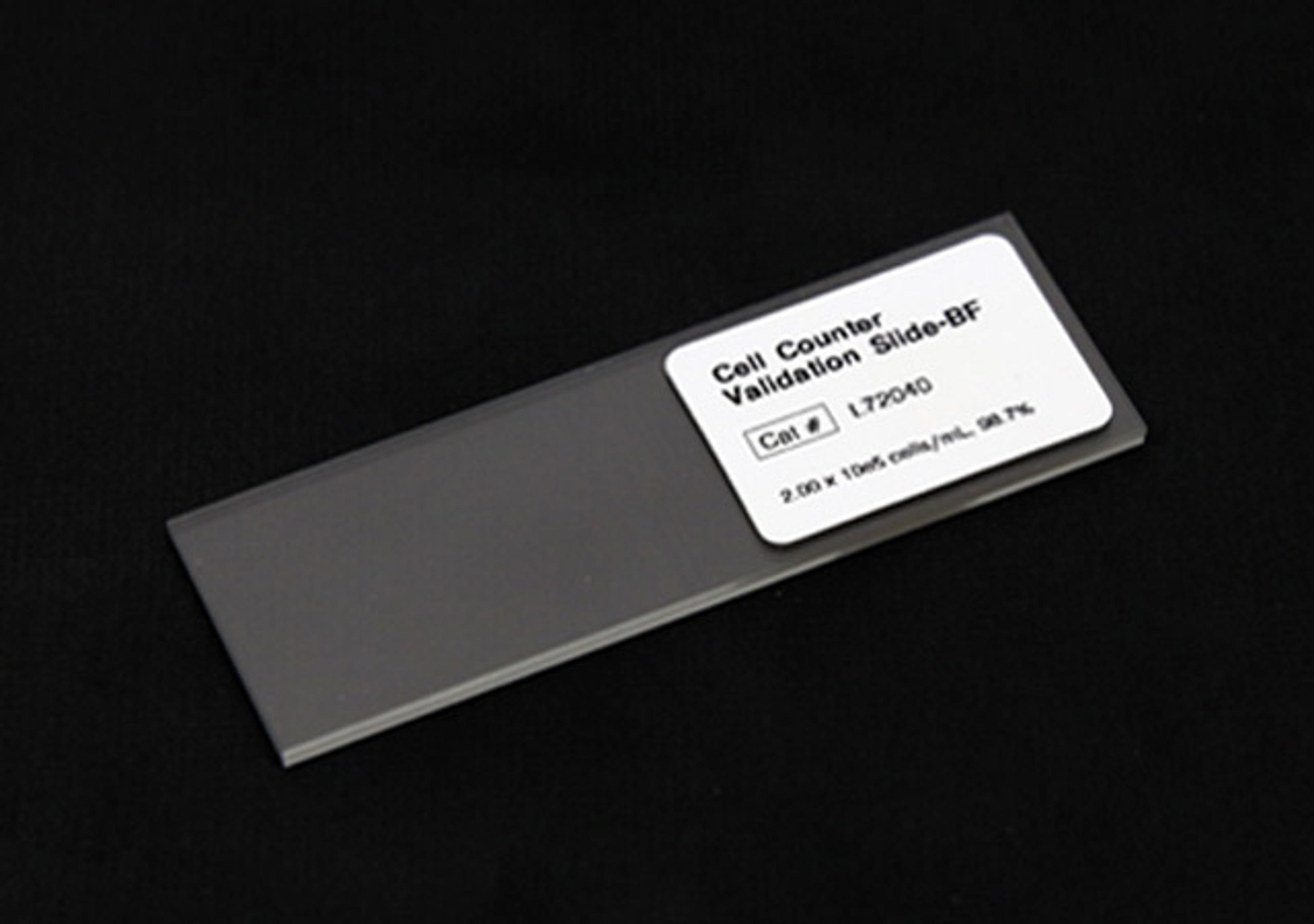
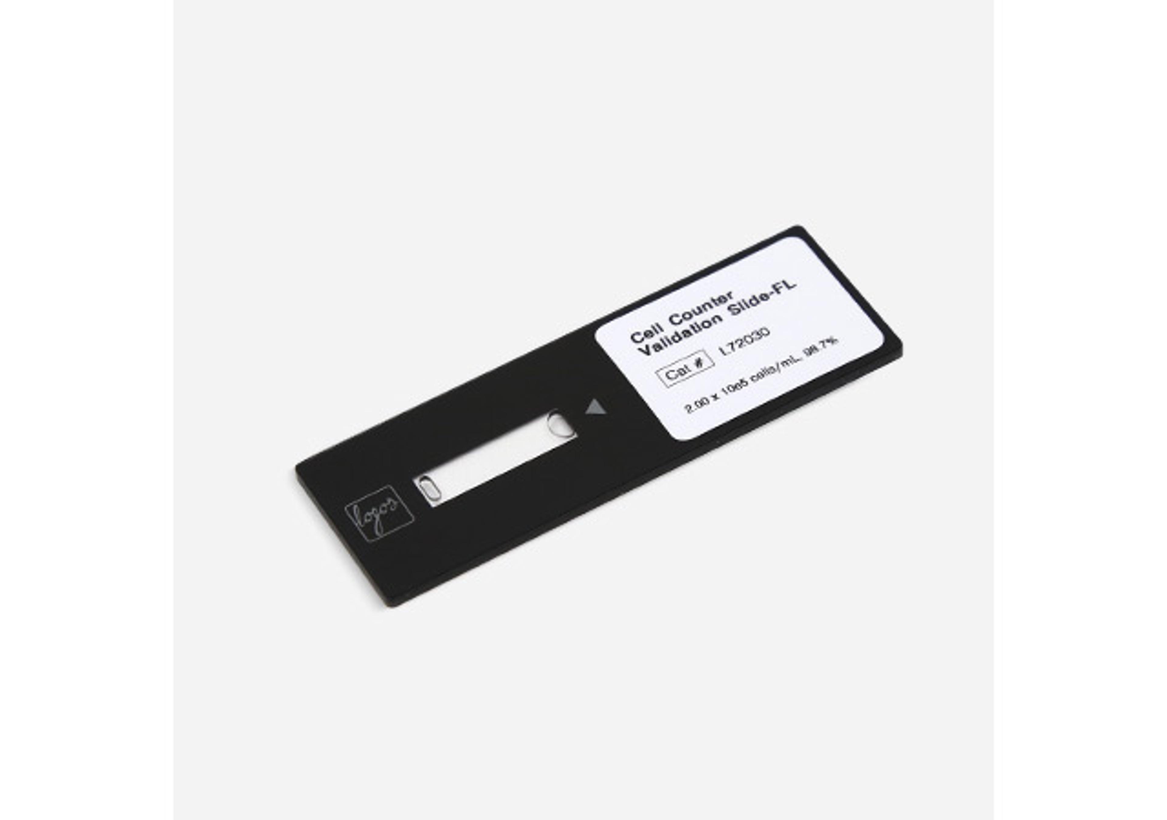
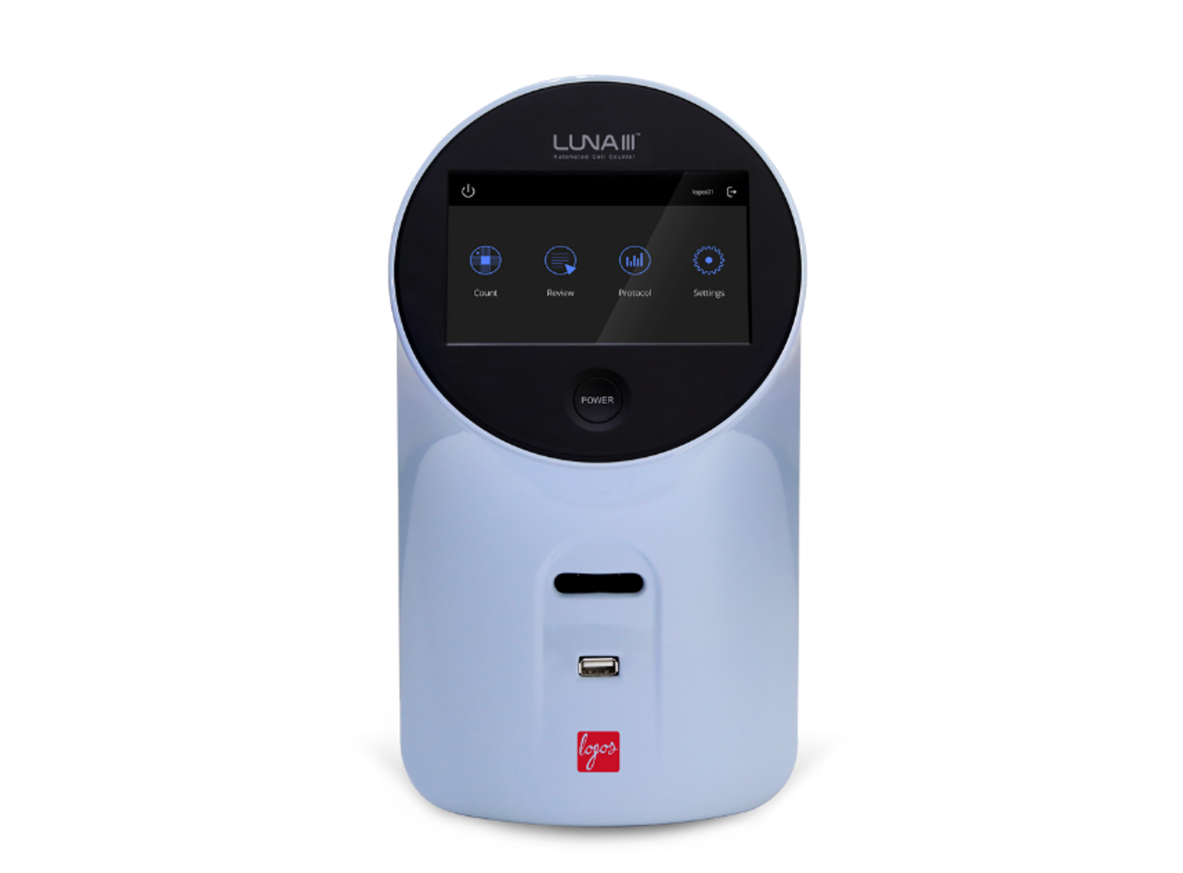
- Automated cell count and viability analysis
- Check cell counts and viability with the onboard cell counter
- Z-stack imaging
- Capture multiple images along the Z-axis with the Z-stack function.
Brochures

CELENA® X:The Complete Solution for High Content Imaging and Analysis
Say hello to the all-new CELENA® X High Content Imaging System, the most affordable solution for rapid high content image acquisition and analysis. Customizable imaging protocols, image-based and laser autofocusing modules, and a motorized XYZ stage simplify well plate imaging and slide scanning. Powerful and flexible software allows you to set up advanced image analysis sequences that can be used to quantitatively analyze numerous cellular features for the simplest fixed cell assays to more complicated, time-lapse live cell assays. With the power to capture and quantify cellular information in both fixed and live cells, the CELENA®X is a valuable tool for life science research as well as drug discovery and development.
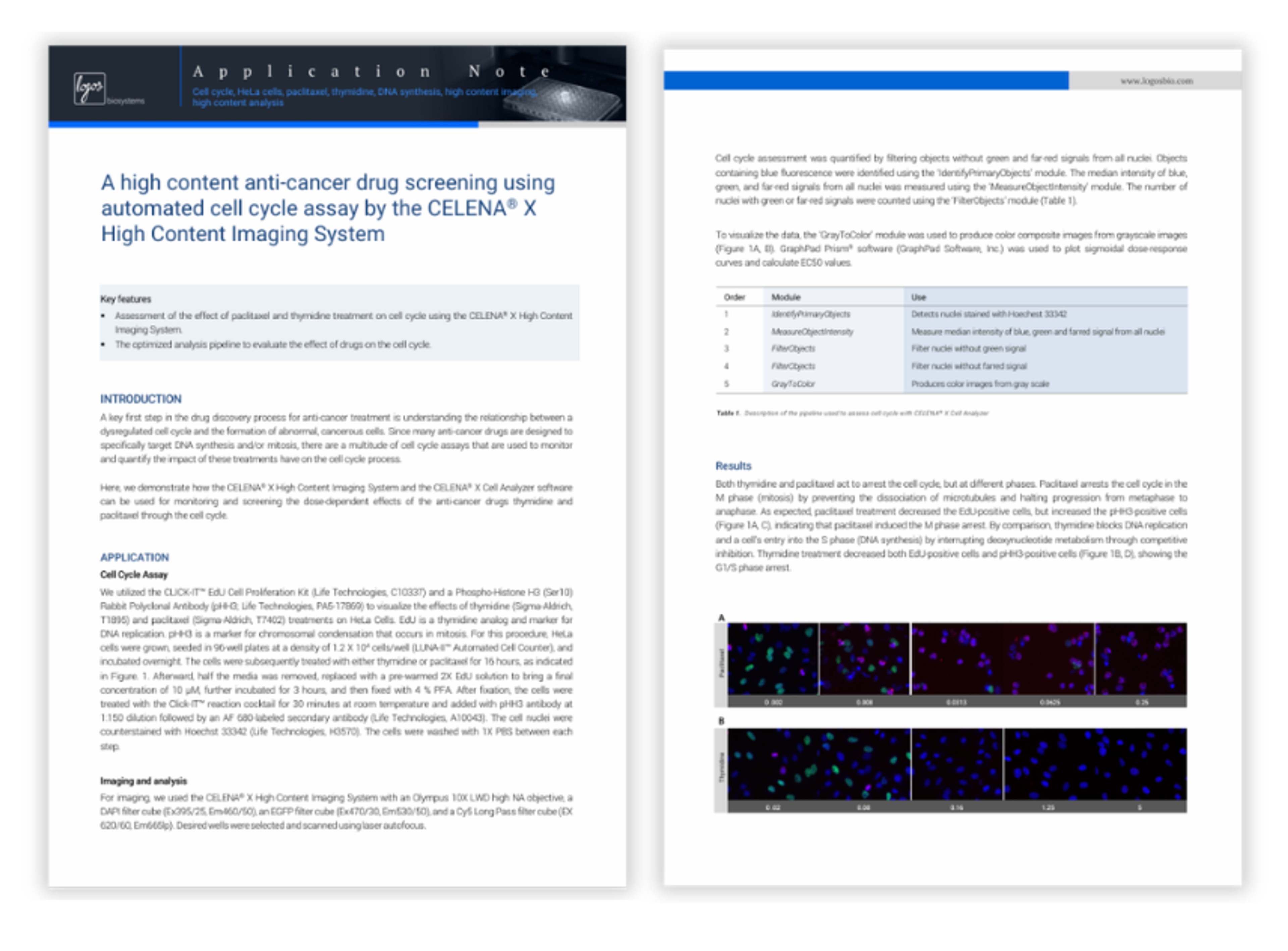
A high content anti-cancer drug screening using automated cell cycle assay by the CELENA X High Content Imaging System
This application note demonstrates how the CELENA® X High Content Imaging System can be used to perform automated cell cycle assays for anti-cancer drug screening. Using fluorescence imaging and analysis pipelines, the system enables evaluation of dose-dependent effects of compounds such as paclitaxel and thymidine on cell cycle progression. The workflow supports high-throughput, quantitative analysis of drug responses, helping accelerate early-stage drug discovery.
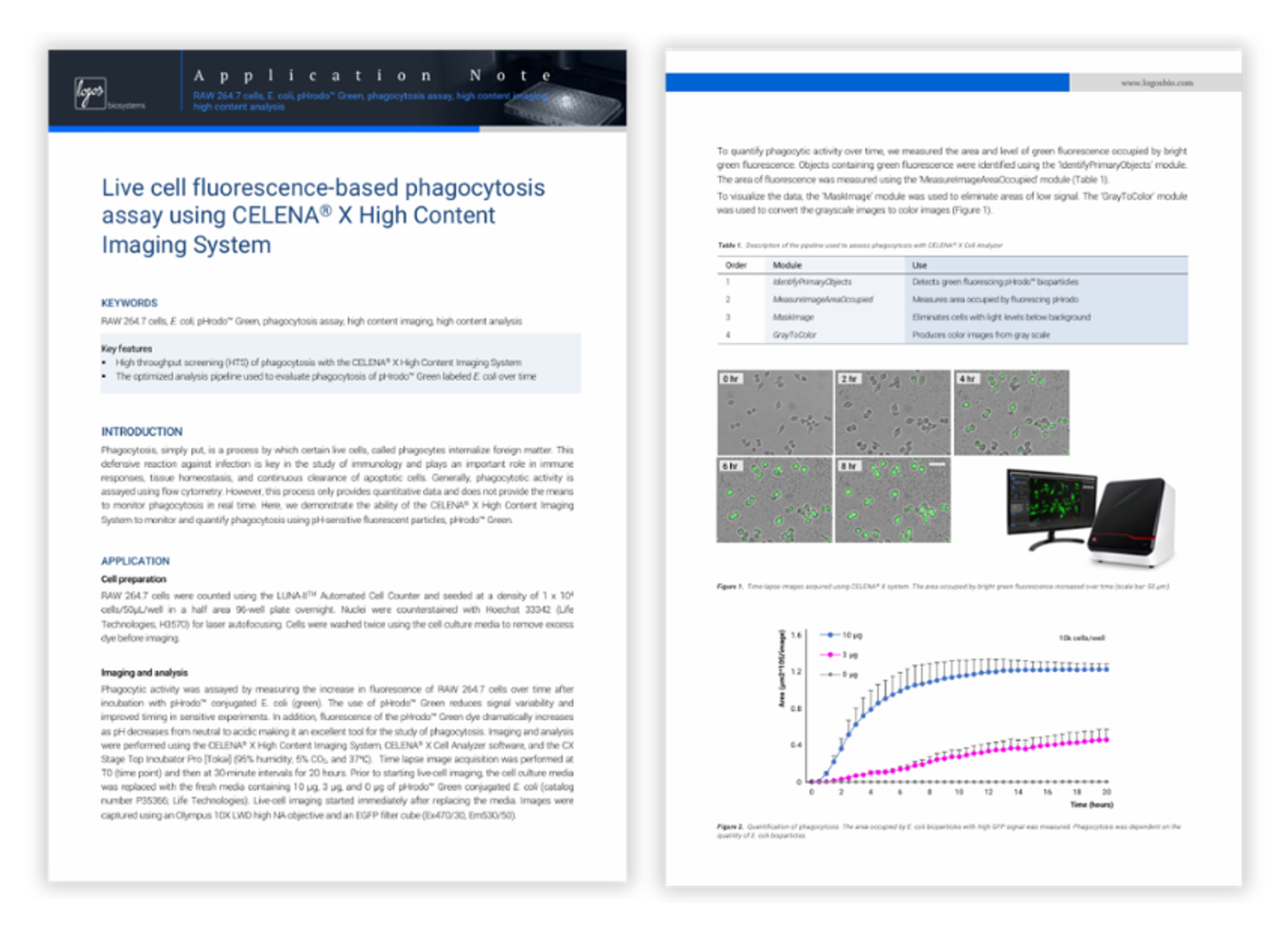
Live cell fluorescence-based phagocytosis assay using CELENA X High Content Imaging System
This application note demonstrates how the CELENA® X High Content Imaging System can be used to monitor and quantify phagocytosis in live cells using fluorescence-based assays. By tracking the uptake of pH-sensitive fluorescent E. coli particles over time, the workflow enables real-time visualization and measurement of phagocytic activity.
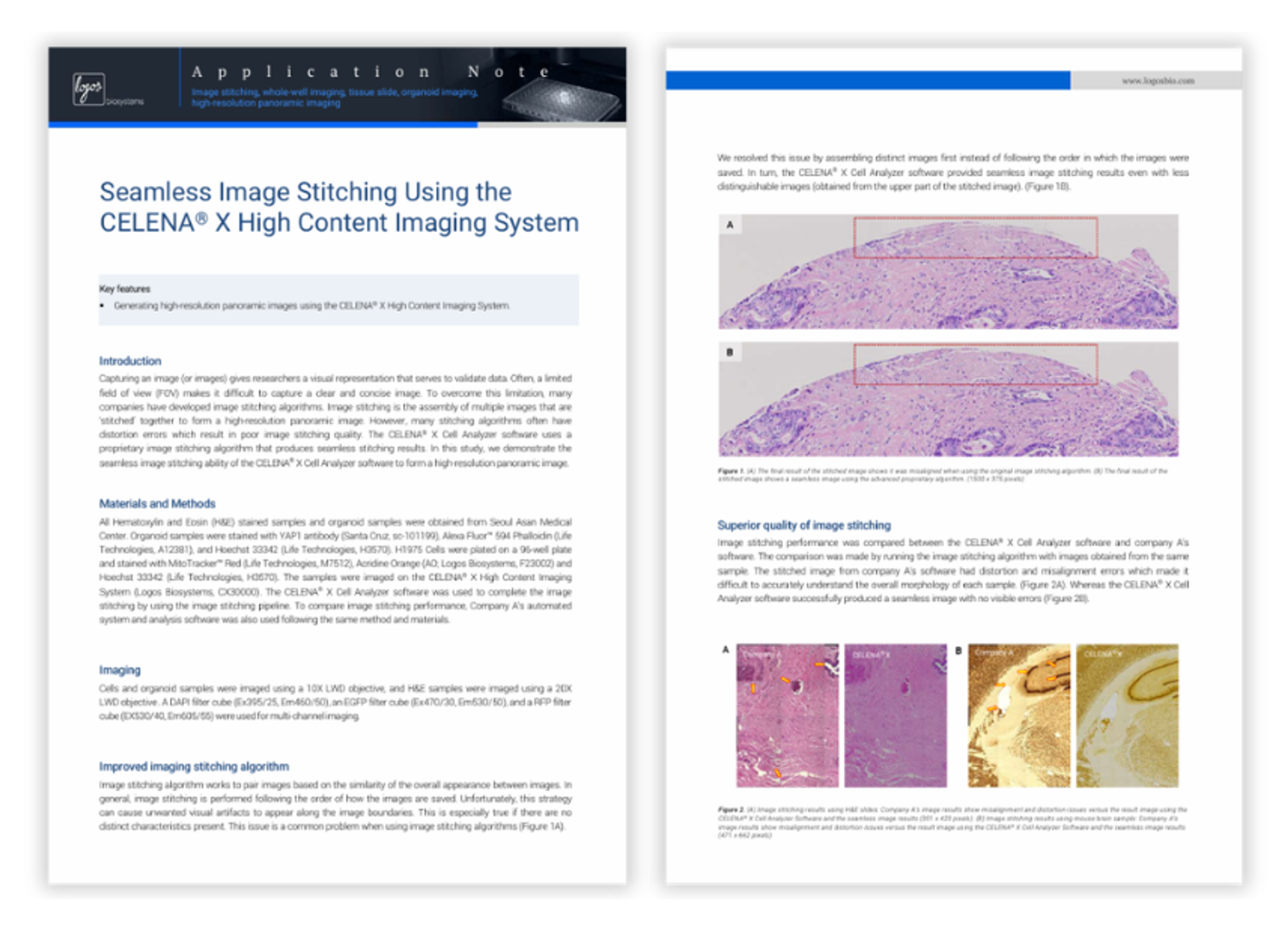
Seamless image stitching using the CELENA X High Content Imaging System
This application note demonstrates how the CELENA® X High Content Imaging System from Logos Biosystems enables seamless image stitching to generate high-resolution panoramic images from multiple fields of view. By using an advanced stitching algorithm, the system reduces distortion and misalignment commonly seen in conventional approaches, improving image quality and interpretability. The workflow supports whole-well imaging and multi-channel analysis, making it suitable for applications such as tissue imaging and organoid studies.
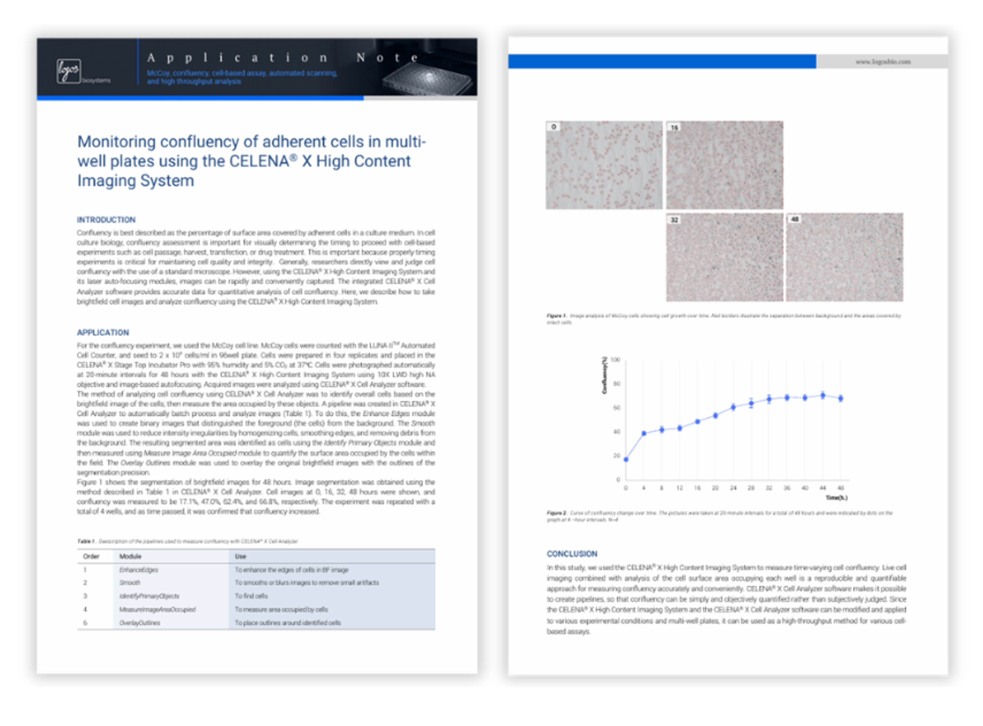
Monitoring confluency of adherent cells in multi well plates using the CELENA X High Content Imaging System
This application note demonstrates how the CELENA® X High Content Imaging System from Logos Biosystems can be used to monitor and quantify cell confluency in multi-well plates. Using automated brightfield imaging and image analysis pipelines, the system enables accurate measurement of cell coverage over time, supporting reproducible and objective assessment of cell growth. Discover how the workflow allows high-throughput analysis and reduces reliance on subjective visual estimation in cell culture experiments.
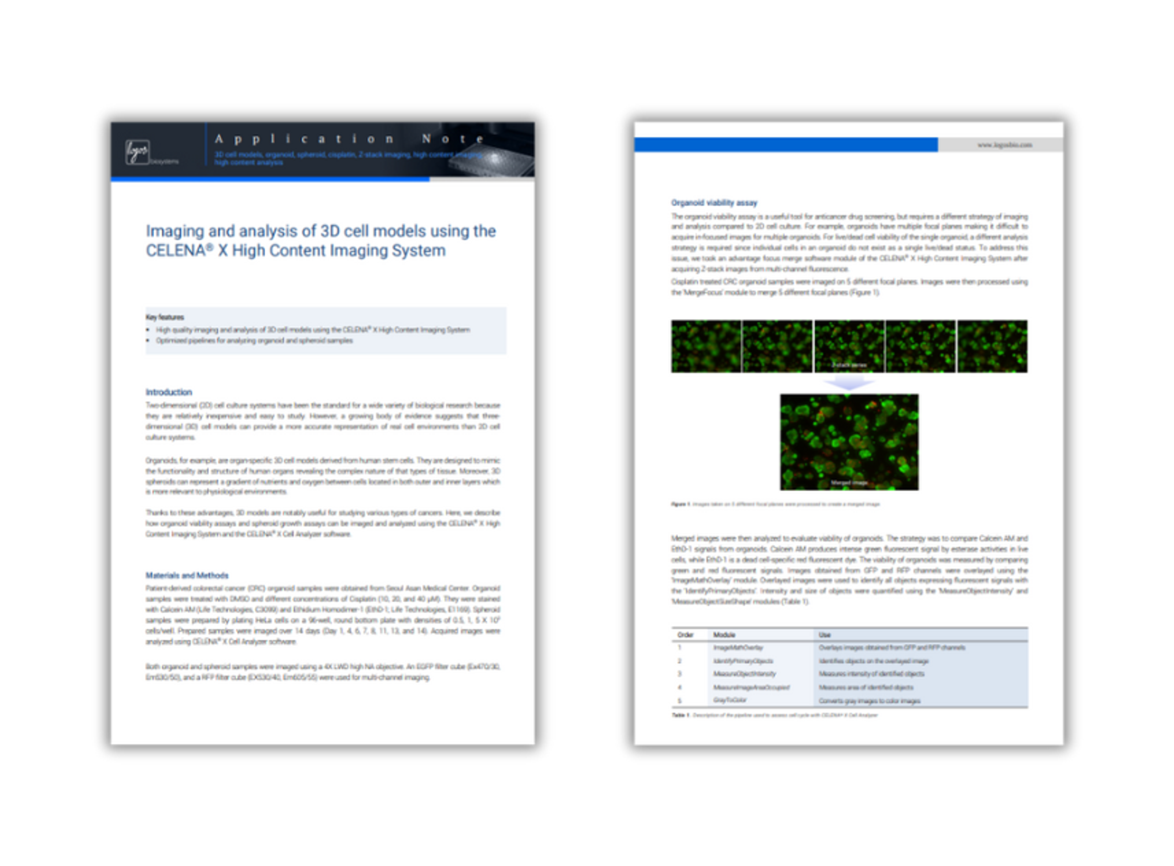
Imaging and analysis of 3D cell models using the CELENA X High Content Imaging System
Two-dimensional (2D) cell cultures have long been used in research due to their simplicity and low cost. However, three-dimensional (3D) cell models offer a more physiologically relevant environment, better mimicking in vivo conditions. Organoids, derived from human stem cells, replicate the structure and function of human organs, while 3D spheroids reflect nutrient and oxygen gradients similar to those in tissues. These models are particularly valuable in cancer research. This article outlines how organoid viability and spheroid growth can be imaged and analyzed using the CELENA® X High Content Imaging System and Cell Analyzer software.
High content imaging analysis for basic and applied research
High content imaging analysis systems have limitations that have prevented smaller labs – including basic research labs – from gaining access to them. Issues with how image analysis software handles very large datasets, large footprints, and prohibitively high costs have all proved problematic.
In this eBook, we take a look at the CELENA® X High Content Imaging System from Logos Biosystems to help you understand how its integrated image analysis software, benchtop capability, affordable cost and versatility can fulfil the high content imaging needs of any small laboratory.
Download this free eBook to learn more about:
- Apoptosis and phagocytosis assays
- Monitoring confluency of adherent cells in multi-well plates
- A high content anti-cancer drug screening using automated cell cycle assay
- Fluorescence-based Calcium Mobilization Assay
- Dose-dependent cytotoxicity of Camptothecin on HeLa cells
- Evaluating transfection efficiency
- Counting adherent cells in multi-well plates
- Wound healing analysis based on image segmentation
- Non-destructive quantification of cytotoxicity in live HeLa cells
Non-Destructive Quantification of Cytotoxicity in Live HeLa Cells
Cytotoxicity assays are a crucial step in screening for and developing therapeutic anti-cancer drugs. Most assays designed to measure cytotoxicity in vitro evaluate cell membrane integrity or metabolic activity after exposure, but are typically based on studying a single time point and require disturbing the growth of cells in culture. This application note demonstrates an automated, non-destructive method to monitor and quantify cytotoxicity based on its effects on confluency.
Automated Cell Counting: Achieving higher throughput and accuracy from the bench to the cleanroom
Implementing automation solutions to carry out time-consuming and error-prone tasks is key to optimizing processes.
In this webinar, Tim Steppe, Ph.D., application specialist at Logos Biosystems will demonstrate how the LUNA-FX7™ Automated Cell Counter can be used in a breadth of applications from CAR-T cell therapy development, to single cell sequencing. Plus, learn how the LUNA-FX7™, with the added 21 CFR-compliant CountWire™ system, can be incorporated into development and production processes that need to be carried out within a regulated environment.
Key learning objectives
- Learn about the versatility and accuracy of the LUNA-FX7™ for disparate cell counting sample types and applications.
- Understand how higher throughput and increased accuracy may be achieved in cell counting.
- Learn how the LUNA-FX7™ can be used in to ensure quality and efficiency in single cell sequencing.
- Understand the ease and simplicity of establishing a cell counting process that is 21 CFR Part 11 compliant.
Who should attend?
- Quality Control/Quality Assurance managers
- Biopharmaceutical/Biomedical/Biotech decision-makers/researchers/scientists
- Discovery developers/Production & Processing Operators
Certificate of attendance
All webinar participants can request a certificate of attendance, including a learning outcomes summary, for continuing education purposes.