The role of automated live cell imaging with the Celloger Pro
Expand horizons in life science research and discover the power of real-time cell monitoring
19 Mar 2024
Product news
In the realm of life science research, live cell imaging represents a transformative approach that allows scientists to observe and analyze cellular processes in real-time, within their natural environment. Unlike traditional fixed-cell imaging techniques, which capture snapshots of cellular structures at a single moment in time, live cell imaging offers dynamic insights into the behavior, interactions, and responses of cells over extended periods.
Live cell imaging holds immense significance across various fields of biology, ranging from developmental biology to neuroscience and beyond. By visualizing cellular dynamics as they unfold, researchers can investigate a multitude of phenomena, including cell division, migration, signaling, and responses to external stimuli. This capability not only enhances our understanding of fundamental biological processes but also provides crucial insights into disease mechanisms, drug responses, and therapeutic interventions.
However, the process of live cell imaging can be inherently complex and labor-intensive, requiring meticulous attention to experimental conditions, sample preparation, and imaging parameters. Moreover, the need for continuous observation over extended periods presents logistical challenges, as manual intervention may introduce variability and compromise data integrity.
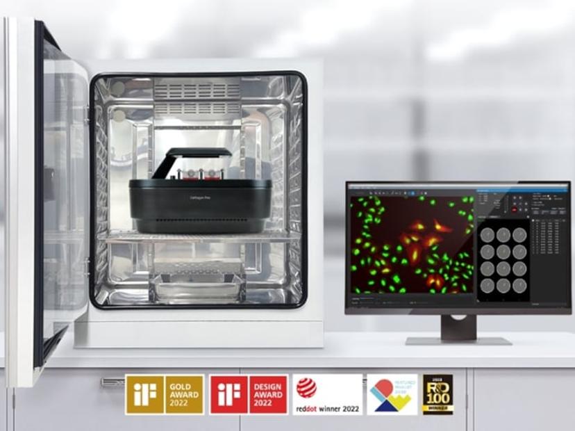
This is where automation plays a pivotal role in revolutionizing live cell imaging. Automated imaging systems, such as the Celloger® Pro, streamline the experimental workflow by integrating automatic camera, imaging technologies, and data analysis algorithms. It has been designed to simplify live cell imaging with its automated capabilities, reducing manual intervention and ensuring consistent and reliable results.
The Celloger Pro promises to streamline real-time cell monitoring within an incubator, minimizing sampling variability and ensuring precise data on the functions and development of live cells. Its dual fluorescence microscopy capability enables high-resolution imaging, allowing for the simultaneous observation of multiple markers. Multi-point time-lapse imaging captures cellular events across different locations, facilitating in-depth feature analysis. With user-interchangeable objective lenses catering to various magnifications, customization is enhanced, and the software is designed with user-friendly tools for data analysis, such as stitching, z-stacking, confluency, intensity graphing, and cell counting functions.
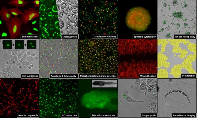
Key applications using the Celloger Pro
- Spheroid formation: Utilizing the Celloger Pro, researchers can observe and analyze the formation and growth dynamics of multicellular spheroids. The ability to continuously monitor spheroid development facilitates studies on tumor biology, drug penetration, and therapeutic efficacy in a more physiologically relevant context.
- Cell proliferation assay: With the Celloger Pro, researchers can track changes in cell confluency and morphology over time, enabling precise measurements of cell proliferation dynamics. The system's automated imaging and analysis features streamline the process, providing accurate data on cell growth rates and responses to various stimuli.
- Wound healing assay: Continuous imaging of wound healing assays allows researchers to understand the cellular and molecular processes driving wound closure and tissue regeneration. This analysis is crucial for assessing collective cell migration under varying experimental conditions, highlighting the Celloger Pro as an indispensable tool in wound healing research.
- Cytotoxicity assay: Assessing the cytotoxic effects of compounds on living cells is essential in cancer research and drug screening. The Celloger Pro enables the continuous monitoring of cellular viability and morphology, providing insights into the potential adverse effects of chemical agents.
- Apoptosis assay: The Celloger Pro allows for the visualization of apoptotic events such as cell shrinkage, membrane blebbing, and nuclear condensation. This capability is crucial for studying programmed cell death mechanisms, understanding disease processes, and evaluating the efficacy of anti-cancer drugs.
- Phagocytosis monitoring: Phagocytosis monitoring in immunology and infectious disease research involves observing how cells engulf foreign particles or pathogens, revealing crucial mechanisms in immune responses. The Celloger Pro facilitates real-time monitoring of phagocytic activity, offering invaluable data for understanding the dynamics of immune responses.
Want the latest science news straight to your inbox? Become a SelectScience member for free today>>
Related Products
Request Quote for All Products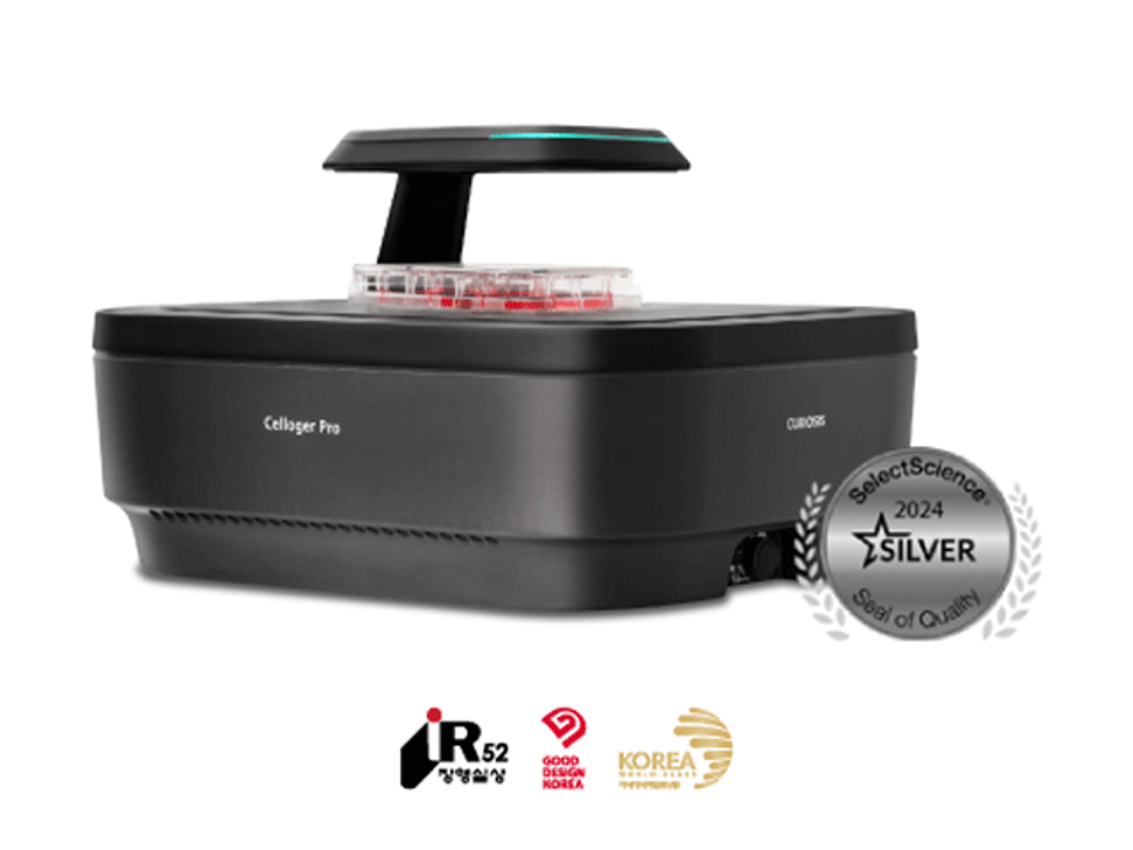
Celloger® Pro, Automated Live Cell Imaging System from Curiosis
CURIOSISWith its streamlined workflow and versatile capabilities, Celloger® Pro is a valuable tool for researchers seeking efficient, accurate, and cost-effective cellular analysis.