Rigaku to Present Nano3DX and CT Lab at XRM 2016
New X-ray microscopy and computed tomography technology to be showcased at conference
7 Aug 2016
Rigaku Corporation, will be presenting its lines of X-ray microscopy and computed tomography instrumentation at the 2016 X-Ray Microscopy Conference (XRM 2016), Monday, August 15, through Friday, August 19, 2016 at the University of Oxford in the U.K.
X-ray microscopy and computed tomography equipment from Rigaku enable nondestructive analysis of large samples at high resolution. X-ray microscopy (XRM) is suited to all kinds of materials, from low-density materials such as biological samples to high-density materials such as ceramics and steels. Computed tomography (CT) reveals, at high-speed, the high-resolution, 3D structure of an object by means of computer-processed combinations of numerous X-ray images taken from different angles.
The International Conference on X-Ray Microscopy will bring together experts in the development and use of X-ray microscopes and will address recent advances in X-ray microscopy and its applications via a dynamic program of discussions and posters. XRM 2016 will be the 13th conference in the X-ray Microscopy series, which began in 1983.
Related Products
Request Quote for All Products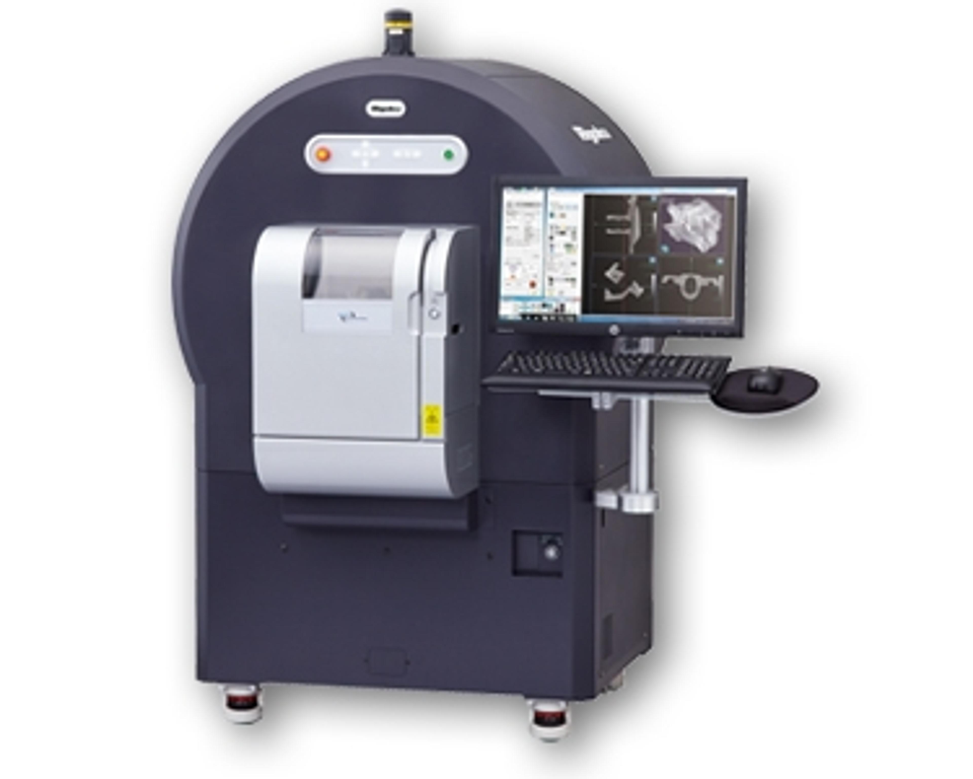
nano3DX XRM
Rigaku CorporationRigaku nano3DX is a true X-ray microscope (XRM) with the ability to measure relatively large samples at high resolution. This is accomplished by using a high powered rotating anode X-ray source and a high-resolution CCD imager. The rotating anode provides for fast data acquisition and the ability to switch anode materials easily to optimize the data acquisition. AVAILABILITY: Japan, Austraila/NZ and North AmericaThe new nano3DX allows you see into many types of samples, including those that have low absorption contrast, for example CFRP, or denser materials like ceramic composites. The nano3DX allows you to achieve this by providing the ability to change the X-ray wavelength to enhance contrast or penetration.In the nano3DX, the magnification takes place in the detector using true microscope elements. This design places the sample close to a high-resolution detector, allowing for a near-parallel beam experiment. This means greater instrument stability and shorter data collection times providing the highest resolution of any X-ray microscope in its class.The nano3DX design is a vast improvement over older implementations that use a small source and a long sample-to-detector distance. This geometric magnification requires a very small source and extreme stability to prevent smearing. Data acquisition times can be quite long because small sources are also low power.nano3DX XRM Features: Ultra-wide field of view, 25X larger volume than comparable systems 3 X-ray wavelengths (Cr, Cu and Mo Ka) to optimize imaging for different sample matrices Parallel beam geometry for high contrast and rapid data collection Auto 5-axis (XYZ and rotation) stage and on-axis imaging system High resolution three dimensional (3D) images High power rotating anode X-ray source High contrast for low-Z materials High-resolution CCD imager