Powerful Analysis in Pint-Sized Packaging from JEOL USA
26 Mar 2019JEOL USA has introduced 4th generation benchtop Scanning Electron Microscope (SEM) that delivers many powerful features of a full-sized SEM in a small package.
The new JEOL NeoScope™ (model JCM-7000) was demonstrated in booth #3035 at Pittcon 2019 in Philadelphia. This benchtop SEM’s advanced technology and functions make it simple for users at any skill level to obtain outstanding SEM images and elemental analysis results in minutes.
The new NeoScope produces high magnification up to 100,000X with large depth of field. It features a large sample chamber, high and low vacuum modes, secondary and backscatter electron detectors, real-time 3D imaging, highly-advanced auto functions and the option to add a fully-embedded EDS with real-time, ‘Live’ analysis.
“Zeromag” seamless navigation
This 4th generation benchtop SEM utilizes “Zeromag” a feature first introduced in full-sized JEOL tungsten SEMs. The microscope seamlessly navigates across a sample to automatically focus on areas of interest and quickly achieve high resolution SEM imaging and analysis.
Live analysis
With our embedded Energy Dispersive X-ray detector (EDS), the EDS spectrum and major elements that comprise a sample are automatically detected and displayed in real time.
Live 3D imaging
In addition to live SEM images, the NeoScope can display live 3D images of the sample surface, including valuable depth information about the sample.
Automatic Operating Features
Automatic condition setting based on sample type and application ensures high-quality results and enhances productivity. Highly-advanced auto functions generate images of exceptional fidelity.to high resolution SEM imaging and analysis.
Automated features include focus, alignment, contrast, and brightness for easy operation and application to a variety of samples.
SEM for Anyone
The NeoScope meets the need for a simple-to-operate small Scanning Electron Microscope, yet offers several key performance features of a high-end instrument. The NeoScope is the ideal teaching tool for schools, a screening instrument for quality control, and a flexible small research microscope.
All the highlights from Pittcon 2019 here >>
Related Products
Request Quote for All Products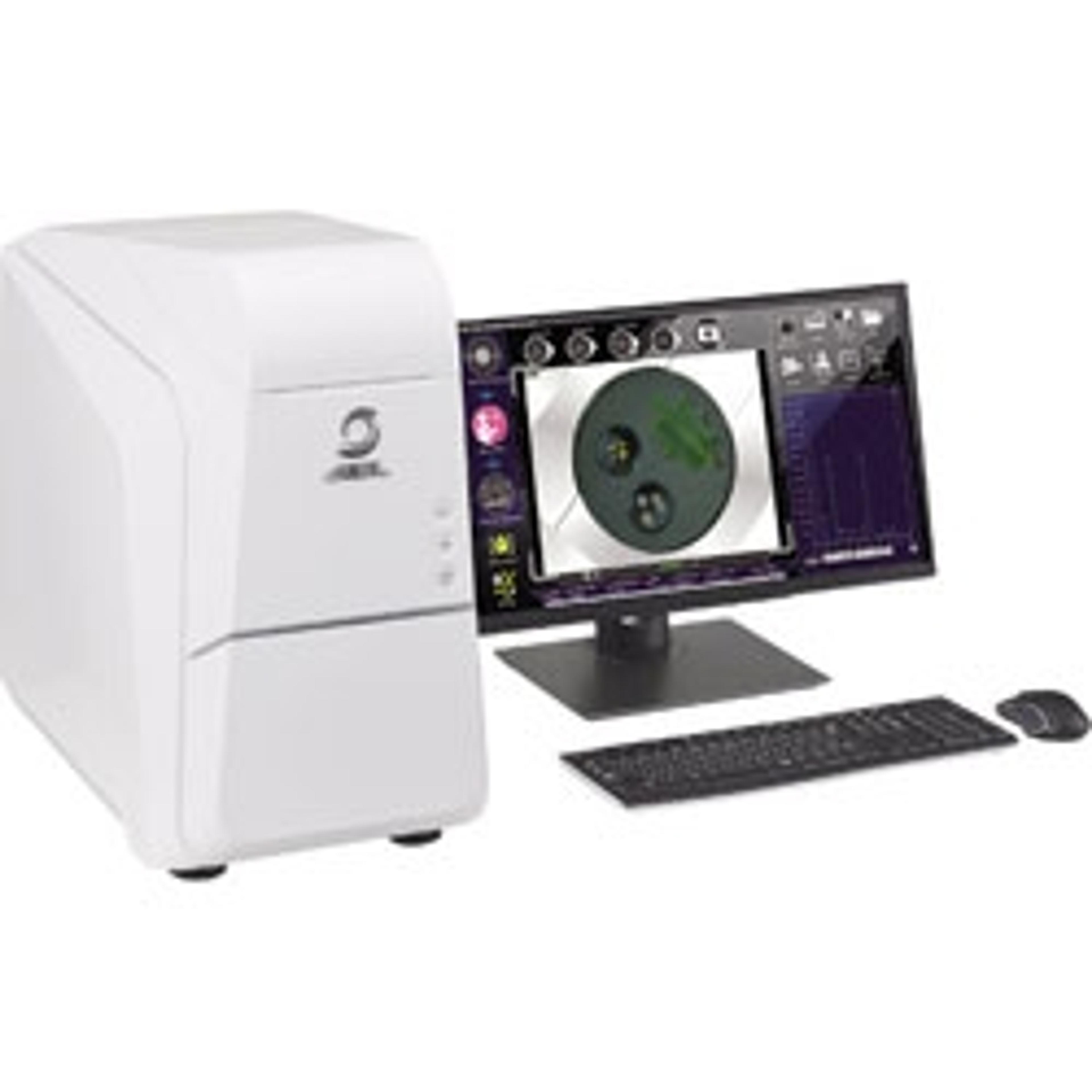
JCM-7000 NeoScope Benchtop SEM
JEOL USAThis 4th generation Neoscope incorporates advanced technology and functions that make it simple for users at any skill level to obtain outstanding SEM images and elemental analysis results in minutes. It is equipped with a large chamber, high and low vacuum modes, secondary and backscatter electron detectors, real-time 3D imaging, highly-advanced auto functions and the option to add a fully embedded EDS with real-time, ‘Live’ analysis. This 4th generation NeoScope™ is SMART-FLEXIBLE-POWERFUL. Smart –The latest innovations built to our benchtop platform make this SEM accessible to everyone. Seamless navigation across the sample allows you to quickly go from an optical image to high resolution SEM imaging and analysis. Automatic condition setting based on sample type and application ensures high quality results and enhances productivity. Highly-advanced auto functions generate images of exceptional fidelity. Flexible – Choose a platform that is right for you. Add options such as our Stage Navigation System (color camera), fully-embedded EDS for elemental analysis and Smile View Map for 3D image reconstruction and surface texture analysis. Powerful – The high resolution W filament source allows magnification up to 100,000X. A benchtop SEM with both secondary electron and backscatter electron detectors, plus high and low vacuum modes allow for the study of a wide variety of sample types. Automated montage is built-in for high resolution view over a larger area. Includes montage X-ray map with EDS option. The BSE detector supports live 3D imaging for intuitive knowledge of sample surface shape. LIVE ANALYSIS Our analytical model includes JEOL’s fully embedded EDS system which provides real time EDS spectra during image observation. With Live Analysis you can: View EDS spectra in real time as you search for the area of interest. Set analysis points, areas, map position and line scan from the live image observation screen. View major elements as displayed on the live EDS window.