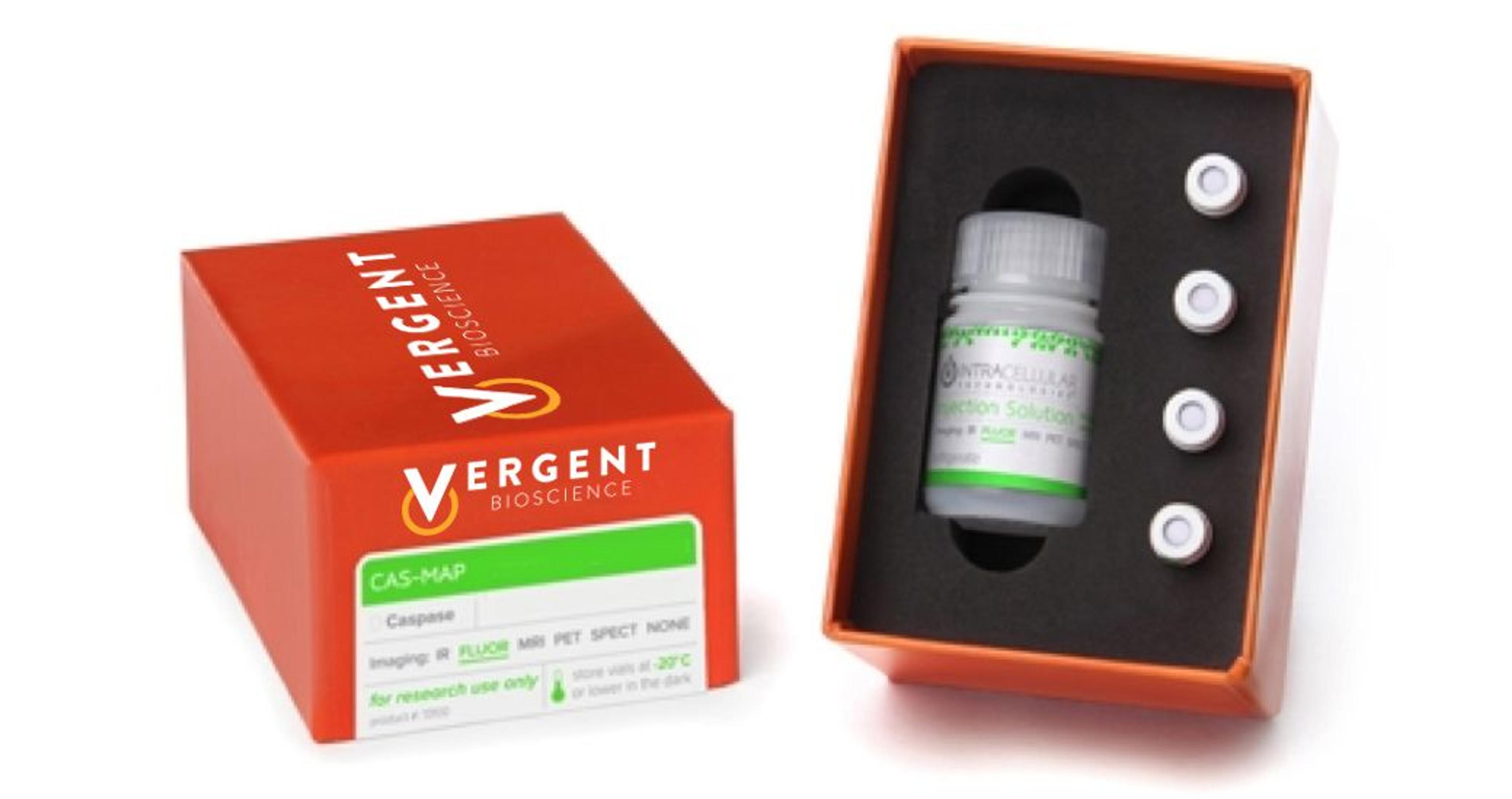
CAS-MAP ProBo Pan Caspase (VAD-FMK) Red Real Time Apoptosis Probe (in vivo)
Vergent BioscienceRed Fluorescent non-invasive imaging probe for in vivo detection of apoptosis (pan caspase)
<i>In vivo</i> imaging systems, including pre-clinical imaging systems and medical imaging systems are used to non-invasively visualize and capture images of live animals and plants. Monitor the natural processes or diseases of your subjects using small-animal pre-clinical imaging systems, including single photon positron emission tomography (SPECT), positron emission tomography (PET), computed tomography (micro-CT), magnetic resonance imaging (MRI), X-ray radiography, ultrasound, fluorescence and bioluminescence imagers. Multimodal systems and software solutions are also available for correlative analysis of organ, tissue, cell, or molecular-level processes. Find the best in vivo imaging products in our peer-reviewed product directory: compare products, check customer reviews and receive pricing direct from manufacturers.
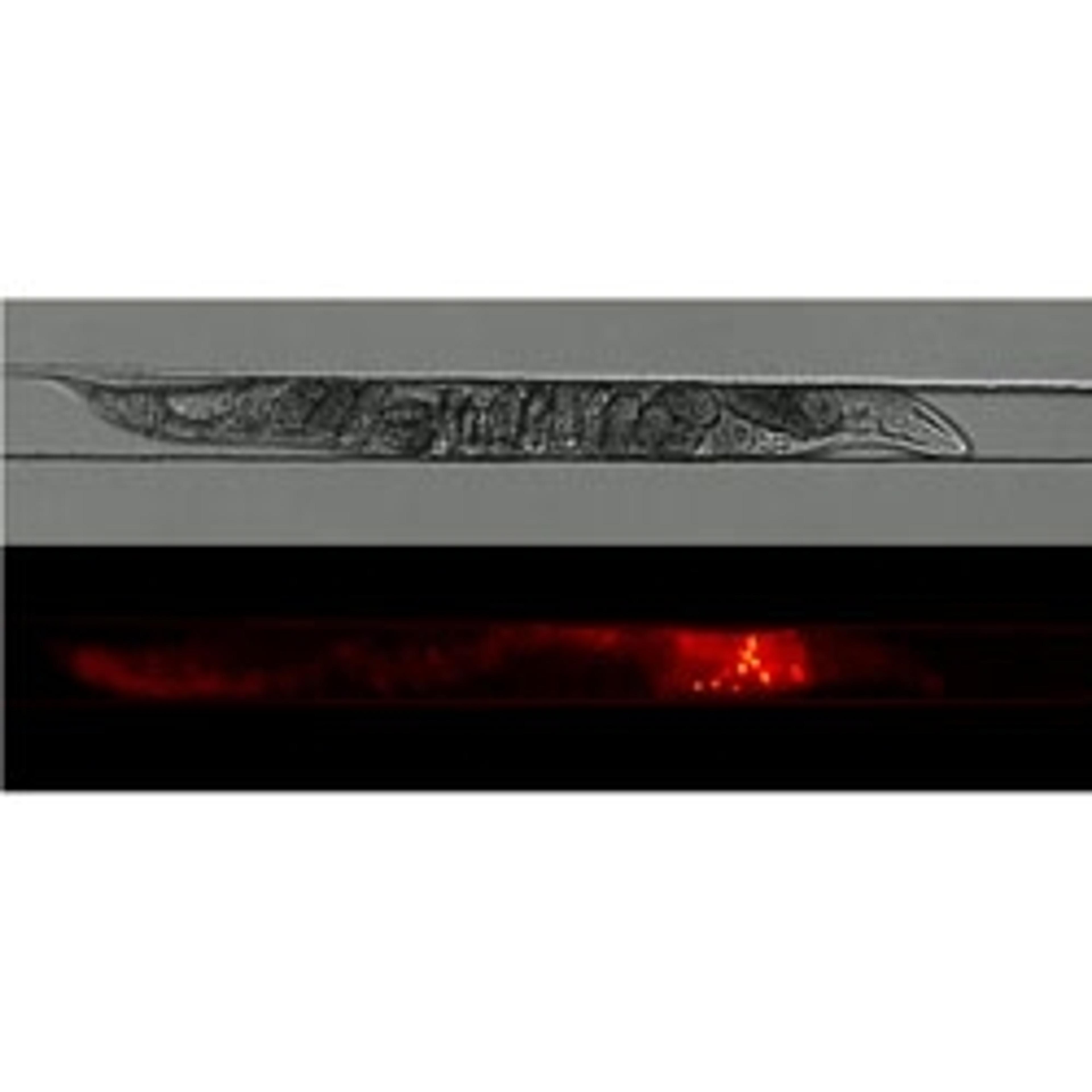
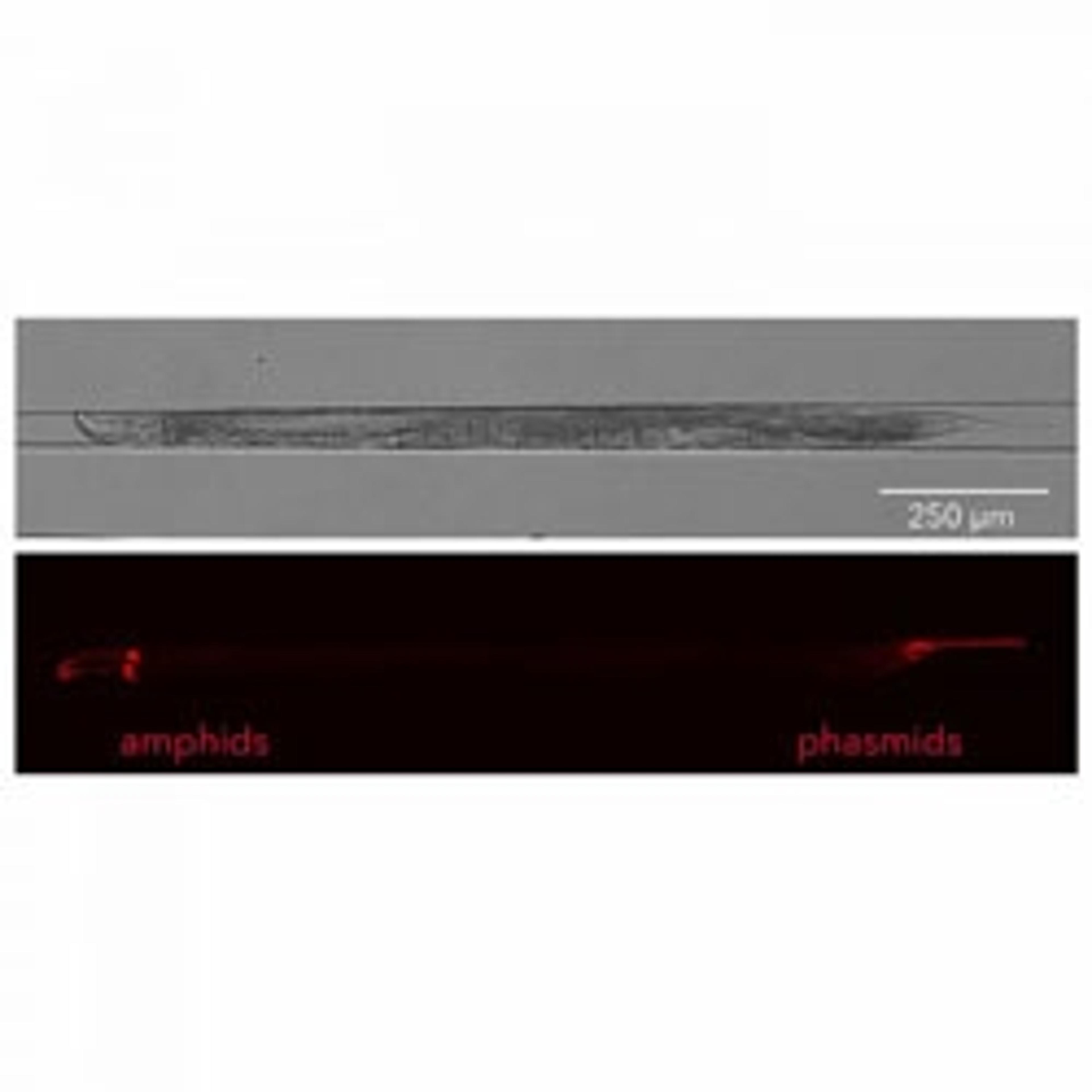
Red Fluorescent non-invasive imaging probe for in vivo detection of apoptosis (pan caspase)
Red Fluorescent non-invasive imaging probe for in vivo detection of cell death (caspase-1)
Red Fluorescent non-invasive imaging probe for in vivo detection of apoptosis (caspase-8)
Red Fluorescent non-invasive imaging probe for in vivo detection of cell death (caspase-1)
Green Fluorescent non-invasive imaging probe for in vivo detection of cell death (caspase-3)
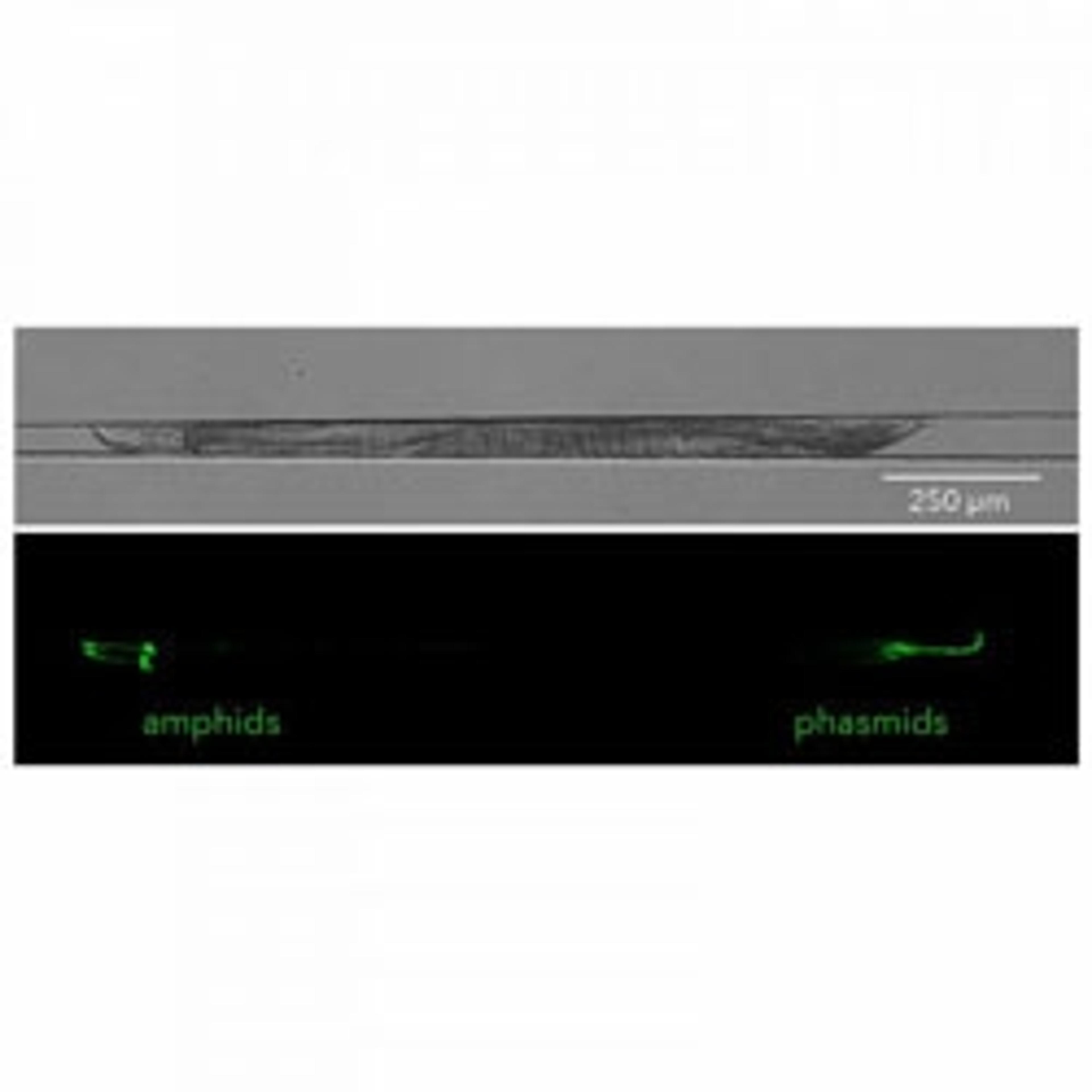
Green Fluorescent non-invasive imaging probe for in vivo detection of cell death (pan caspase)
Green Fluorescent non-invasive imaging probe for in vivo detection of cell death (caspase 1)
Green Fluorescent non-invasive imaging probe for in vivo detection of cell death (poly caspase)
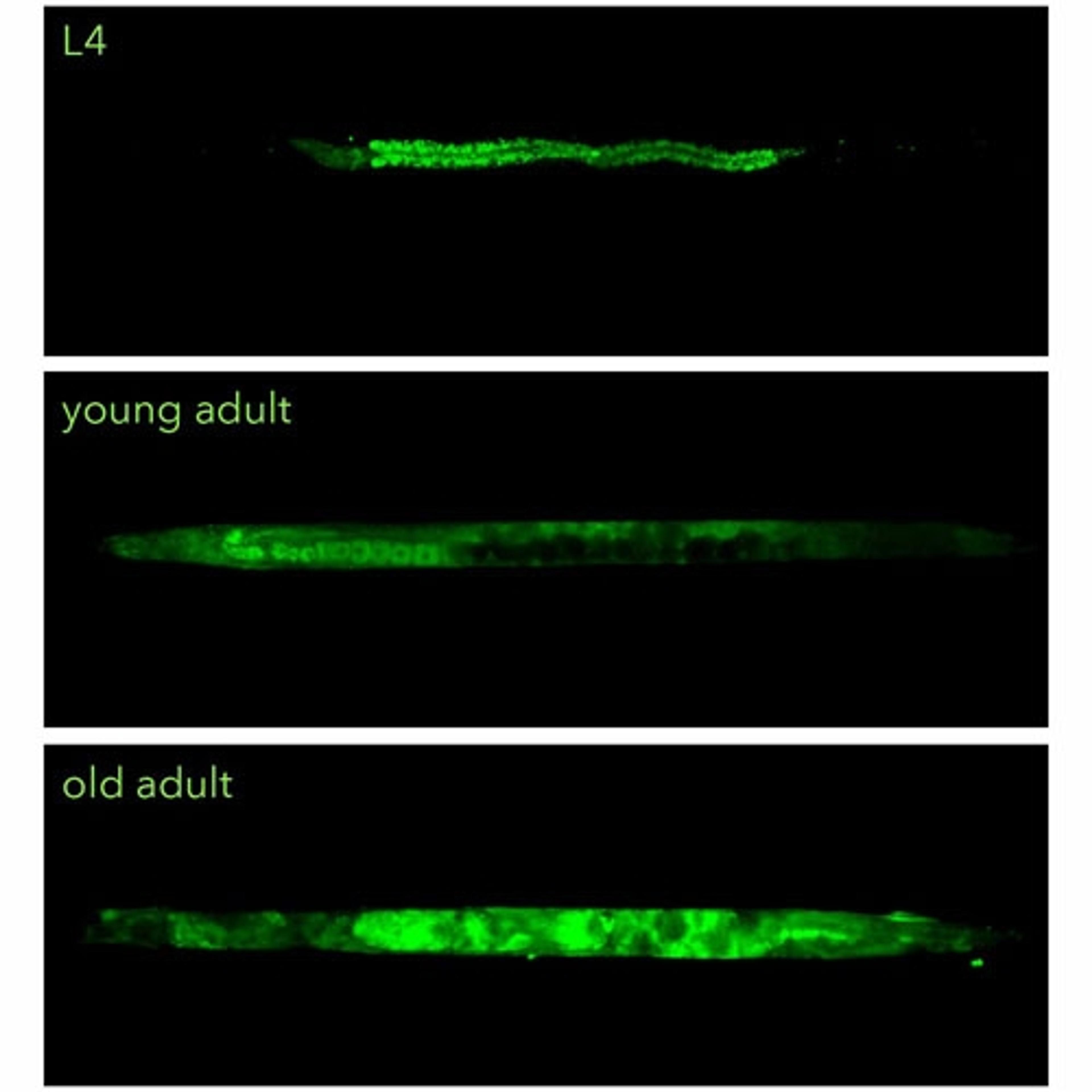
Green Fluorescent non-invasive imaging probe for in vivo detection of apoptosis (pan caspase)
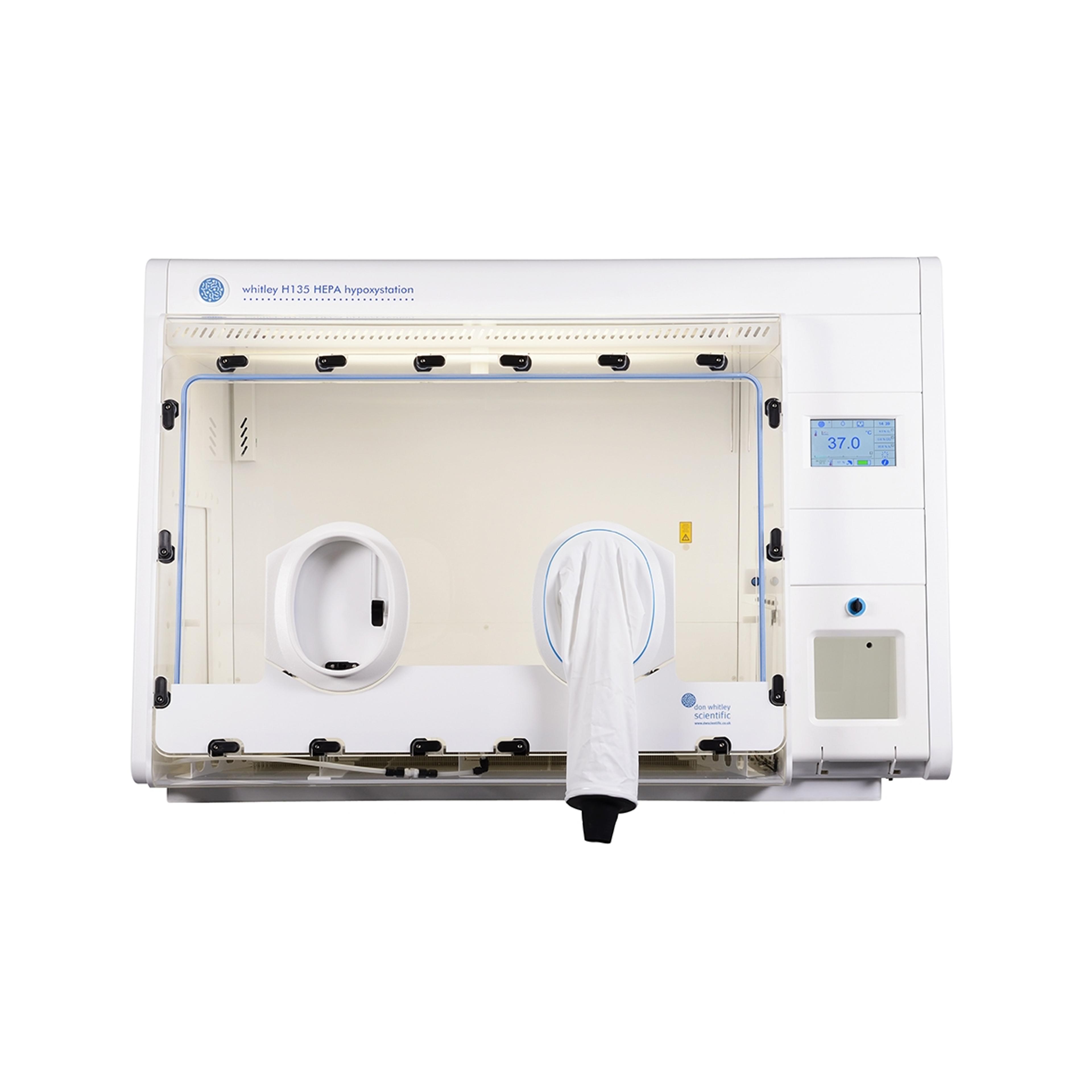
The tall, wide, deep hypoxic chamber from the Whitley range
Cutting edge brain research in awake mice without the biases introduced by anesthetics.
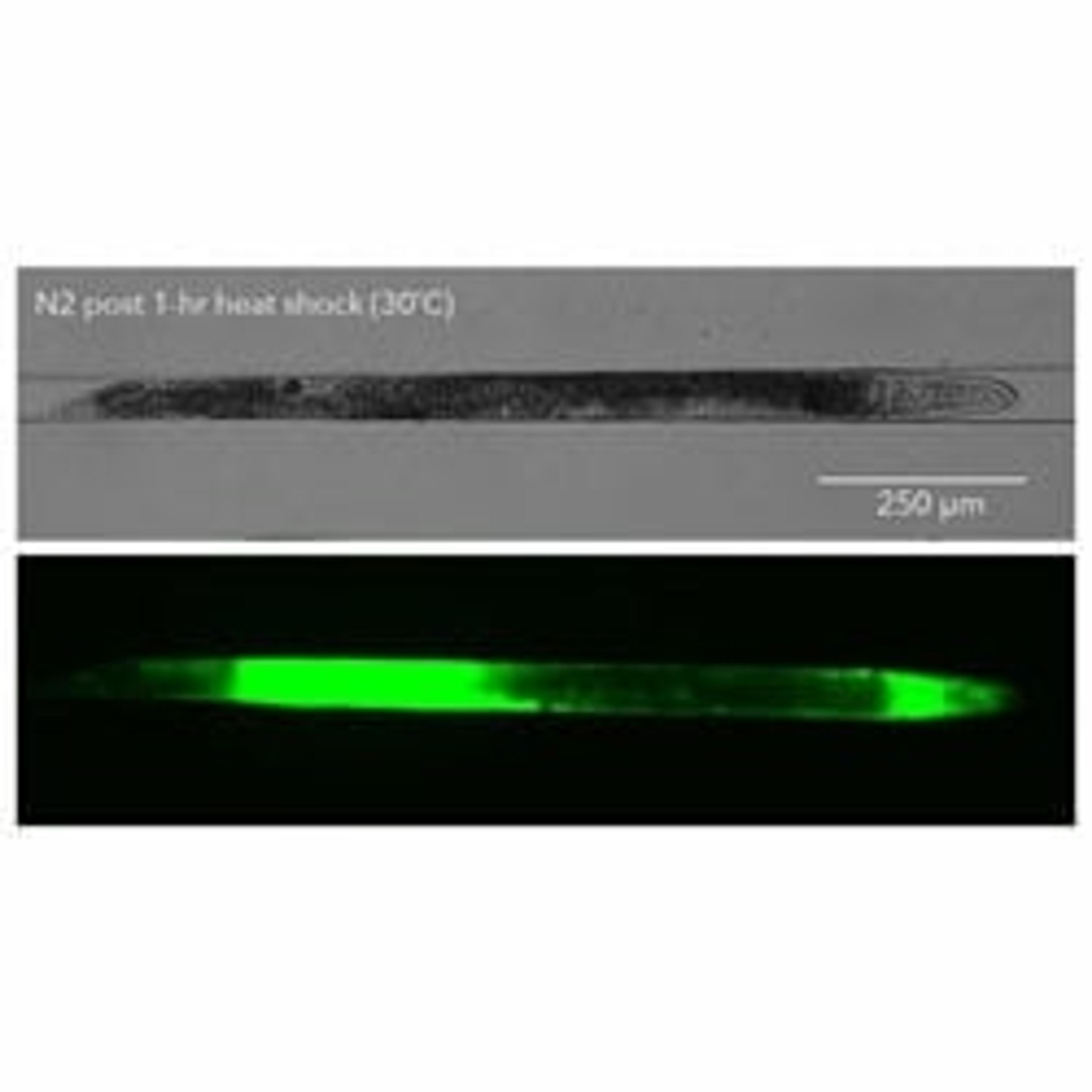
Stain apoptotic corpses in the C. elegans gonad in live worms
Stain amphid sensory neurons in live C. elegans
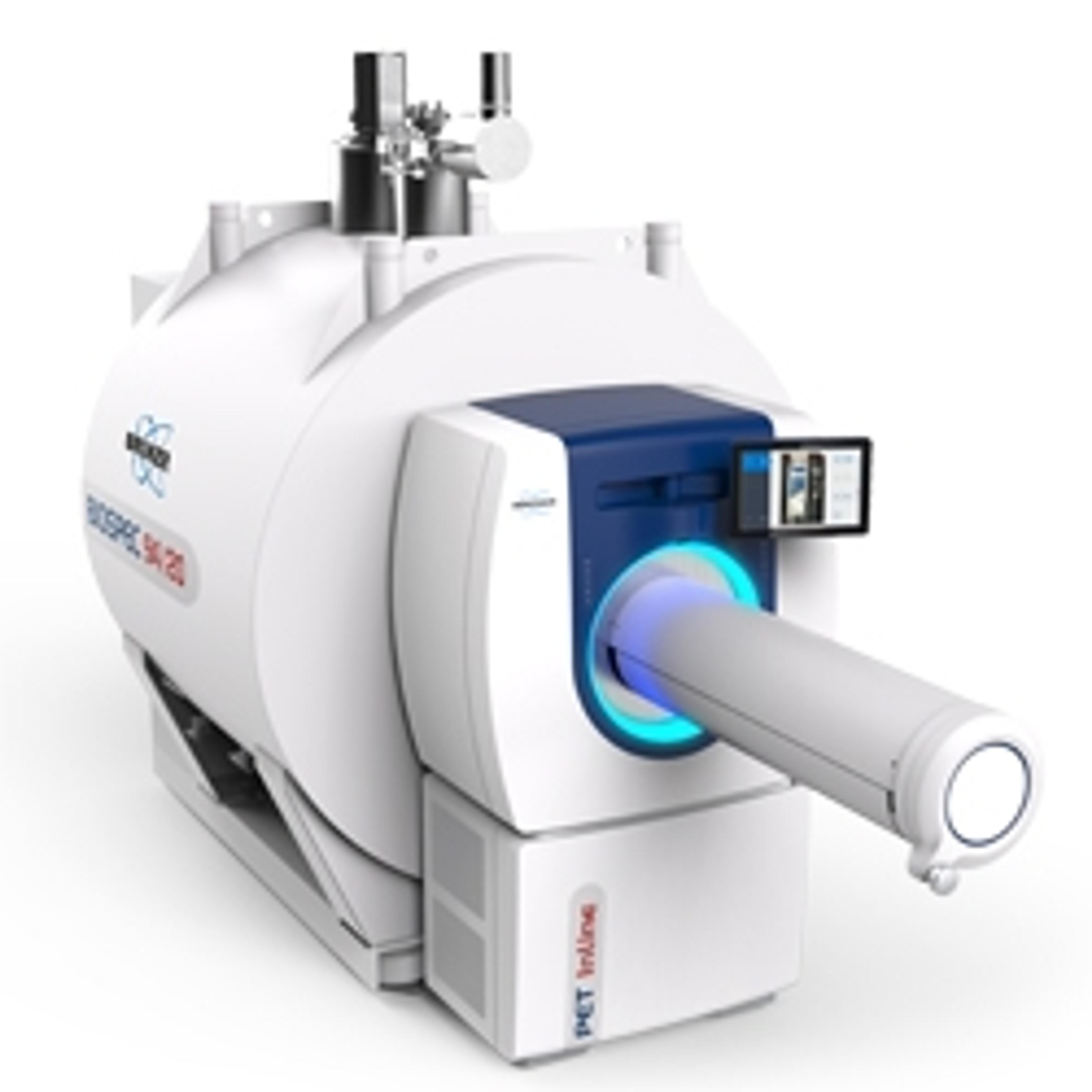
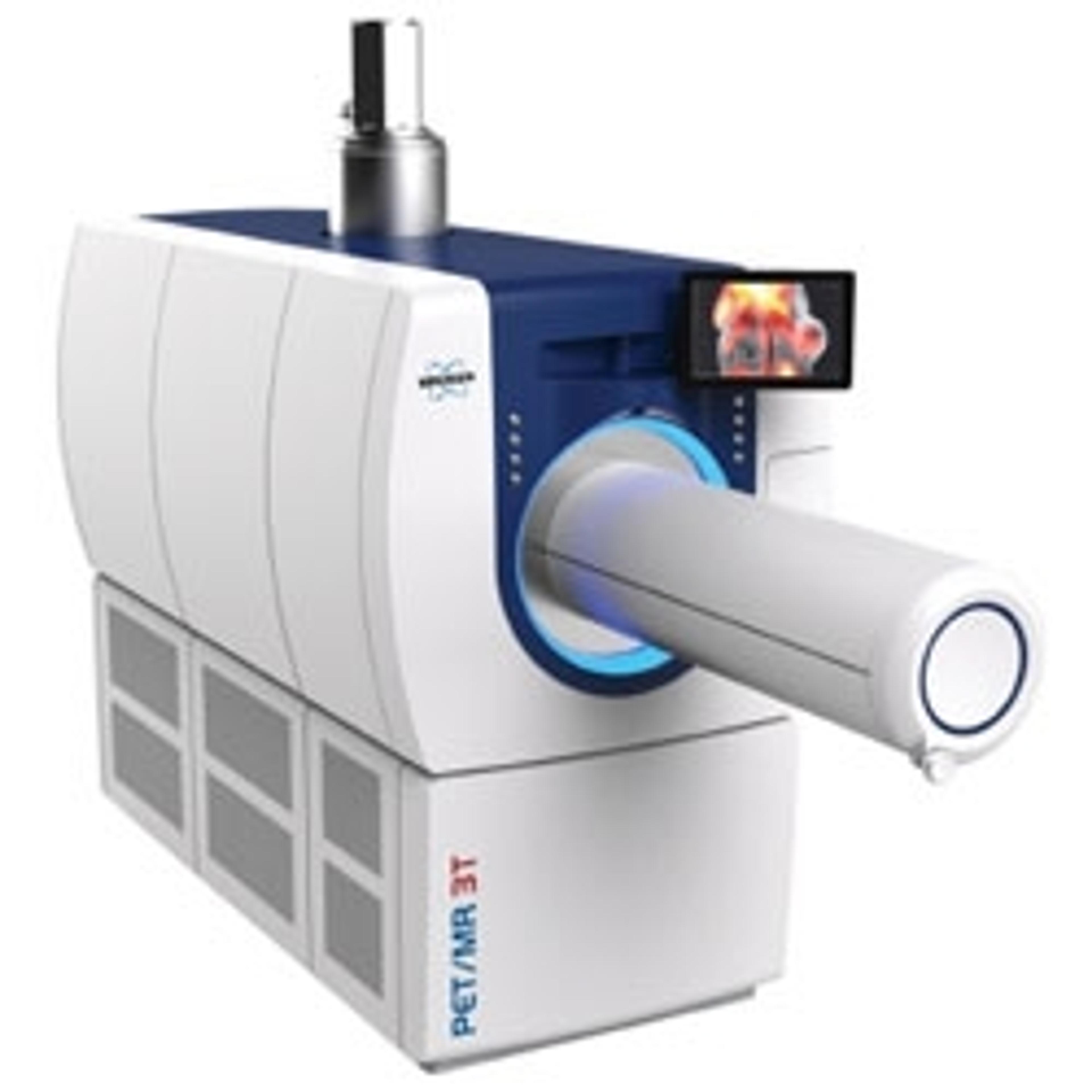
Highest molecular sensitivity combined with excellent MRI contrast and resolution
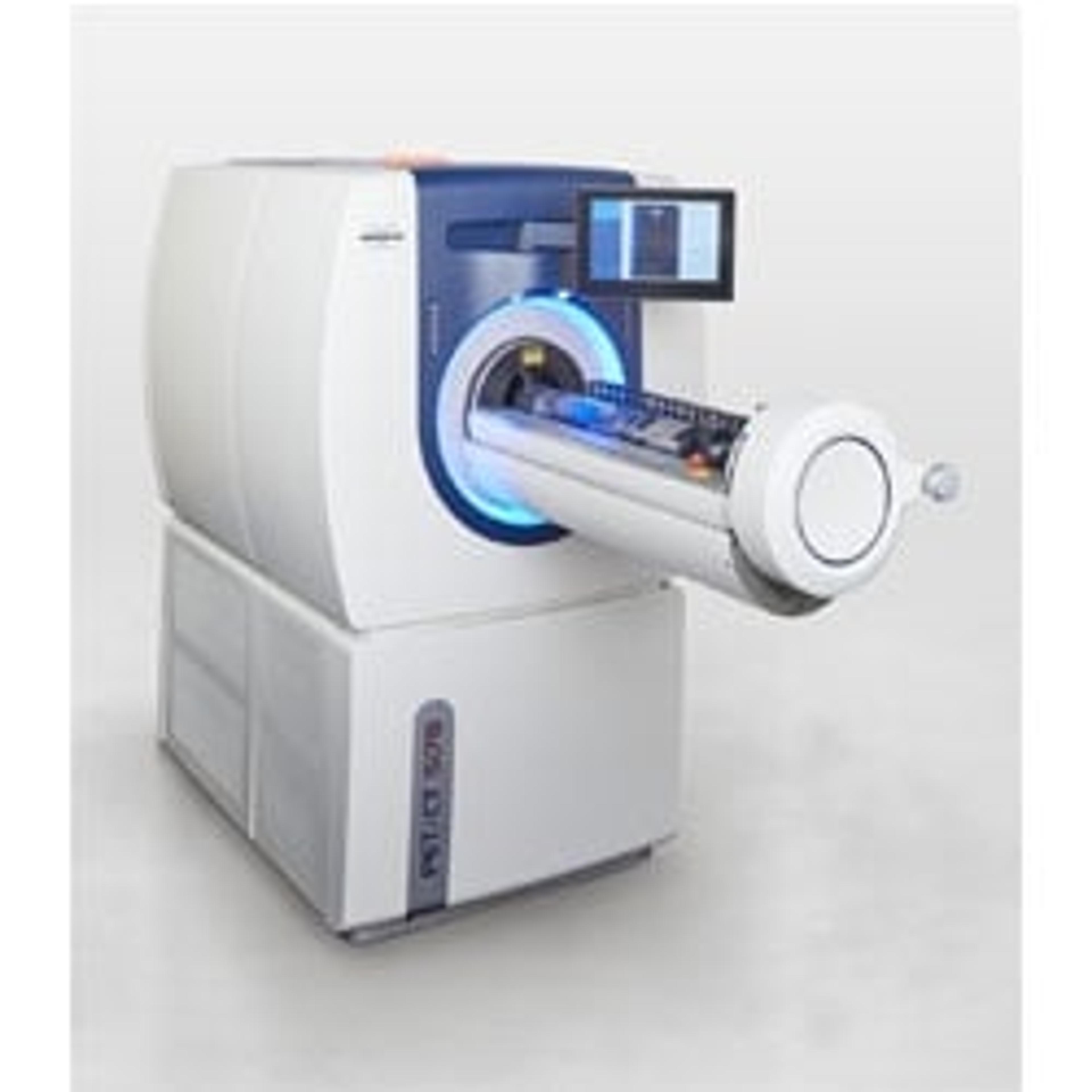
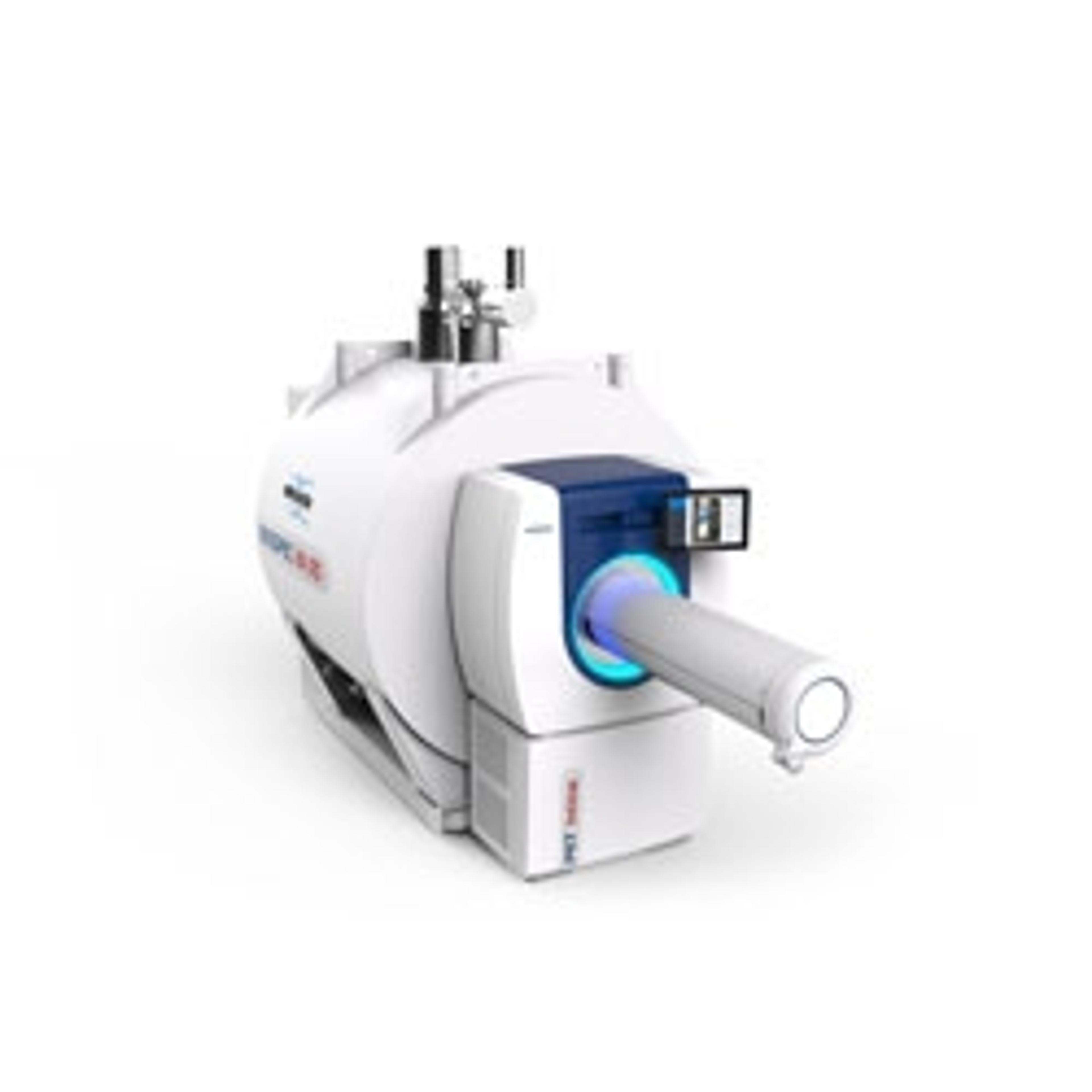
Integrated best in class PET and CT imaging technologies, combined with the simplified workflows of gold-standard preclinical imaging software packages ParaVision 360 and PMOD, the Bruker PET/CT Si 78 is a high-end solution designed for outstanding 24/7 imaging services at predictable costs. The Si 78 provides seamless integration into nuclear molecular and medical imaging research labs and preclinical imaging core facili…
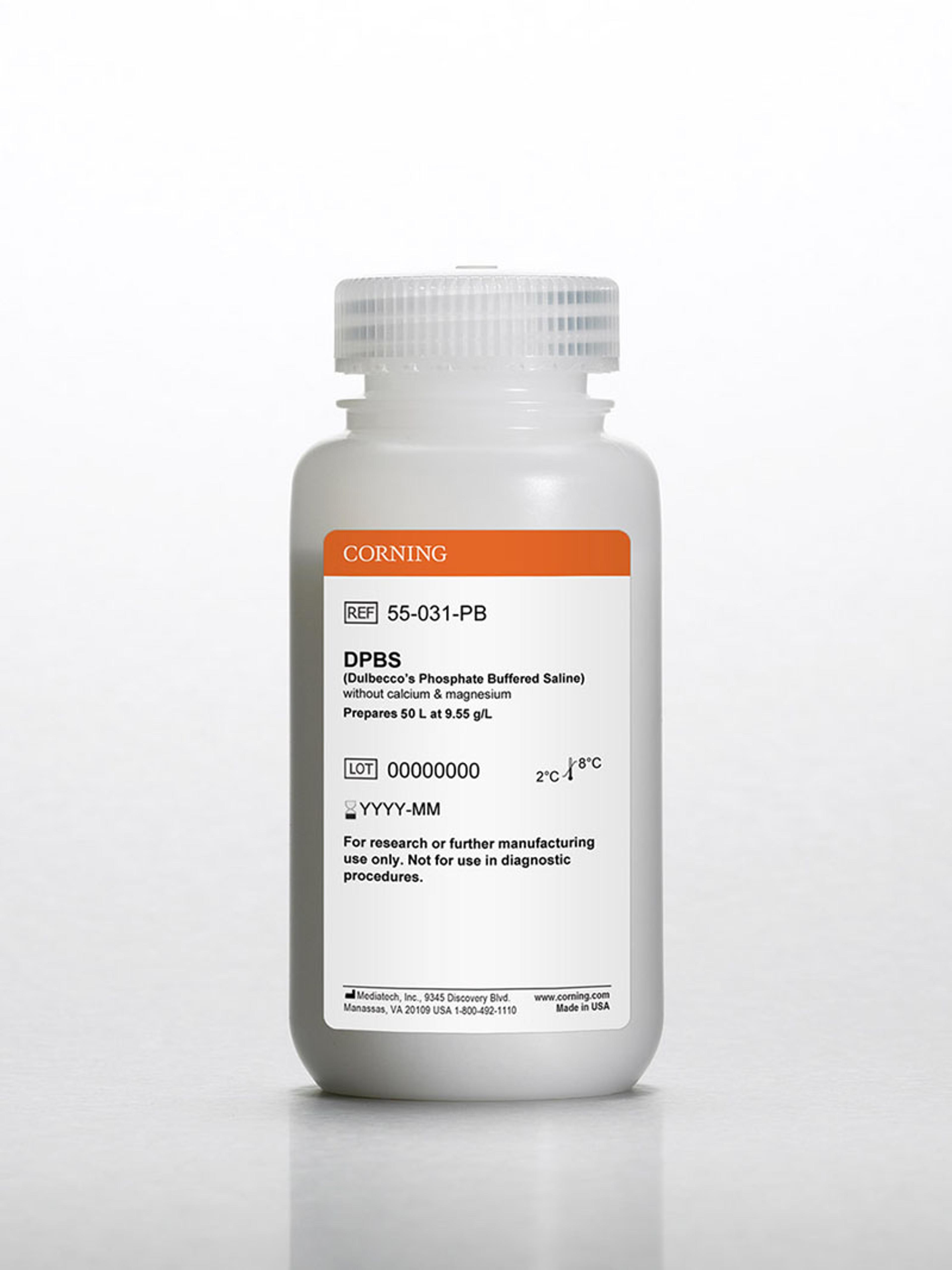
Dulbecco's Phosphate Buffered Saline (DPBS) is a buffer solution which can be used to maintain cell culture media in the physiological pH range of 7.0 to 7.6. It can serve as an irrigating, transporting or diluting fluid while maintaining cell tonicity and viability for a limited period of time. This formulation does not contain calcium and magnesium.
Compact yet unlimited PET/MR research possibilities at translational field strength - ready to be installed in small spaces using Maxwell magnet technology.
Bruker’s established solution for simultaneous multi-mouse PET/MR is extended for small bore systems, now supporting bore diameters from 17 to 40 cm and field strength from 3T to 9.4T. Have a look in 3D – and measure in 4D.
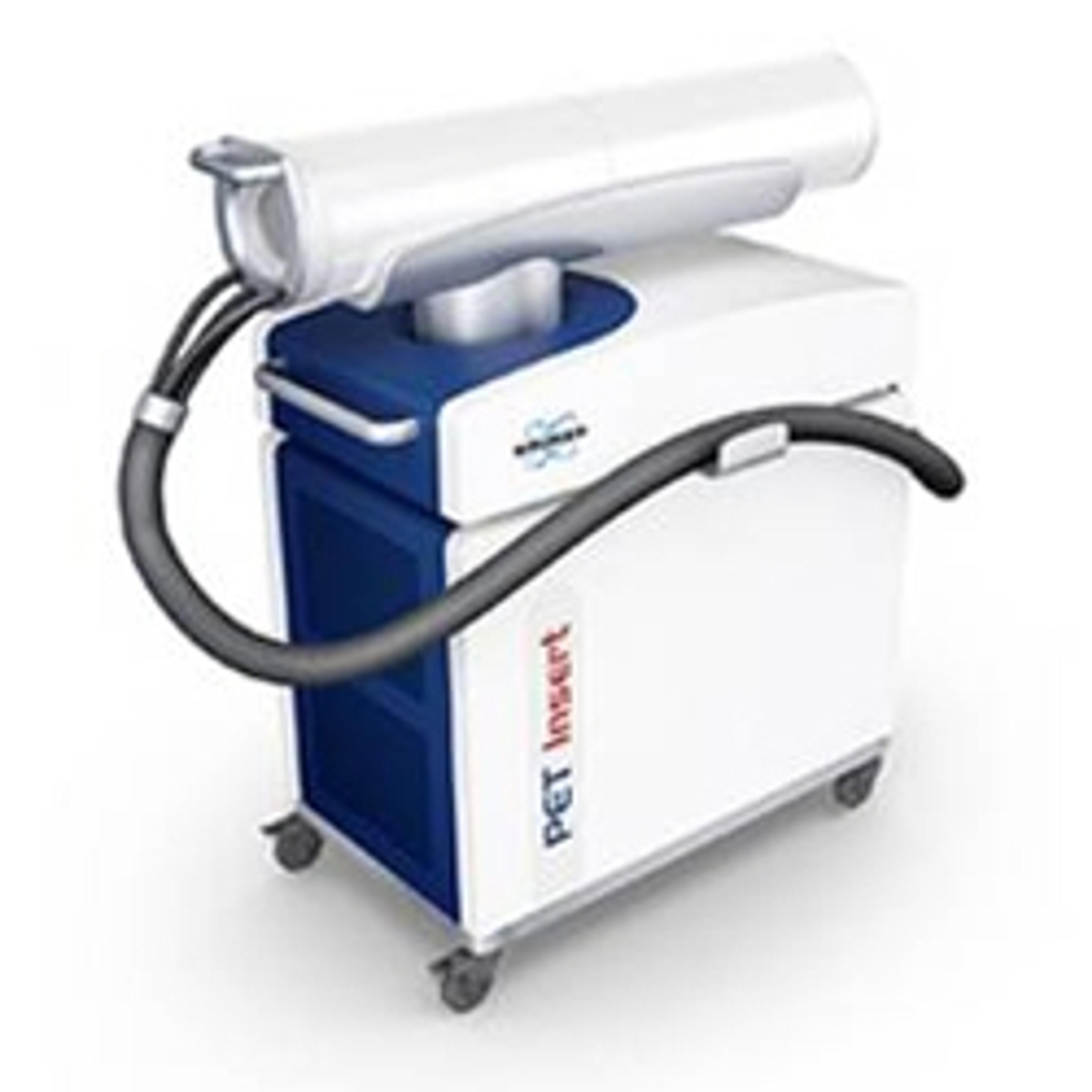
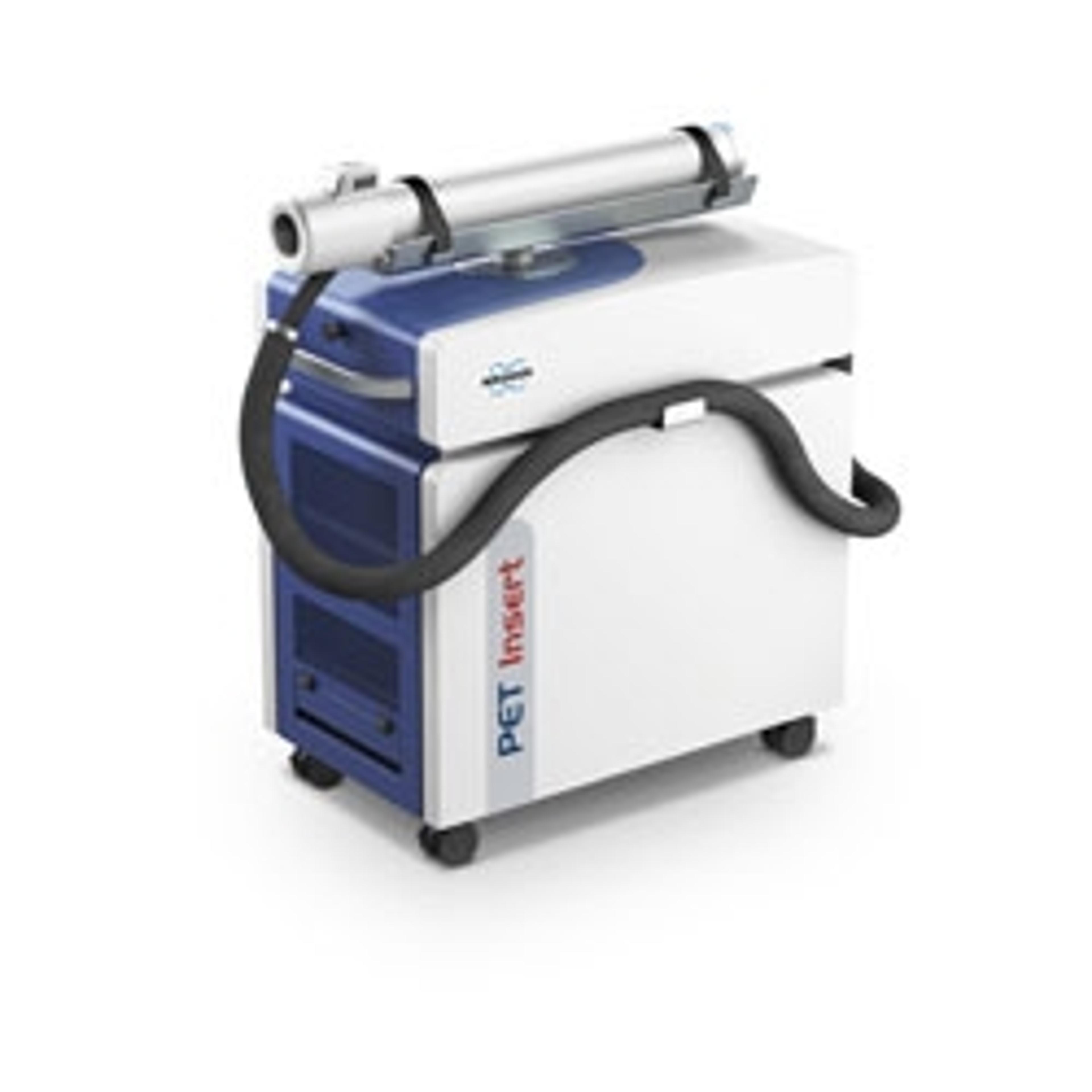
The new PET Inline Module unfolds the power of functional molecular imaging with multiparametric molecular imaging beyond PET quantification based on excellent morphological contrast.
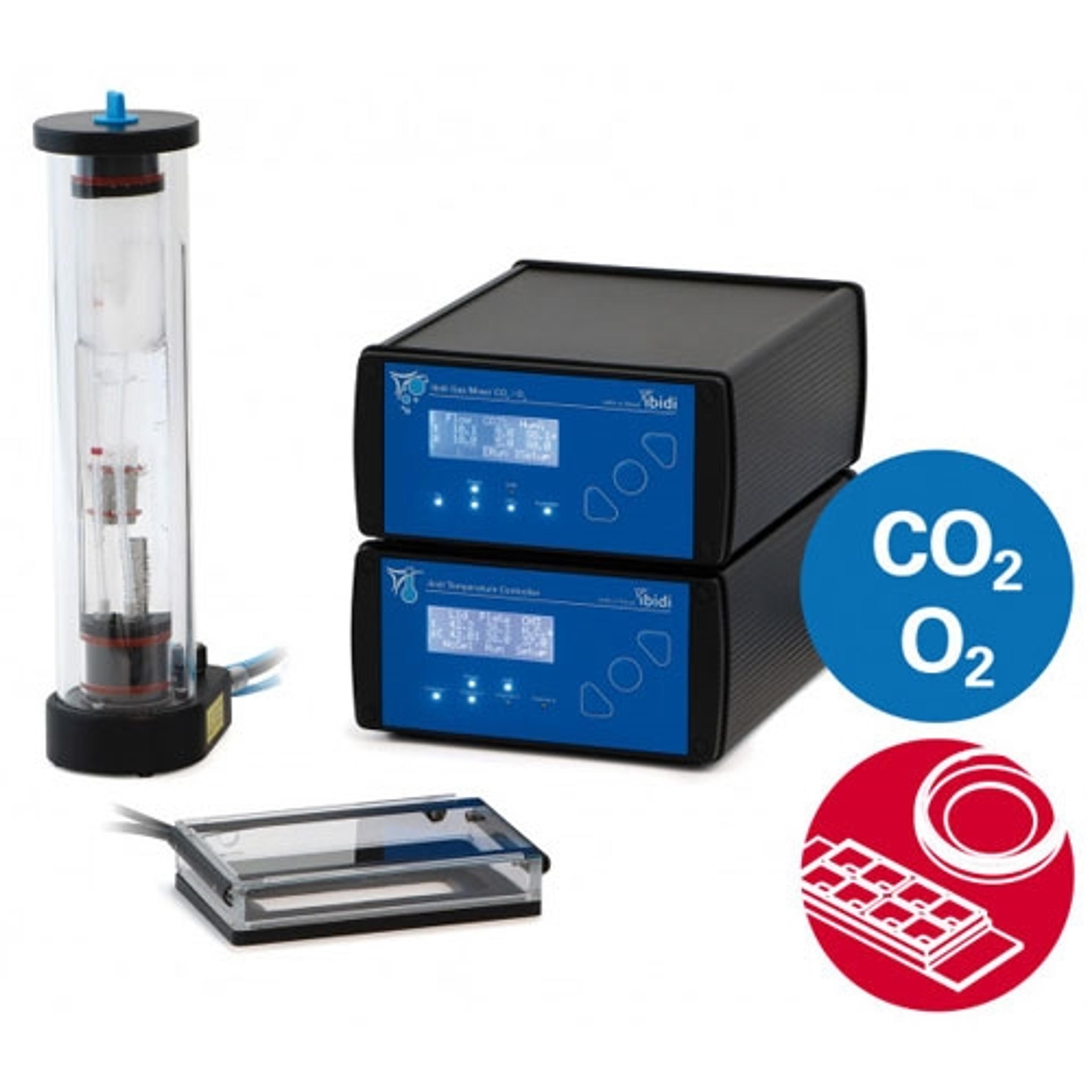
Easy setup directly on your microscope: perform live cell imaging in dishes, slides, or multiwell plates. The ibidi Stage Top Incubation Systems fit every standard inverted microscope and include CO 2 and O 2 control as well as actively controlled humidity. They are ideal for all live cell imaging applications and available for single slides and dishes as well as for multiwell plates.