How to accelerate nanobody discovery workflows with advanced flow cytometry
On demand webinar looks at nanobodies and the use of high-throughput flow cytometry to increase the throughput of nanobody screening campaigns
29 Nov 2019

While antibodies are set to be increasingly important as therapeutic options for cancer and other diseases, the discovery of fully functional antibodies that lack light chains in Camelidae (camels and llamas) has opened the door to an exciting new generation of therapeutic antibodies known as nanobodies.
Unlike conventional antibodies, the heavy-chain antibodies of Camelidae contain a single variable domain (VHH) and two constant domains (CH2 and CH3) only. Using these functional antibodies as a base, nanobodies have been created and represent the next generation in antibody-derived biologics.
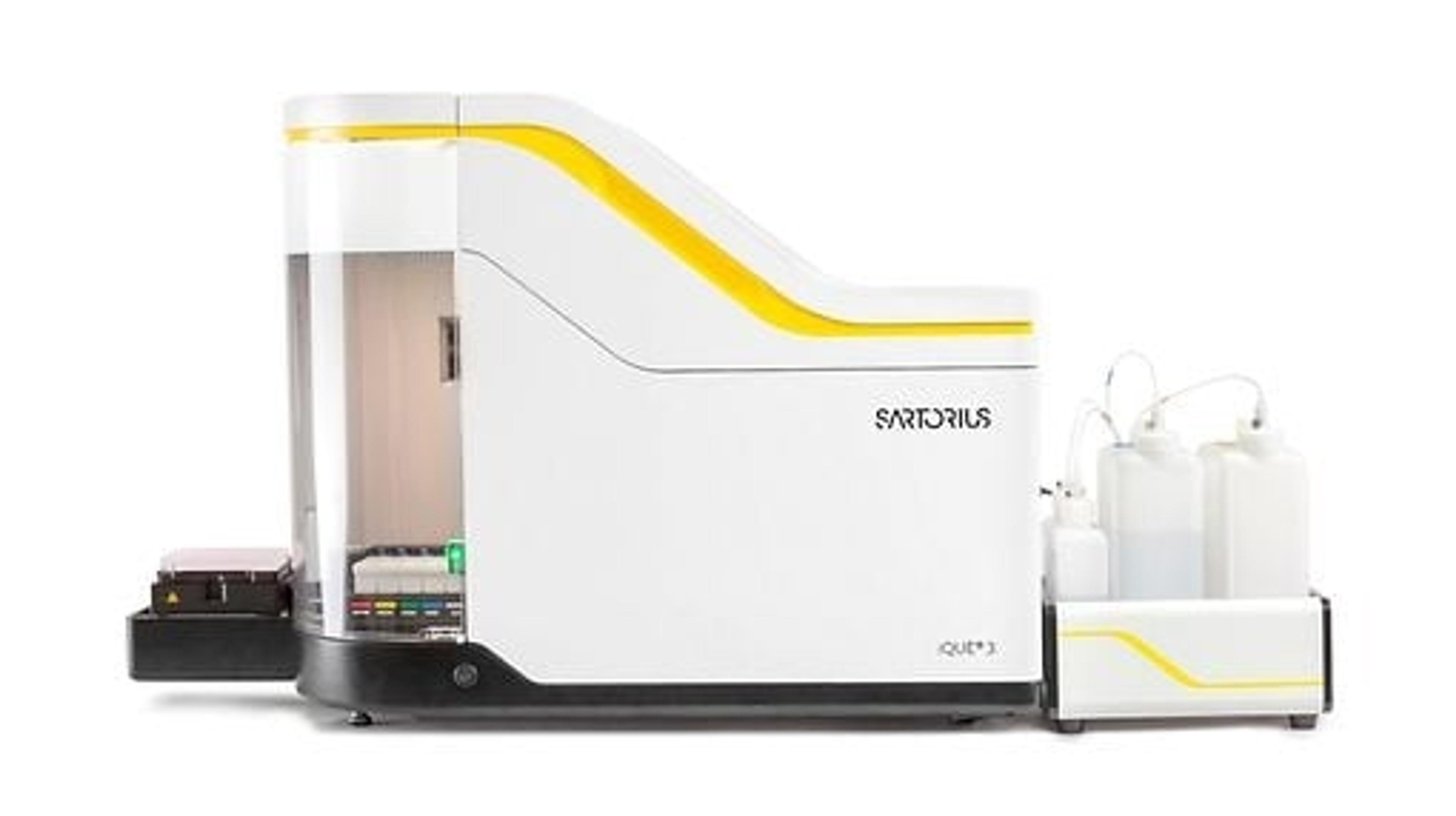
In a webinar, now available on demand, Dr. Pieter Kennis looks at nanobodies and how high-throughput flow cytometry is being used to accelerate the screening process.
Watch NowWatch the webinar to learn about:
- Efficient labeling of multiple cell lines for simultaneous analysis
- Multiplexed nanobody screening, assay setup and analysis to screen for compound binding (with examples from nanobody discovery campaigns)
- Nanobody characterization – semi-automated data analysis to determine EC50 values from dose response binding
Below are some highlights from the live webinar Q&A session:
Q: Do you see a difference in results between fresh and frozen cells?
PK: In general, we don’t see a lot of difference between fresh versus frozen cells. The main difference that we observe is in viability, sometimes after freezing certain fragile cells, there is a drop in viability.
Q: Which dead stain would you recommend?
PK: We use PI, TO-PRO-3 as standard and since the recent purchase of the iQue platform we also use the DAPI stain. For some purposes, it is necessary to use a fixable dead stain, for those situations we tend to use Zombie dyes.
Q: For multiplexing using PKH26 and CFSE, which dead stain do you use and why don't you compensate the data?
PK: Back in the day it was quite difficult to compensate the data. For the CFSE and the PKH, we were able to separate the cell lines sufficiently without the compensation. Since we only had the standard iQue Screener with four colors available, we would use one color for the CFSE, one for the PKH26 and one for nanobody detection. A good option was PI as it was combinable. However, if we had a bright dye and the intensity of the CFSE was too bright, it would make it difficult to separate the PI from the CFSE-labeled cells. For that situation, in the single-cell line controls, we would do a viability stain with a compatible dye.
Q: Do you screen for intracellular proteins and, if so, do the anti-tag antibodies permeate well?
PK: Since our nanobodies don't have the need to target intracellular proteins, there is no need to detect the nanobodies to intracellular proteins. However, we did some testing for intracellular proteins and we observed that they could permeate through the cell membrane.
Q: Did you try combining different dyes to increase multiplexing? For example, FL1 and AS6 or 7 in different concentrations?
PK: That is on my to-do list for further optimizations. We have FL1 and FL4 MultiCyt staining dyes and we use both in our experiments, but if we combine them, we would be able to have a multiplex for the cell lines of 25, so it would be something to evaluate.
Q: Is there a standard linker sequence or length for making multivalent nanobodies?
PK: There's not really a standard linker. But we have the possibility to use a variety of different linkers for the nanobodies.
Q: What could be improved in the FACS workflow, technology, or data analysis to make it better suited for high-throughput screening and analysis?
PK: In our current protocol, there are still a few manual parts. The washing of the cells is done by centrifugation and sticking the plate upside down. We are looking at a way to semi-automate this by using plate washers or another alternative. That may be a good way to improve the workflow. We are also evaluating the feasibility of the bleach being together with the cells to increase the plaques. We could also generate standard parental cell lines that already express a certain intensity of dye and use those to transect our target protein, which means we wouldn’t need to stain big batches of the cell lines every time.
Q: How many clones do you test per target or phage display round?
PK: That depends on your projects. For some projects you don't have a lot of output and you need to screen a lot of clones but for others, it's really straightforward and you only need to screen, say, 1,000 clones for example.
Q: Do you typically normalize nanobody concentrations before doing comparative screening?
PK: We do not normalize our nanobody concentration since we separate the concentration of the nanobodies and this is a yes or no answer. So, we don't see the need to know the exact concentrations.
Find out more on this topic by watching the full webinar on demand>>
SelectScience runs 3-4 webinars a month across various scientific topics, discover more of our upcoming webinars>>
Related Products
Request Quote for All Products
Incucyte® Live-Cell Analysis Systems
Sartorius GroupIncucyte ® , Empower Live-Cell Analysis Inside Your Incubator