How to streamline development of a cytotoxicity assay for high-content compound screening
Watch this on demand webinar to learn how to speed up your assay development process
17 Mar 2021

High-content screening and morphological profiling is becoming increasingly popular in the characterization of new compounds and their mode of action in pharmaceutical and academic drug development. Using highly sensitive technologies, morphological changes can already be detected in sub-cytotoxic concentrations. For the generation of morphological profiles, a precise investigation of cytotoxicity with respect to concentration and time is therefore indispensable.
In this on-demand webinar, Dr. Christopher Wolff, FMP Berlin, presents a fast and reliable method to develop a cytotoxicity assay for high-content screening (HCS) using Tecan’s Spark® Cyto multimode reader with imaging capabilities. The imaging-based apoptosis assay enables a precise determination of the apoptotic state in treated cells. The Spark® Cyto provides highly sensitive and precise characterization of compound-induced cytotoxicity, while multicolor image analysis allows for rapid and reliable detection of early and late apoptotic cells. The assay performance was found to be comparable to dedicated high-content screening imagers, enabling a rapid integration of this assay into large-scale high-content screens.
Watch on demandRead on for highlights from the live Q&A session or watch the webinar at a time that suits you.
Q: Is the cell painting assay limited to compound screening or are there other possible applications?
CW: The cell painting assay can be used for different things. You could use it to characterize genes using RNA interference and it would work roughly the same way. You incubate your cells with RNAis, perform the cell painting assay and then analyze the morphological changes in the cells with regard to gene interference.
You could also use the cell painting assay to identify specific cell types, for example, if you stain cells without any incubation of compounds you could create a database of morphological profiles for each cell. Knowing the morphologic profiles of specific cell lines lets you could compare these to other cell lines and identify cells in co-cultures. If you are doing IPS cell differentiation, you could check how good the differentiation is and how close you are to the original cell line.
Q: Since morphological profiles show distinct concentration dependency, are they also influenced by the incubation time?
CW: Yes, the profiles are influenced by the incubation time, but in a different manner. The longer you incubate your compounds, the more compounds will produce distinguishable profiles. However, when you increase the incubation time, you also get less specific feature changes. If you test your cells at an earlier incubation time, you get a more specific profile for this compound, which will give more information on the mode of action of the compound.
Q: Can you explain how the cell painting analysis is done and how the heat maps are generated?
CW: We get all our feature data from the microscope. We use Harmony® to detect the features in the different compartments of the cells and channels. This produces a multiparameter table containing several hundred features per cell which can be saved as a text file. We import this file into KNIME to perform the averaging of the single-cell data to well data and as well as z`- score normalization.
KNIME also offers the option to produce heat maps. In summary, the microscope does the image analyses, and we use KNIME, a free software, to do all additional analyses.
Q: Are there any alternative dyes available for the apoptosis assay, with regards to the channel selection?
CW: Annexin V would be a good apoptosis marker, but as it's a membrane marker, it's not as good for object detections compared to other dyes. For the mitochondrial membrane potential marker, you could use rhodamine-123 which can be visualized in the green channel; but is otherwise very similar to tetramethylrhodamine. Another alternative would be a caspase 3/7 stain, which is offered by several different companies, for example, the CellEvent™ Green from Thermo Fisher, but it is more expensive.
Q: Why not use Annexin V and PI for early apoptosis assays?
CW: It’s true that Annexin V is better for early apoptosis detection. However, since it's a membrane staining, it's more difficult for object detection in the image analysis. I would refrain from using Annexin V for this.
Q: Can cell painting analysis be performed under hypoxic conditions?
CW: Hypoxic conditions are critical because the mitochondrial membrane marker are quite sensitive for the membrane potential which can be greatly influenced by the hypoxic conditions. I don't think it would be optimal to perform this under this condition.
Q: Is it possible to add two different cell types by coloring the cell of interest beforehand to check for apoptosis of the cell?
CO: It will be quite difficult looking at the capabilities of the system, as you would need an additional marker or more than the three channels that you can measure simultaneously. The system offers four individual fluorescence channels, but only three can be measured or imaged in parallel. You would need to have a morphological marker to differ between different cells. Our software is not powerful enough to do that discrimination just from the morphological profile of two individual cells.
Q: What's the magnification for whole-well imaging of a 384-well plate?
CO: For the whole-well imaging of a 384-well plate, the system automatically picks the 4x objective. You could also use the 10x objective with more than one image per well to cover the whole 384-well area. Both are possible, but the 4x is the default and that means the system will just shoot one single image of the whole well.
Q: How long does it take for 384-well analysis in the Spark® Cyto?
CO: The Spark® Cyto offers real-time analysis capability, meaning you can acquire the images and they are directly analyzed. If you measure two channels with a whole-well view applying the 4x objective, the complete plate is imaged in around 30 to 40 minutes, depending on exposure time and other factors.
Q: How many plates can be imaged in parallel with the Spark® Cyto for live-cell imaging assays?
CO: The Spark® Cyto can hold one plate per time. Tecan offers ways to increase the throughput, such as the Spark Motion concept, where we connect the Spark® Cyto to an incubator with a robotic arm, a solution to increase the throughput up to a maximum of 44 plates.
Q: Is it possible to image cells that are in suspension or growing in a 3D environment?
CO: The system is tailored to analyze 2D monolayers, we can also image spheroids and suspension cells. When it comes up to image analysis, you would need to transfer these kinds of images to third-party software as our analysis software is exclusively tailored to 2D cells grown in a monolayer.
SelectScience runs 10+ webinars a month across various scientific topics, discover more of our upcoming webinars>>
Related Products
Request Quote for All Products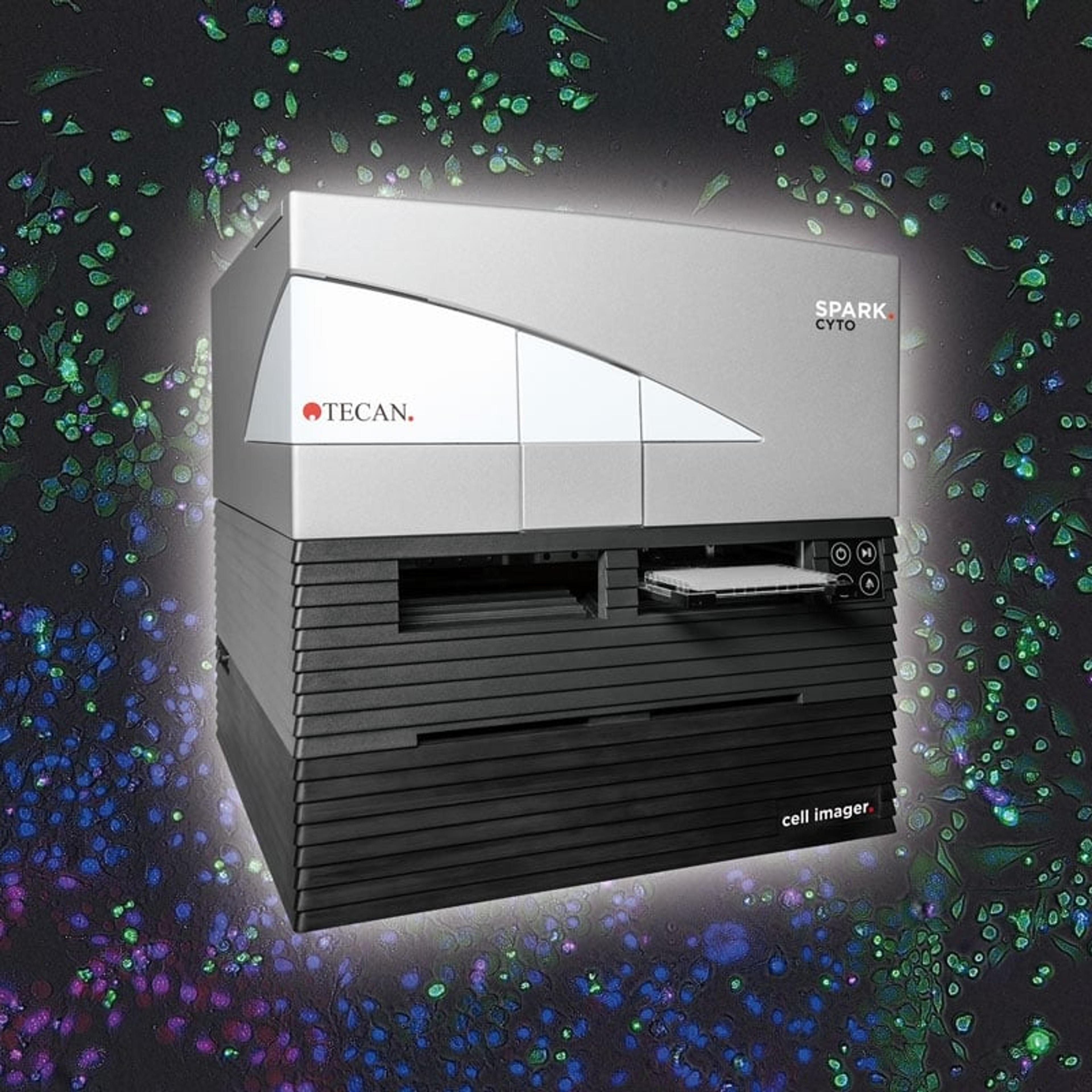
Spark® Cyto
TecanSpark Cyto is the first live cell plate reader to detect biological, chemical or physical events – capturing the maximum amount of data possible from each plate well, at the same time and under the same conditions.