Using Molecular Spies to Understand Changes in Cell State in Health and Disease
SelectScience® spoke to Thomas Deerinck, National Center for Microscopy and Imaging Research at University of California, San Diego, about how correlative microscopy is helping scientists understand disease states
26 May 2016

SelectScience® spoke to Thomas Deerinck, National Center for Microscopy and Imaging Research at University of California, San Diego, about how correlative microscopy is helping scientists understand disease states
European Molecular Biology Laboratory (EMBL) Founded in 1974, EMBL is Europe’s flagship laboratory for the life sciences – an intergovernmental organization with more than 80 independent research groups covering the spectrum of molecular biology.
The National Center for Microscopy and Imaging Research (NCMIR), based at the University of California, San Diego (UCSD), aims to develop technology to help understanding of biological systems, bridging anatomical and molecular scales. SelectScience visited the main Heidelberg site of the European Molecular Biology Laboratory (EMBL) to attend the workshop ‘From 3D Light to 3D Electron Microscopy’ (EMBL 3D), run in conjunction with ZEISS Microscopy. There, we spoke to NCIMR researcher Thomas Deerinck about the innovations enabling his work on understanding cell states in health and disease.
“Our overall goal is to try to understand how cells work in both health and disease”, Thomas explained, working with “two very talented teams of researchers from UCSD and Massachusetts Institute of Technology (MIT)”. Working with Nobel-laureate Roger Tsien’s group from UCSD, Thomas is developing “molecular spies” to study the structure and function of cells.
“When you look at cells through a microscope”, Thomas explained, “it’s very difficult to see what’s going on”. Cells are made up of a wide range of proteins which carry out “the functions that give rise to life”. However, when you look through a microscope, “it’s difficult to see them in space and time, and how they’re reacting”. Using molecular biology, chemical spies that can be “fused to the proteins” have been developed, allowing the proteins to be seen with both light and electron microscopy.
Caught in the act
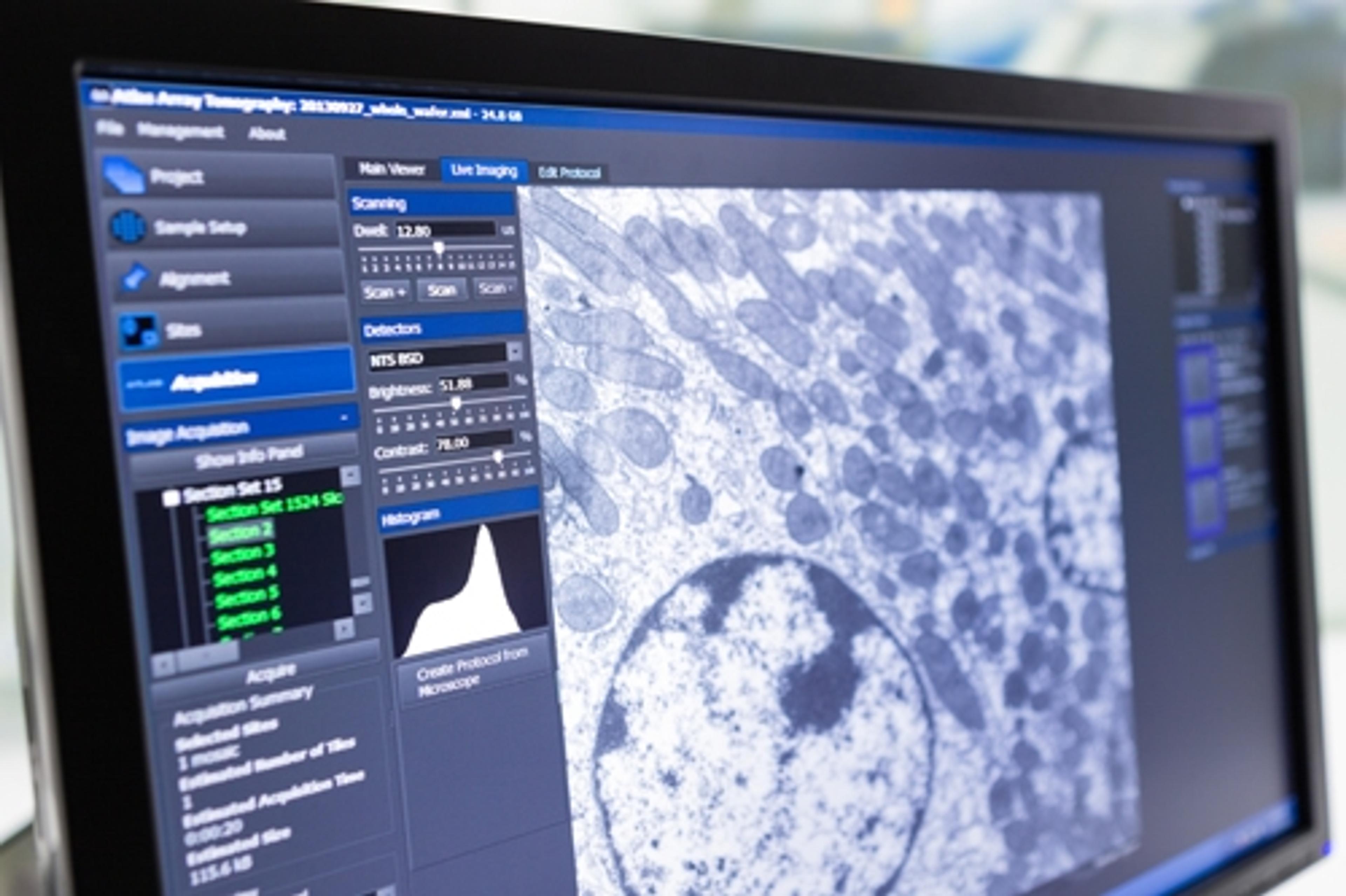
Correlative microscopy is a “powerful technique” – it allows researchers to “study live cell dynamics and watch different things happen”, before “freezing the action” and analyzing the samples using electron microscopy. “Using techniques such as volume imaging with serial block face electron microscopy, we can see structures of the cells at nanometer scale resolution”, Thomas revealed.
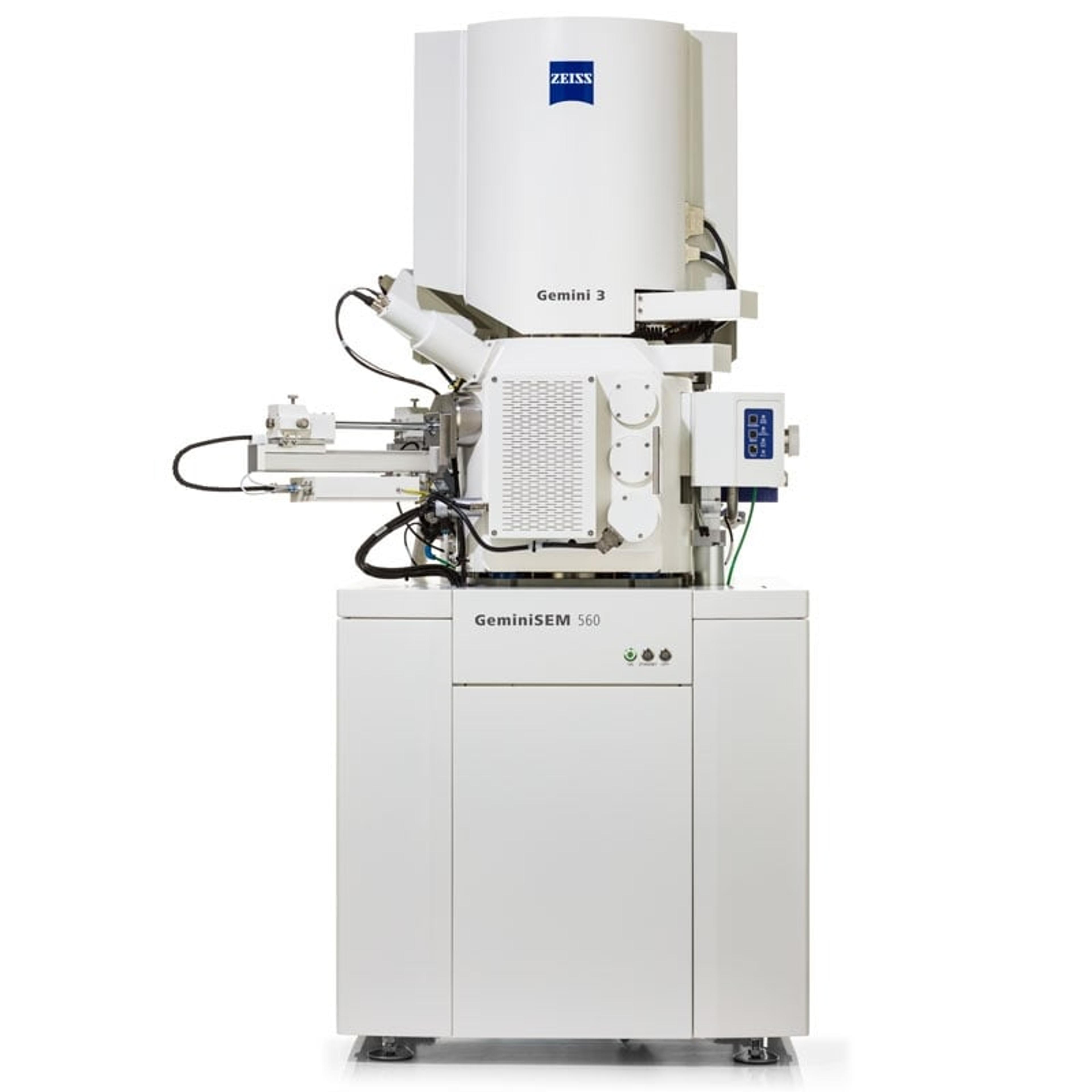
3D imaging of samples is now possible with a “very powerful, relatively new technique called Preview ”. Essentially a “miniature microtome”, it is placed inside the chamber of a scanning electron microscope (SEM) and removes a layer of a sample, such as “a piece of brain tissue”, as the SEM images it until “you form a whole stack of images” which are combined to give a 3D result.
At EMBL3D, Thomas presented “two new sets of tools for biomedical researchers”. One, developed in the laboratory of Dr Alice Ting at MIT, is a “bio-complementation tool to study protein-protein interactions”. The other, Click-EM, a “labeling technique for correlative light microscopy and EM imaging of non-protein biomolecules”, was developed at UCSD. Thomas hopes “these new tools will go a long way to aid researchers” doing correlative microscopy and large scale volume imaging.
Find out more about the other speakers and presentations from EMBL 3D, or learn more about the applications of Correlative Microscopy.
Watch our interview with Thomas here.