The power of parallel processing: A vision to achieve 24-hour biopsy diagnosis with confidence
Find out why optimized tissue preparation and processing is crucial for rapid, accurate, and flexible histopathology workflows
3 Jul 2022
Tissue processing is a crucial part of the in vitro diagnostics workflow in histopathology laboratories around the globe as all downstream steps rely on accurate processing for a correct diagnosis to be given.
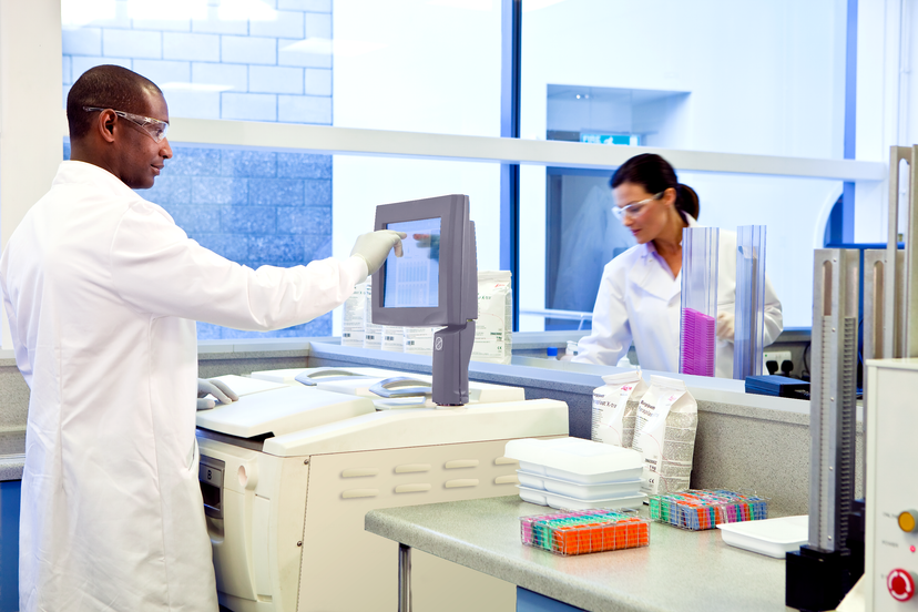
Currently, clinical labs are facing several challenges with biopsy analysis due to the range of samples that can be received - from tiny needle biopsies containing small, delicate tissues, to larger samples such as whole bowel resections or breast mastectomies. Each sample must be treated differently depending on its size and composition. The diverse tissue makeup of each sample adds an additional layer of complexity, as a wide array of tissues can be present in a single specimen. Additionally, the lack of control that many labs have over the time a specimen is received drives further challenges in tissue management. Aside from receiving the tissue, there are several considerations around managing the tissue, grossing the tissue, and processing the tissue that exacerbate management issues.
The key challenge is to ensure optimization of the tissue processing protocol to suit the tissue, but also to get the tissue processed as fast as possible for a timely diagnosis. A compromise is to put multiple samples into one run, resulting in a longer protocol which may be suitable for bigger tissue, but not for smaller samples. This also creates bottlenecks as the retort - the chamber in which processing of the tissue sample occurs - is being used for more time-consuming protocols.
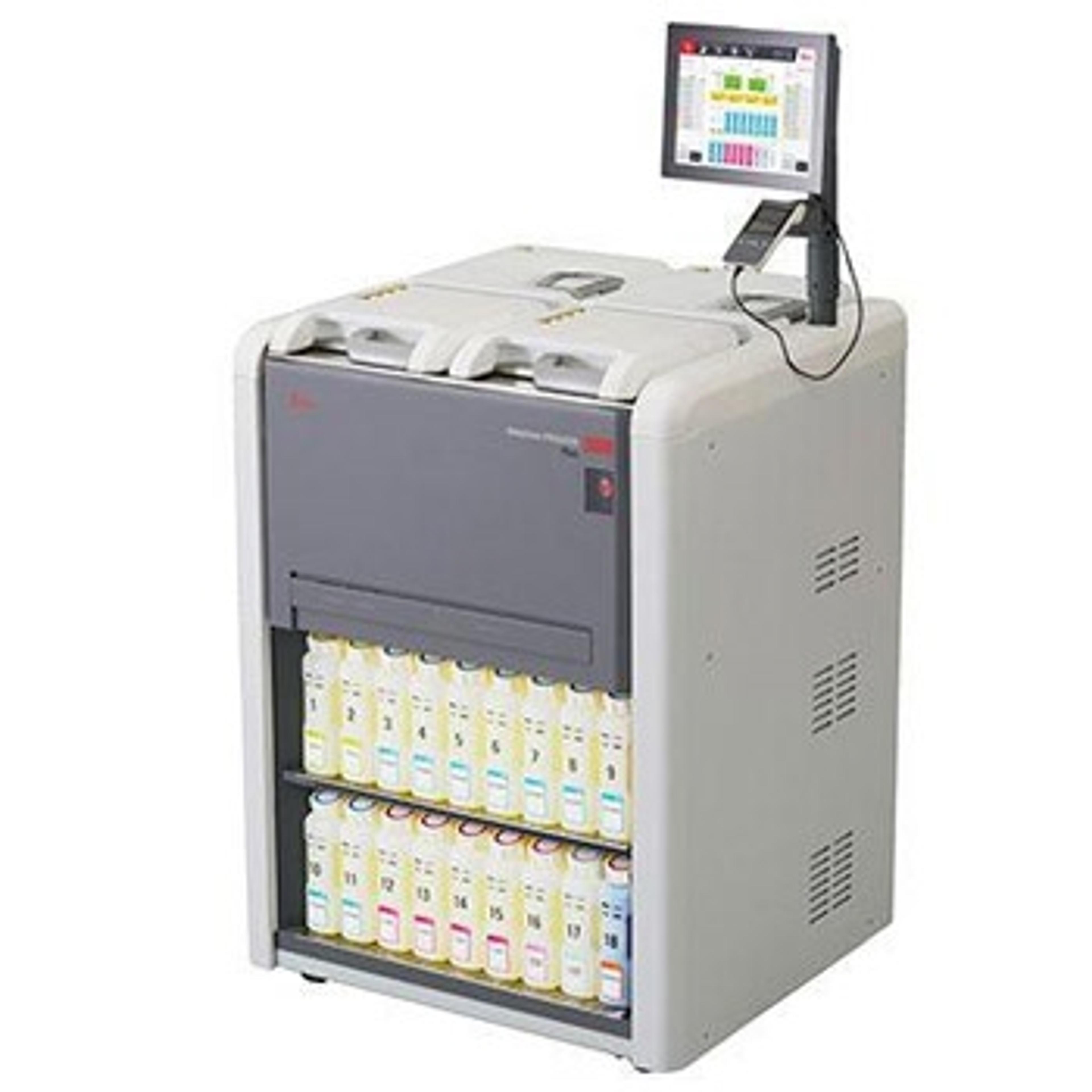
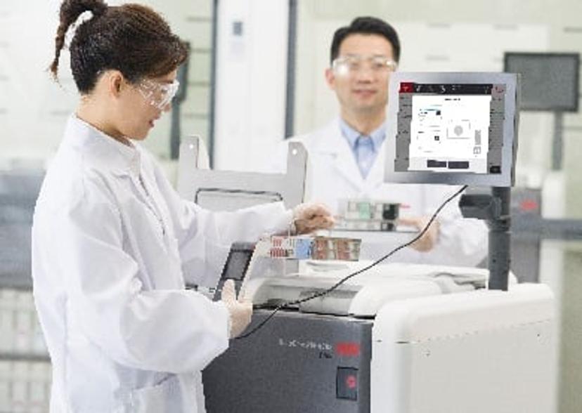
Challenges such as these can have consequences for the efficiency of clinical workflows. That’s why Leica Biosystems have developed dual-retort technology, as found in the HistoCore PEGASUS Plus Tissue Processor, in which tissues can be processed in parallel. This facilitates the flexibility of optimizing your protocol to suit a unique specimen within one instrument. The ability to speed up tissue processing, without having to compromise on optimum processing conditions, facilitates confidence in results and reduces need to reprocess under or overprocessed tissue.
This more optimized tissue processing allows downstream steps such as specialist referral or further diagnostic tests, such as molecular and biochemical techniques, to progress, enabling clinicians to efficiently provide patients a highly confident diagnosis in less time. This efficient turnaround time is revolutionary, providing patients with accurate diagnoses as fast as possible.
Here, we speak with Fiona Tarbet, senior global product manager within the tissue processing arena at Leica Biosystems, about the company’s mission to provide rapid histopathology workflows, and how the PEGASUS Plus is the ideal parallel processing solution.
What is the overall mission of Leica Biosystems?
FT: The overall vision of Leica Biosystems is what we call VISION24, and that is to provide a 24-hour diagnosis for the patient. We want to achieve this by improving the quality of the sample tissue through tissue processing, offering integrated solutions, and optimizing efficiency. We apply this to all our instrumentation, software packages, and digital imaging to provide a comprehensive portfolio of instrumentation and software that has this aim at its core. From the patient point of view, all of that is just a black hole; they have had a biopsy taken and must wait for the results. This is why its really important to us that clinical labs have the technology necessary to reduce waiting time for patients.
Within the laboratory, there are different aspects of histology that the tissue must go through. We hope that by streamlining this as much as possible we can quickly get results back to the patient. Tissue processing is the limiting time factor in biopsy diagnostics, so we want to optimize these processes and get a highly confident result as fast as possible. This enables the immunohistochemistry or molecular studies, if required, to be completed within that 24-hour period. That is our goal.
As part of our portfolio, we also have active flow cassettes, a set of reagents, and the Paraplast wax, so, it is all an integrated system. We have xylene and xylene-free options, so that we are offering a greener alternative to xylene that is compatible with the instrument and different reagent substitutes. It is important that a lab can look to incorporate greener solutions as well as focusing on efficiencies.
How does the HistoCore PEGASUS Plus help provide 24-hour diagnostics?
FT: The HistoCore PEGASUS Plus is the newest offering within our tissue processing portfolio. We already have HistoCore PELORIS 3, which is our premium tissue processor that is really targeted for the larger, high-performance laboratories. However, we wanted a solution that made this dual-retort tissue processing technology accessible for mid-range processes, so we created the HistoCore PEGASUS - a scalable, affordable solution to enable smaller laboratories achieve that 24-hour end diagnosis.
The key function of HistoCore PEGASUS Plus is to process tissue, and by offering the dual retort technology, the tissue processing protocols can be optimized. There is also flexibility within the system as customers can create their own protocols within guidelines of the instrument.
Track and trace reports are also provided on the HistoCore PEGASUS. From customer feedback, we have learnt that labs want complete visibility of the workflow: what happened to each sample, records of when reagents were changed, the lot numbers of the reagents, which technician was operating the instrument, and when. These kinds of metrics are required within clinical laboratories, and often that is done on a paper base. This is now automated with our tissue processing range. There are individual user logins and, when you perform a critical function, your name goes against that function, and that is all recorded in a user action report. There are reports that provide details of what happened on the instrument during a particular protocol run, and these can all be exported so that you have a permanent record to answer those critical questions.
So, the HistoCore PEGASUS provides tissue processing optimization, an increased throughput, validated protocols, and traceability.
What do you see for the future of this kind of technology and its impact on a diagnostics lab?
FT: There are always improvements to be made, but we are setting a standard for optimized tissue processing. It is important that compromises that impact the quality of the sample are not made as this could have repercussions on subsequent analyses, such as molecular diagnostic techniques. To ensure we have tissue samples that can be analyzed by both current and newly developed diagnostics techniques of the future, we need to have a tissue process that is of the optimum quality.
The glass slide is one consumable that is critical for digital imaging and shared pathology reporting. Shared images should be available to pathologists in different sites around the world, so that opinions and referrals can be readily given. In order to achieve this, you also need to have tissue that is processed so well that it is not distorted in any way, so that you don't need a subjective assessment.
Pathologists consult frequently because not every single tissue is the same as another tissue. There is a degree of interpretation, and that's why pathologists can train for over a decade to learn how to be a pathologist and make a diagnosis.
Overall, I anticipate a lot of changes in the future as we continue to move into a more digital age - towards AI, for instance - and as we see further advances within molecular techniques. To make this possible, it is crucial to start with a good tissue preparation that is well processed, beautifully stained, and is ready to give those answers.