Resources to Improve Your Methods in Cell-Based Assays
6 ways to improve and accelerate your methods in cell-based assays - a SelectScience editorial special feature
15 Oct 2018
Cell-based assays form the core of life sciences research. Testing cellular health in response to treatments, under different conditions, helps get one step closer to understanding their behavior in vivo. Starting from the basic step, i.e. cell counting, moving on to designing cell culture experiments to test toxicity and finally moving onto complex 3D spheroids, this special feature is dedicated to accelerating your cell-based methods.
We've curated useful resources that can improve methods and keep you informed in the ever-evolving field of in vitro biology.

How can I improve my experiments?
1. CELL-MEDIATED CYTOTOXICITY: High-Throughput, Radioactive-Free Assay
Discover a sensitive, specific and high-throughput assay to capture apoptosis, cell permeability and proliferation in a single well. Learn more about this radioactive-free assay in this downloadable method. Using this approach, you can quickly screen through different compounds and conditions, building a detailed understanding of molecular events occurring in each well.
2. CELL COUNTING: 10 Most Common Errors Made in Cell Counting
Manual cell counting, although easy, is error-prone. In this useful list, Corning has compiled the 10 most common errors, which include not suspending the sample properly and confusing debris for cells. Which one do you think is the most common bottleneck for your experiments?
3. CELL COUNTING: There's an App for That
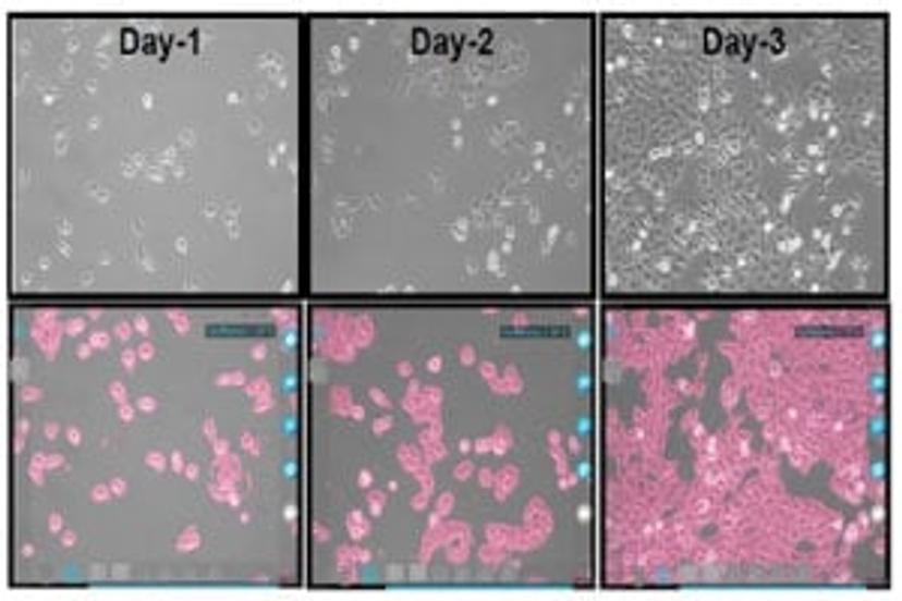
There’s an app to help you determine confluence, cell counts, and transfection more effectively. Accurately calculate total cell count in an automated fashion with the new InCellis cell imager and its smart cell culture app, designed to improve single cell-based assay normalization.
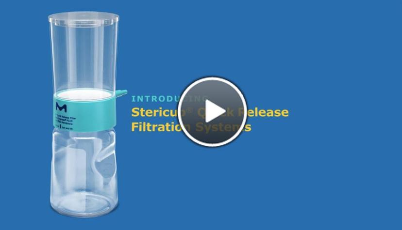
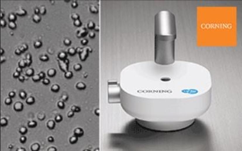
4. STERILE FILTRATION: Vacuum Filters Cleverly Designed for Ease of Use - Thanks to You
The Stericup Quick Release sterile filtration system is designed with recommendations from you. You can now write on its surface, ensure secure closure with the click seal confidence cap and use the peel-and-stick label for part/lot number.
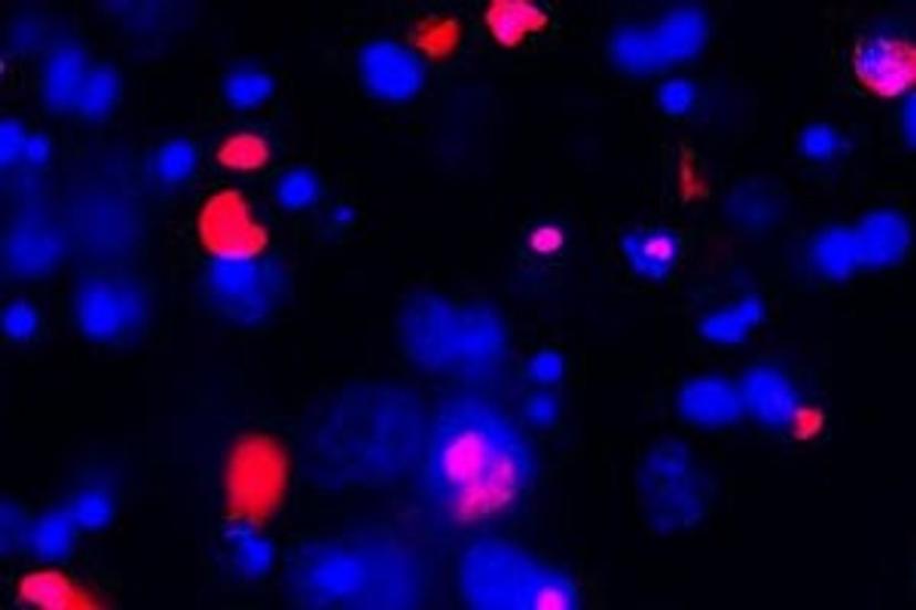
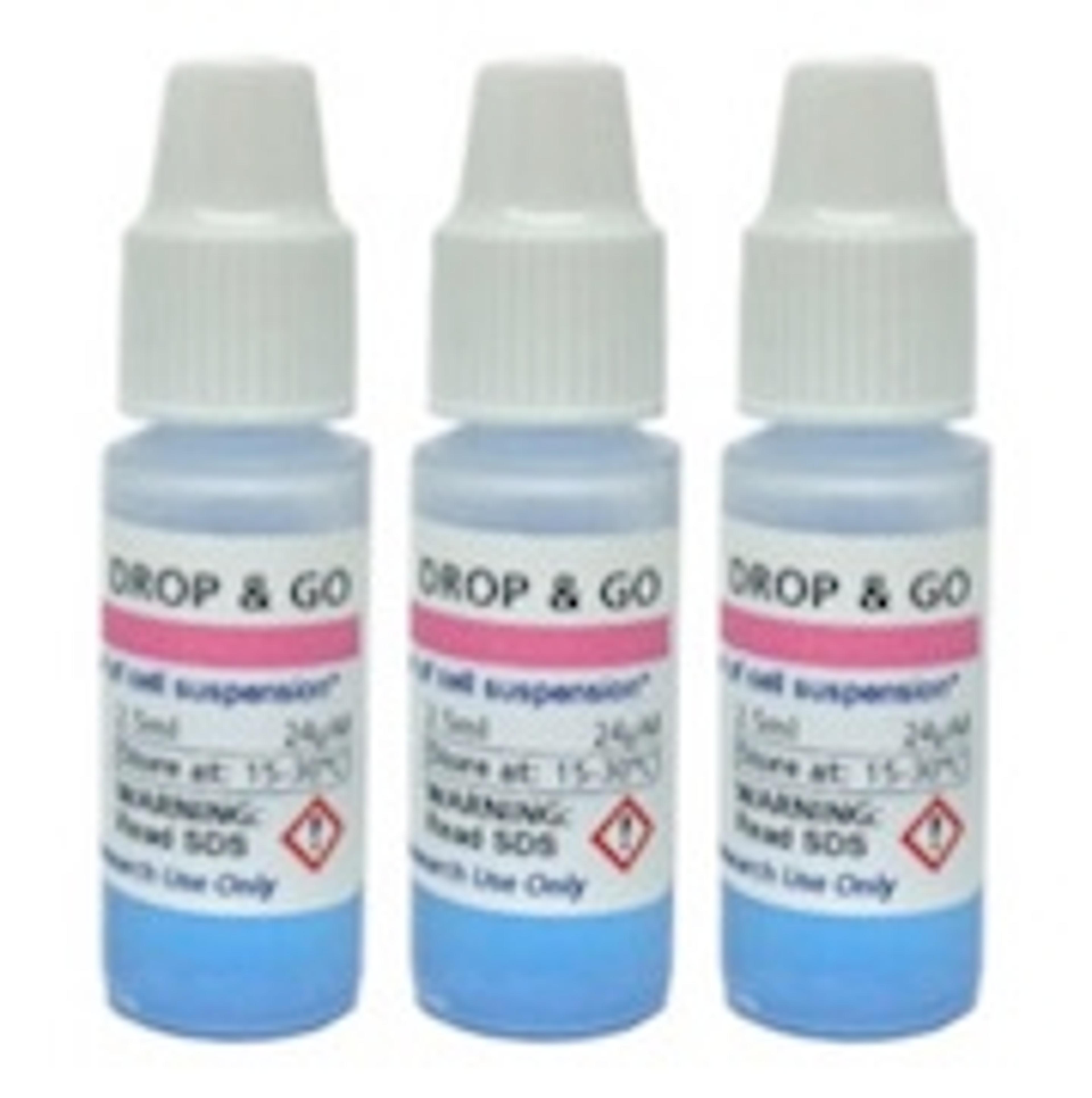
5. CELL HEALTH ASSAYS: Identify Membrane-Compromised Cells with a DNA-Binding Viability Dye
The far-red DNA-binding viability probe, DRAQ7TM by Biostatus, avoids spectral overlap with visible range and UV-excited probes, making it a great candidate for long-term real-time cell monitoring.
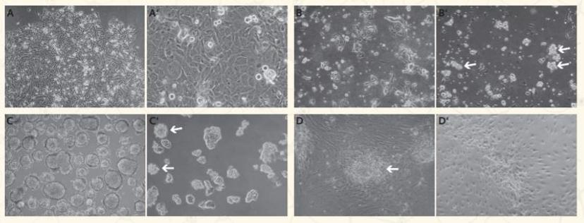
6. 3D TUMORSPHERE MODEL: Selectively Proliferate Primary Cancer Cells
Proliferating malignant cells from a mixed culture is challenging. A new method developed by PromoCell selectively supports the growth of malignant cancer cells, using unique single-cell properties as cues for exclusion selection.
Download Poster
Useful resources: webinars and video
1. An Introduction to Primary Cell Culture
Gain an introduction to cell culturing techniques, and learn about the appropriate steps to take when your cells arrive, to ensure sterile and healthy cultures.
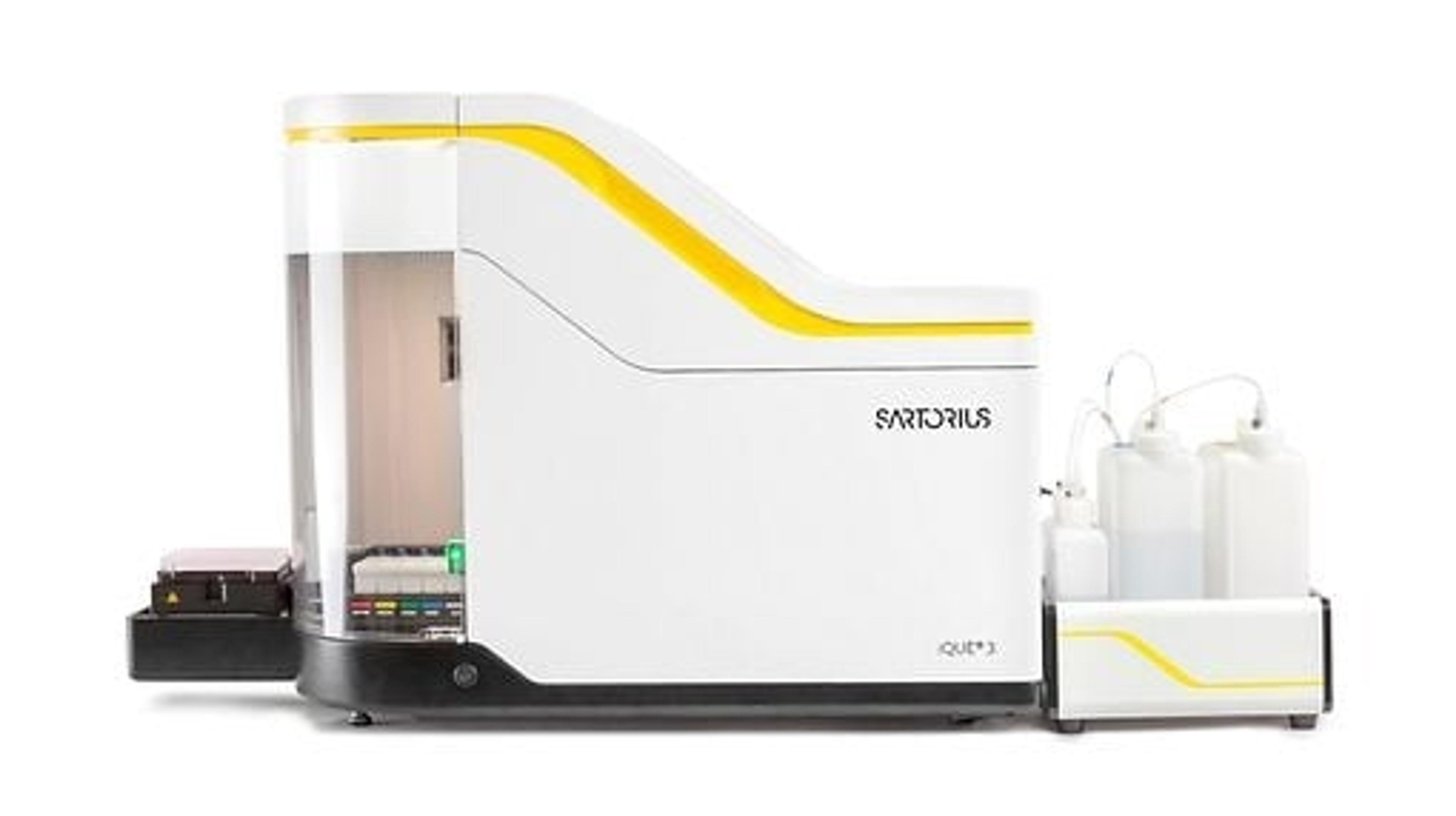
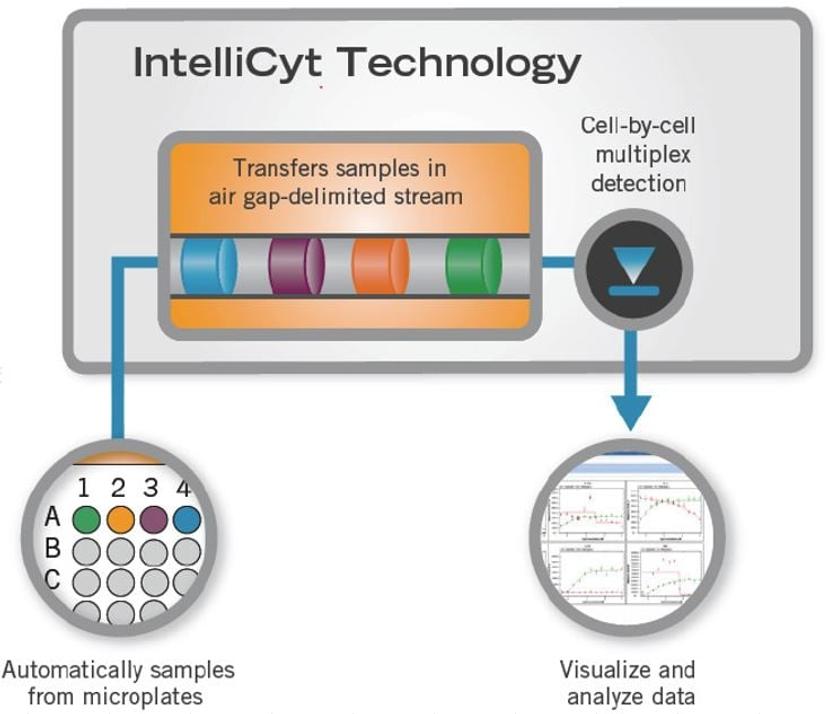
2. Live-Cell Analysis of Cell Subsets and Heterogeneity
In this webinar, learn how cell-by-cell analysis enables the monitoring and quantification of dynamic phenotypic changes in heterogeneous populations.
3. True 3D High-Content Analysis – A New Solution to Improve Quantitative Results from Your 3D Samples
In this webinar, learn how true 3D measurements can improve quantitative results over sub-sampling data sets.
4. Gaining Critical Insights for Drug Discovery Applications Using High-Content Cell Analysis Platforms
In this webinar, learn how to access cell proliferation, confluence and transfection efficiency using longitudinal live-cell imaging.
Help your peers by leaving a 2-minute expert review here.
Related Products
Request Quote for All Products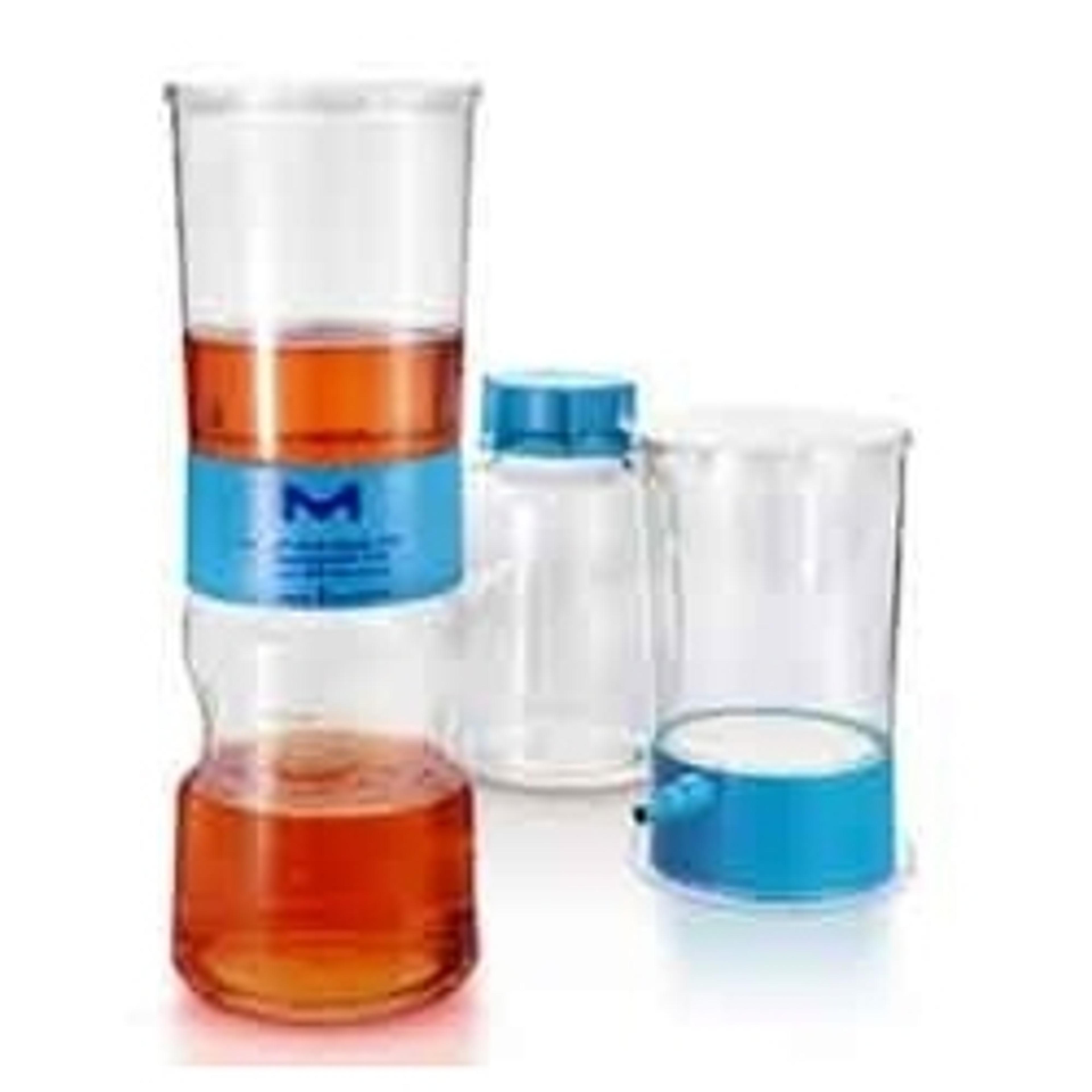
Stericup® Quick Release Filtration Systems
MerckThe Stericup® Quick Release 500 ml vacuum filter is a filter bottle system ideally suited for sterile filtration of cell culture media, buffers and reagents, that includes a Steritop® fiter unit with a re-designed Stericup® Quck Release receiver bottle. It is the next generation of the original Stericup® Sterile Vacuum Filtration System- a staple in labs around the world for decades.