From 3D Light to 3D Electron Microscopy: Highlights from the EMBL Workshop
Discover the latest news from this conference on correlative microscopy
28 Nov 2016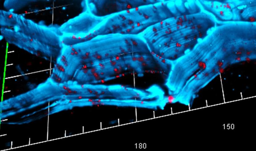
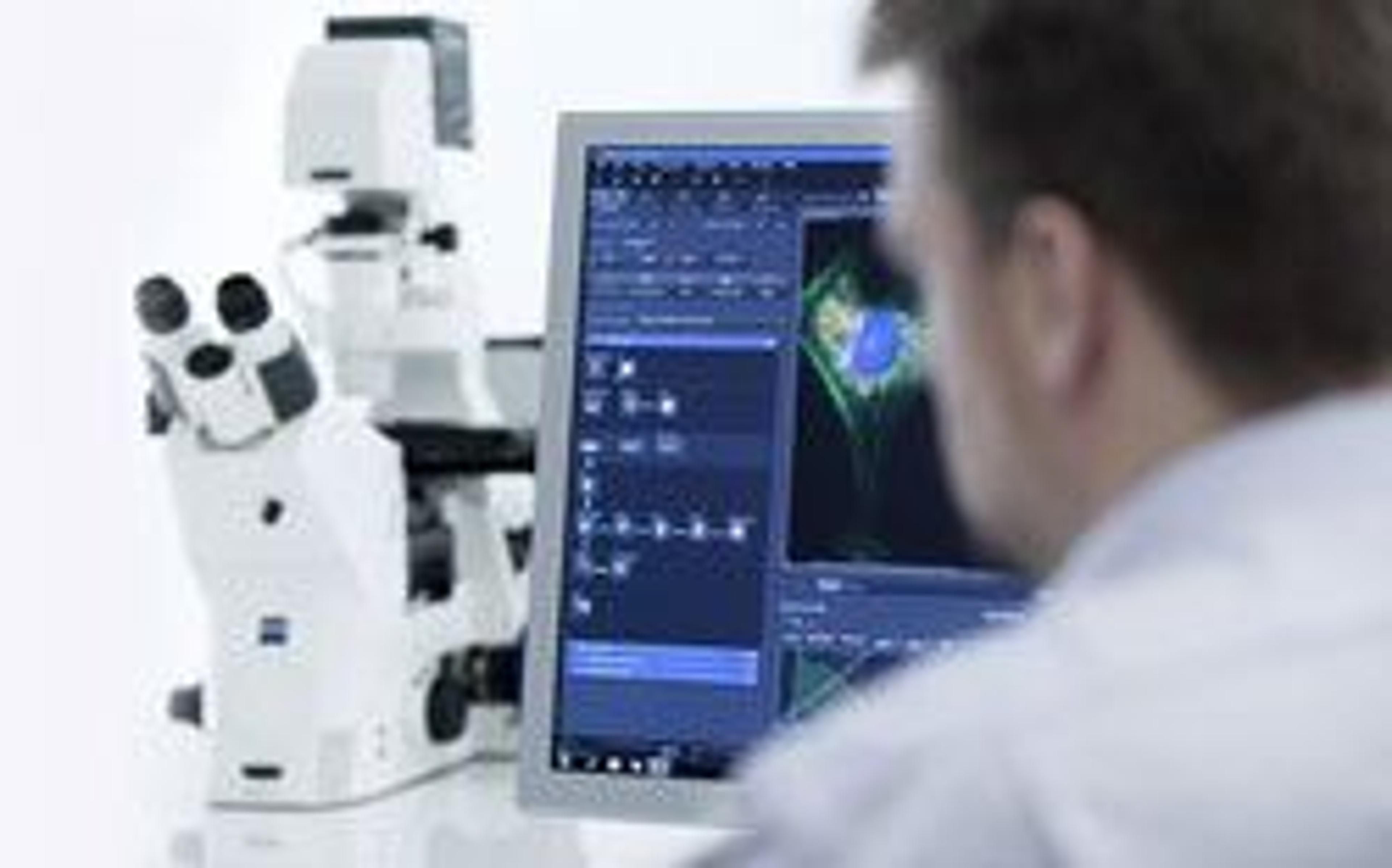
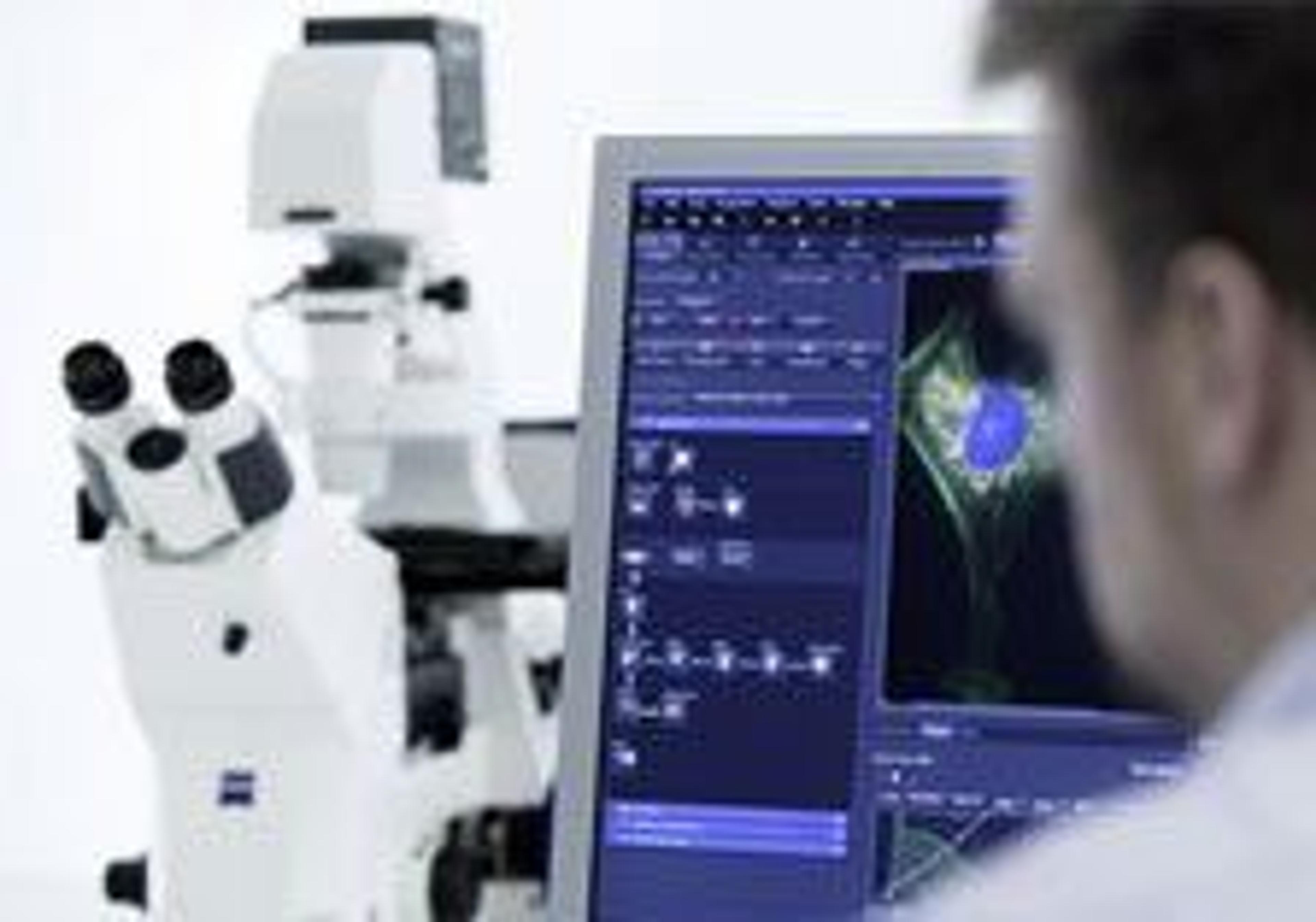
From 13 – 16 March 2016, the European Molecular Biology Laboratory (EMBL) and ZEISS Microscopy hosted a conference entitled “From 3D Light to 3D Electron Microscopy” in Heidelberg, Germany. EMBL is one of the world’s leading research institutions, and Europe’s flagship laboratory for the life sciences.
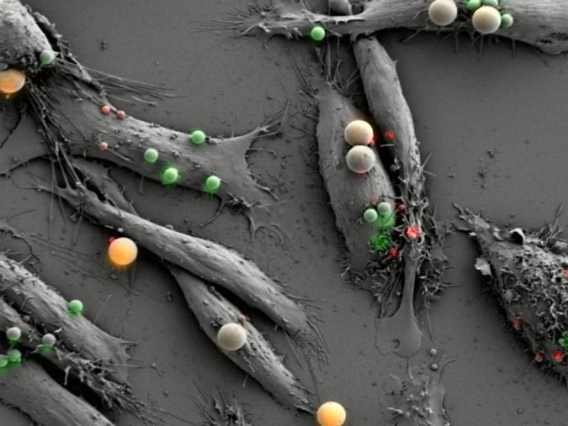
Three-dimensional (3D) imaging is becoming increasingly important in the life sciences as a way to better understand the functions and processes within cells, and techniques such as volume imaging and 3D correlative microscopy are developing fast.
Researchers of 3D correlative light and electron microscopy presented a mix of lectures and practical workshops with a focus on automated serial imaging by scanning electron microscopy from the keynote speakers Dr Winfried Denk and Prof. Dr Fred Hamprecht.
Read on for some of the interviews recorded by SelectScience® exclusively at the conference, and the latest application notes and methods for Correlative Microscopy.
Leaders in Correlative and Volume Microscopy Share Their Research at EMBL Heidelberg
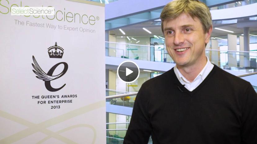
SelectScience spoke to Dr Yannick Schwab, EMBL, to discuss how correlative light and electron microscopy connects different areas of biological study, how these techniques could be used in the future, and how the conference benefits the correlative microscopy community. Watch the video>>
Software Guide to ZEN 2 (Blue Edition): The Shuttle & Find Module
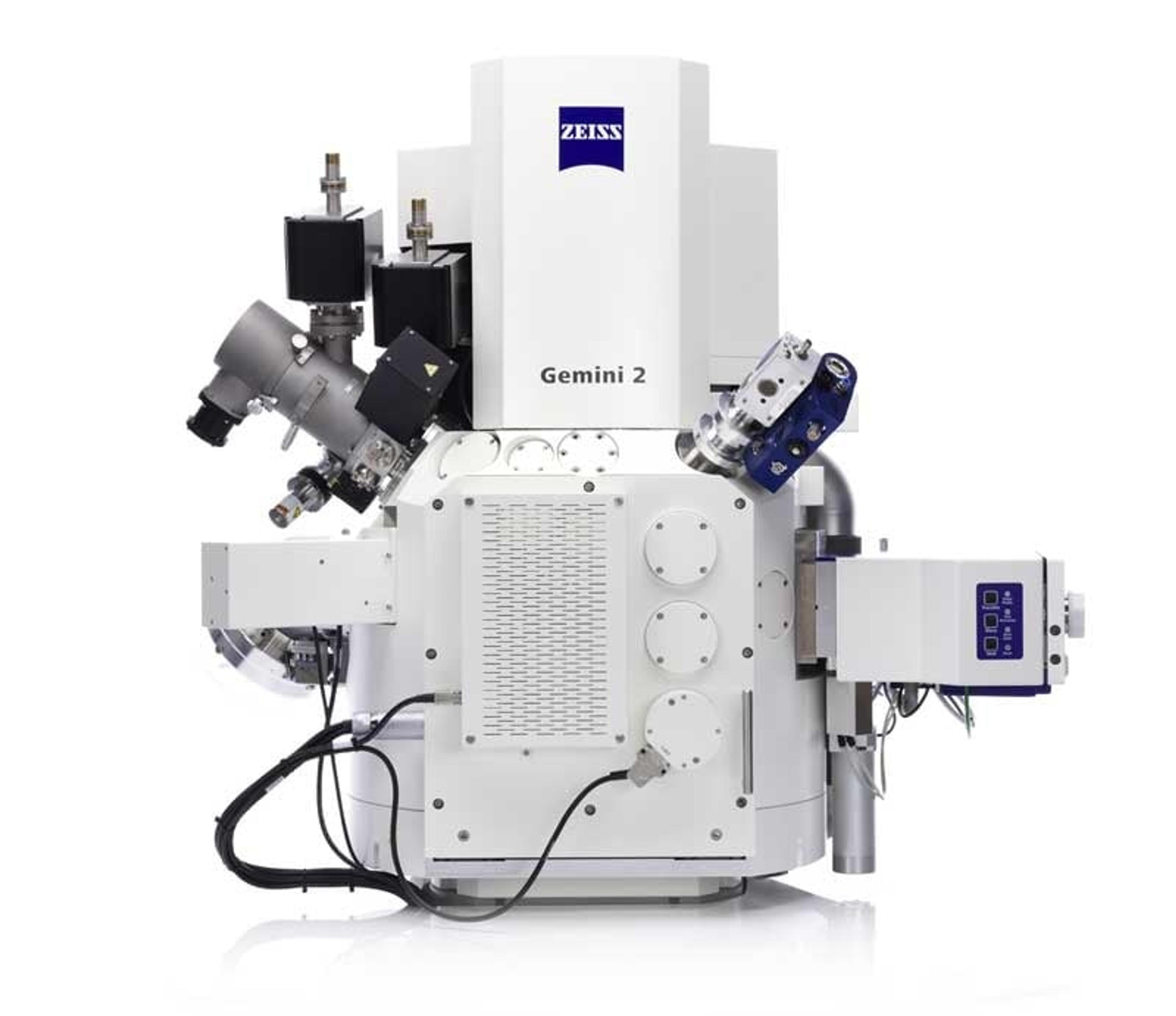
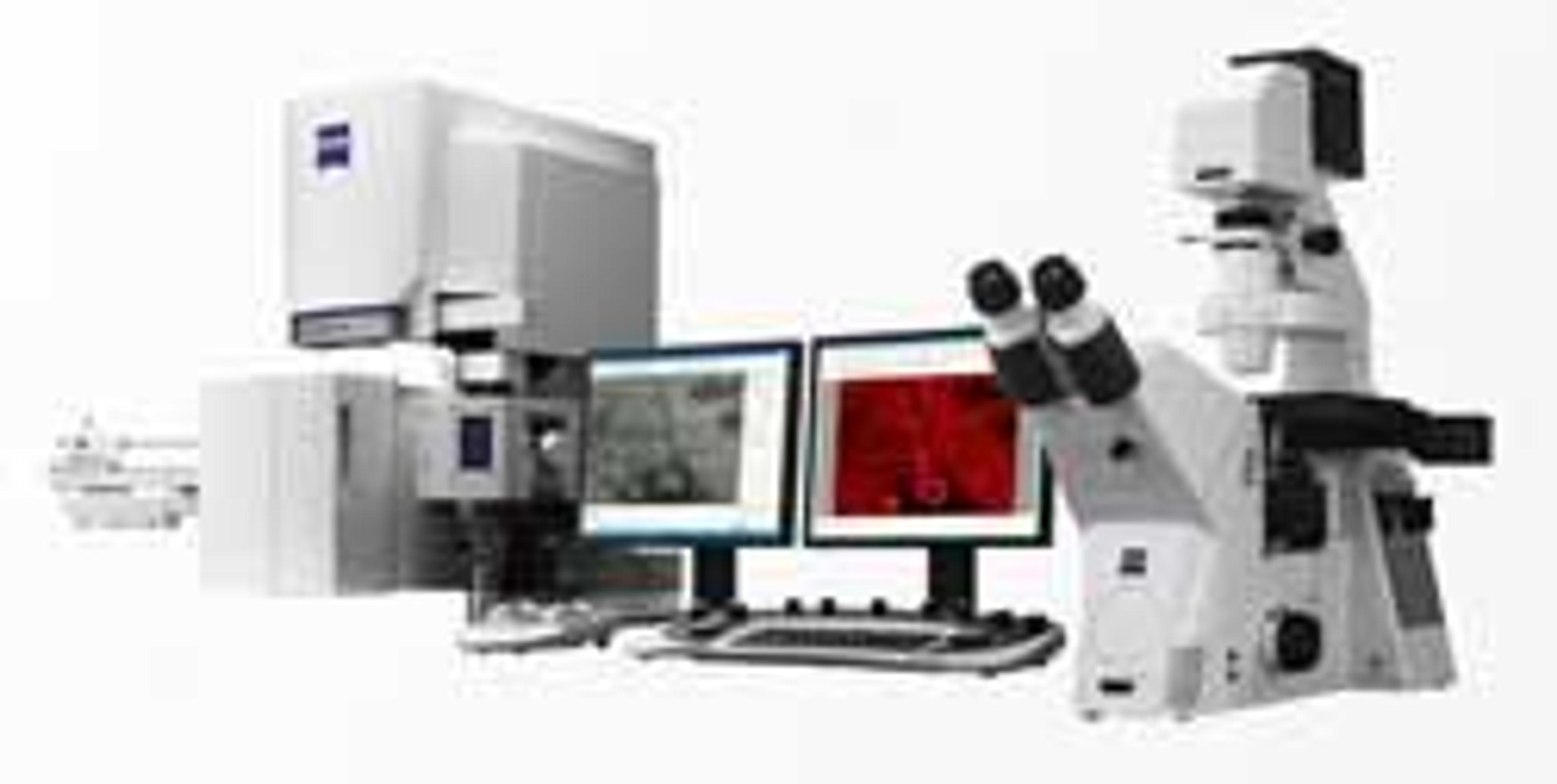
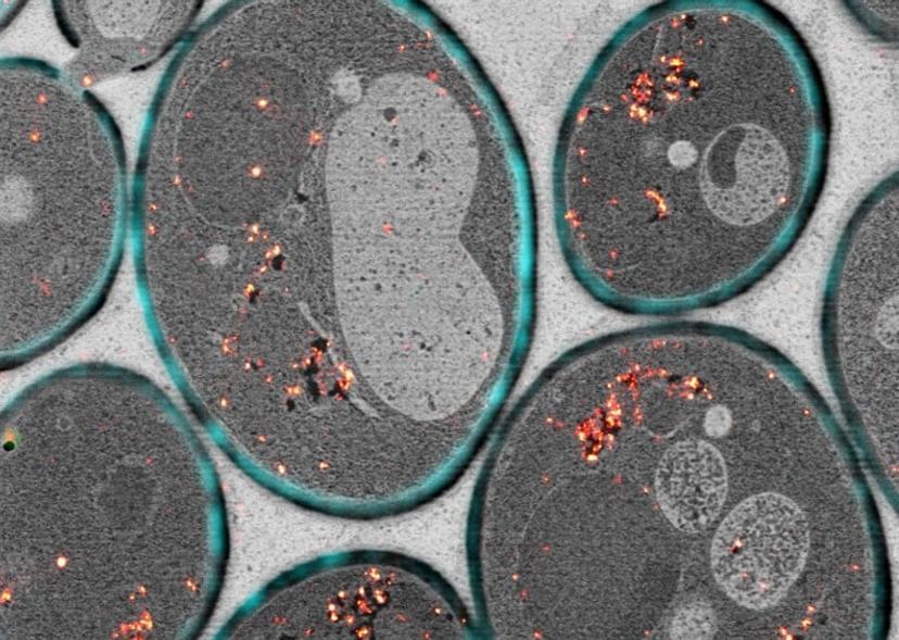
Correlative microscopy is used to combine scanning electron microscopy and light microscopy to produce one image. This software guide describes how to use the Shuttle & Find Module from ZEISS Microscopy to optimize your imaging for biological and material samples using correlative microscopy. Download the guide>>
Dr Peter O’Toole, University of York: Making Light Work of 3D Bioimaging
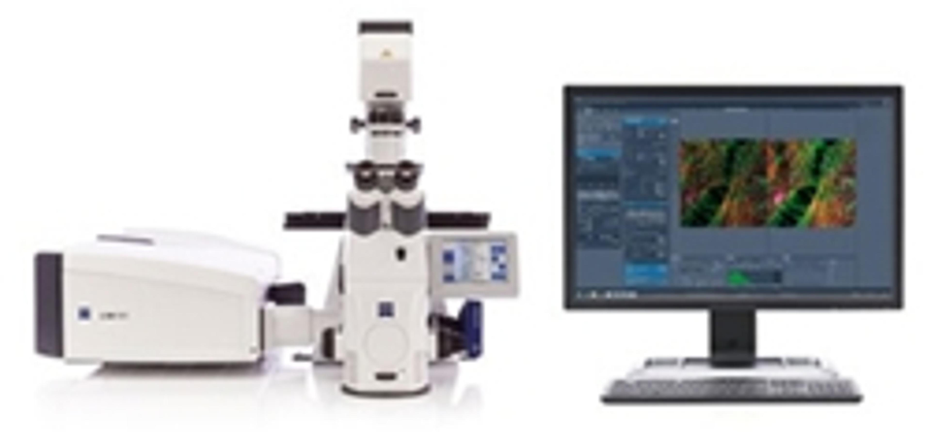
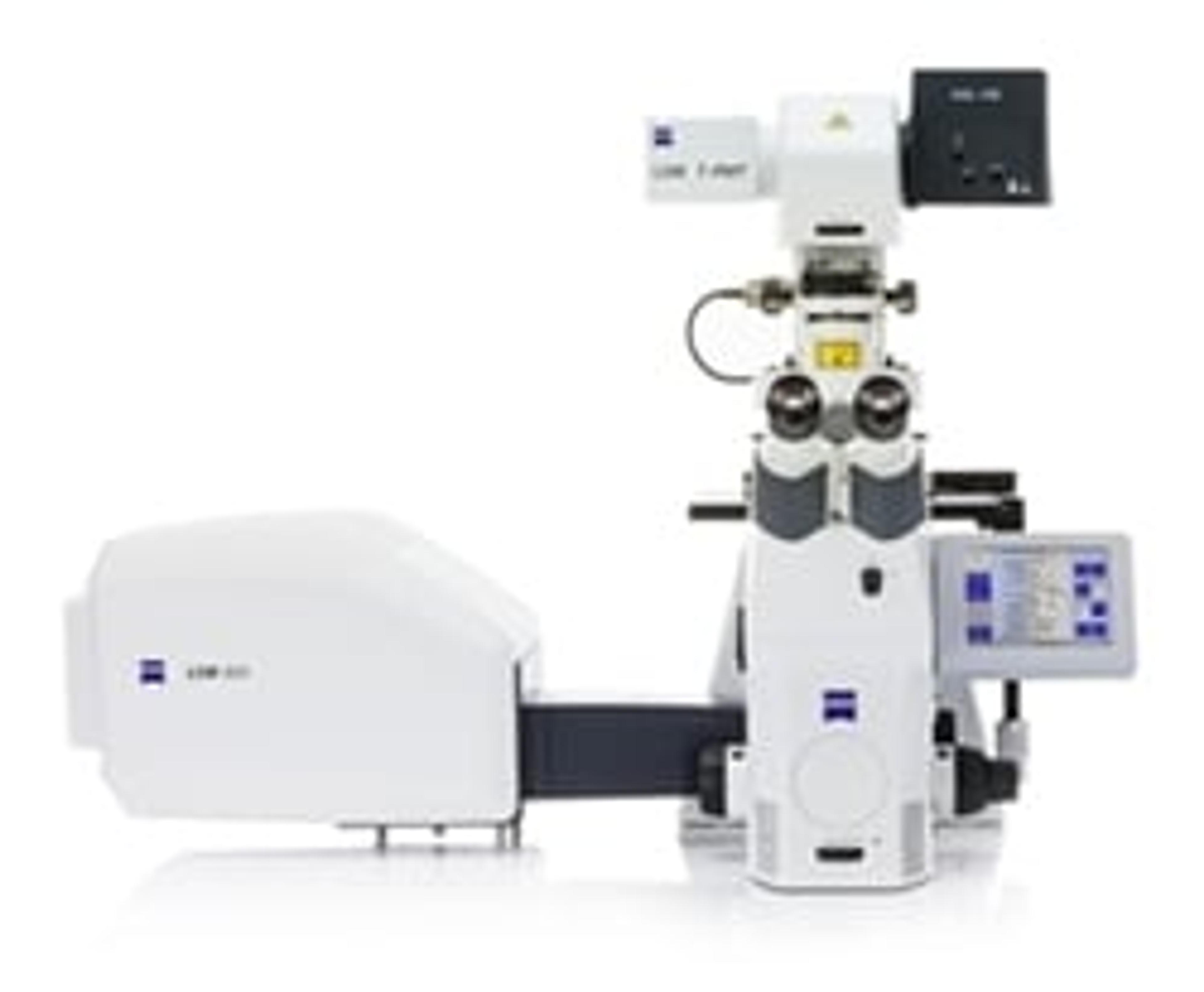
In this presentation, Dr Peter O’Toole, from the University of York, discusses the benefits of microscopy for biologists and his experience as a beta-tester of the ZEISS LSM 880 with Airyscan. Learn about the benefits of light sheet microscopy for in vivo 3D imaging and hear how Dr Peter O’Toole hopes to implement correlative microscopy techniques to understand genomic, metabolomics and biochemical data. Watch the presentation>>
Correlative Microscopy Protocols: A Reference Guide to Correlative Sample Preparation
This guide provides an overview of existing sample preparation know-how for correlative microscopy. Many existing protocols for TEM are typically applicable to the modern technique of correlative microscopy with little or no modifications. Download the guide>>
Optimizing Energy Conversion and Storage Materials Using Correlative Microscopy at HZB
Professor Silke Christiansen, from the Helmholtz Center for Materials and Energy, discusses using correlative microscopy in her research into nanomaterials for solar cells and energy storage. She is studying new ways of converting solar energy into other forms of energy as well as finding efficient storage for the energy. Watch the video>>
To learn more about the conference and watch more exclusive interviews, visit our EMBL 3D Microscopy Special Feature page.
Do you use any of the products mentioned in this article? Is there a product you couldn't do without? Share your experiences with our worldwide community and write a review today.
Image:anyaivanova/Shutterstock