Trailblazing earlier diagnosis of multiple myeloma for better patient outcomes
Guest editorial by Dale Powner, Country Manager for the UK, Ireland and Nordics at Binding Site, part of Thermo Fisher Scientific
28 Aug 2025Editorial article
Multiple myeloma (MM) is the second most common hematological malignancy in the UK, resulting in approximately 3,000 deaths every year.1 It primarily affects individuals over the age of 65, and is two to three times more prevalent among Black and Asian populations than in Caucasian groups.1,2 MM develops when plasma cells in the bone marrow become malignant and produce large amounts of monoclonal proteins (M proteins) – commonly known as paraproteins – which lack the ability to defend against infections.3 The proliferation of these cancerous plasma cells suppresses the production of healthy blood cells, leading to low counts of red blood cells, other white blood cells and platelets,2,3as well as complications such as anemia, increased susceptibility to infections, and organ damage. As the disease progresses, more serious complications – including renal failure, hypercalcemia and bone lesions – may appear.
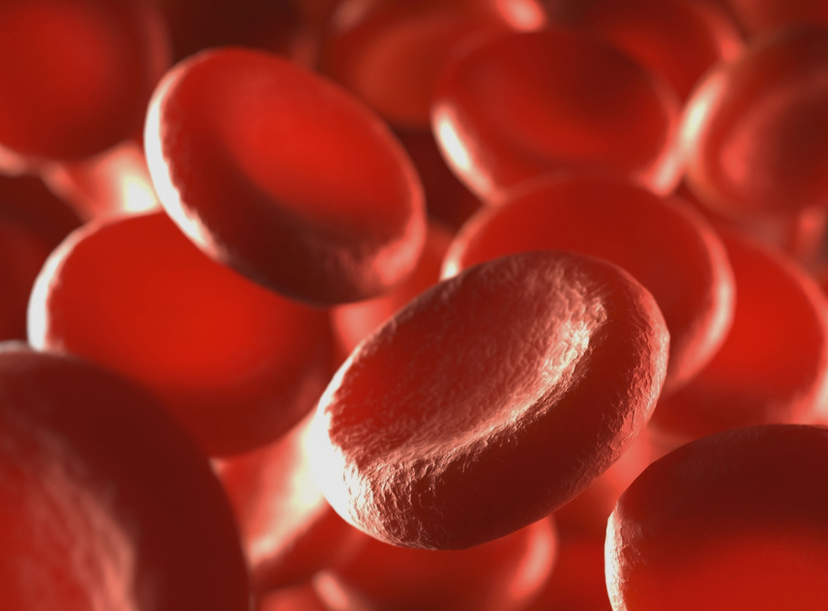
Discover how early diagnosis transforms outcomes in multiple myeloma in this guest editorial. Image copyright: ktsdesign / 123rf
The most common types of myeloma – IgG, IgA and IgM – account for over 80 percent of all cases and are characterized by the overproduction of a monoclonal intact immunoglobulin. In almost all cases, this is accompanied by an overproduction of kappa (κ) or lambda (λ) monoclonal serum free light chains (sFLCs). There are various other disease subtypes, and rare forms of MM that involve the overproduction of less common immunoglobulins, including IgD and IgE myelomas, also exist.4 These variants are often more aggressive than the more common IgG and IgA types, and require specific diagnostic approaches.5,6
Non-secretory myeloma is another unusual subtype affecting one to two percent of patients, and is characterized by the production of little to no detectable paraproteins by traditional methods.7 Conversely, in light chain-only myeloma, only fragments of the immunoglobulin structure – known as light chains – are produced in excess.8 Light chain-only myeloma accounts for 15 to 20 percent of all myelomas, but FLCs – often referred to as Bence Jones proteins when detected in the urine – are secreted by most patients with intact immunoglobulin myeloma. FLC secretion is associated with a high risk of kidney complications such as cast nephropathy, or as in AL amyloidosis.7
Delayed diagnosis
The treatment landscape for MM has undergone a significant transformation over the last two decades, thanks to the development of proteasome inhibitors, immunomodulators and monoclonal antibodies. These advances have dramatically extended median survival times from just three years to up to 10 years. However, overall survival rates for MM still remain low, at least partly due to late/delayed diagnosis, which can be partly attributed to a lack of awareness of symptoms and diagnostic testing guidelines among GPs and other non-hematologist specialists where affected patients invariably present in the early stages of the disease. As a result, initial symptoms could be mistakenly linked to more common conditions, delaying referrals to specialists for additional tests and treatment until more serious health issues develop.9
Budgetary constraints within the NHS further complicate efforts to improve early diagnosis by making it harder for laboratories to adopt newer, more accurate diagnostic tests, despite being more financially sustainable in the long run. These diagnostic challenges can be exacerbated by cultural and demographic disparities in disease risk and health literacy between communities.10,11 For instance, awareness of MM and its early symptoms remains low in higher risk communities, such as Asian and Black ethnic groups, leading to delays in seeking medical advice and receiving a diagnosis.11
Benefits of early disease detection
Early detection is likely to improve outcomes in MM, as it gives clinicians more opportunities to intervene before complications such as renal failure or vertebral fractures arise. In addition, studies have shown that early-stage MM is less genetically diverse than advanced MM,12 making it more responsive to treatment. This means that treatment can begin with less aggressive strategies, reducing the potential for harmful side effects. From a healthcare economics perspective, timely diagnosis also reduces the need for expensive emergency treatments, such as vertebroplasty for spinal collapse or dialysis for renal failure.13 It is likely that preventing these complications and therefore the associated costs, will offset the marginal additional cost of implementing the guideline recommended testing panel.14 These factors highlight the current need for timely diagnosis to support more effective disease management, improve patients’ quality of life and promote long-term survival.
Methods of MM diagnosis
The International Myeloma Working Group (IMWG) established clear diagnostic criteria for MM and its precursor condition, monoclonal gammopathy of undetermined significance (MGUS), in 2003, updated in 2014.15,16 In 2009, the IMWG also recommended that all myeloma investigations should include sFLC testing in place of urine analysis.17In 2016 and 2021, NICE NG3514 and the BSH18 respectively, published similar recommendations for the use of sFLC testing in place of urine analysis. MGUS is a condition in which plasma cells produce abnormal immunoglobulins or paraproteins, but without causing symptoms or organ damage.19It is usually only detected incidentally, when routine blood tests reveal slightly elevated paraprotein levels. Diagnosis is then confirmed when the level of paraproteins in the blood is less than 30 g/l, the proportion of abnormal plasma cells in the bone marrow is below 10 percent, and there are no signs of organ damage.16 Around one percent of patients with MGUS progress to MM annually,20 so regular monitoring of patients diagnosed with MGUS is essential for the early detection of progression to symptomatic disease.
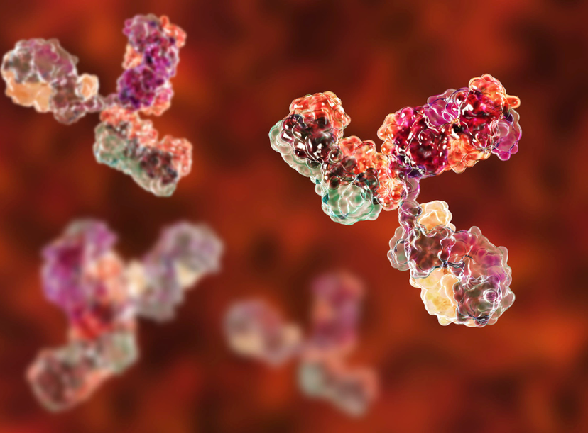
IgG, IgA and IgM myelomas are characterized by the overproduction of a monoclonal intact immunoglobulin and in most cases, accompanied by an overproduction of kappa (κ) or lambda (λ) monoclonal serum free light chains (sFLCs). Image copyright: drmicrobe / 123rf
Active MM is identified in the presence of ≥10 percent clonal plasma cells in the bone marrow or a biopsy-proven plasmacytoma, plus one of the SLiM or CRAB diagnostic criteria. SLiM biomarkers are ≥60 percent bone marrow plasma cells; a serum-involved: uninvolved FLC ratio of 100:1 or greater where the involved sFLC has a concentration of at least 100 mg/L; and the presence of at least one MRI-detected focal lesion measuring 5 mm or larger.16 The acronym CRAB is used to summarize types of organ damage consistent with active MM, and refers to calcium elevation (serum calcium >11 mg/dL), renal dysfunction (serum creatinine >2 mg/dL), anemia (hemoglobin <10 g/dL) and bone lesions.16
Serum and urine protein electrophoresis have traditionally been key tools for detecting monoclonal paraproteins, but urine electrophoresis is limited by low sensitivity and dependence on patient compliance, compromising its reliability. Unlike urine tests, sFLC assays use blood samples, which are collected in all patients suspected of having MM. Monoclonal FLCs are present in the serum in the vast majority of patients with MM (97 percent),21 but only found in the urine in a subset of patients due to the effects of renal filtration. This makes sFLC a more specific biomarker for MM than detection of Bence Jones proteins in urine. sFLC assays also eliminate the logistical barriers associated with urine collection, reducing issues of patient non-compliance.
sFLC tests are particularly useful in identifying cases of light chain myeloma or non-secretory myeloma, where traditional diagnostic methods may fail to detect low levels of circulating paraproteins due to lack of sensitivity.22 sFLC assays quantitatively measure the levels of free κ and λ light chains in the serum to identify abnormal κ/λ ratios, a key indicator of clonal plasma cell activity. Abnormal sFLC ratios have been observed in up to 70 percent of non-secretory myeloma cases, even when paraproteins are absent or only present in trace amounts using other methods, making sFLC assays an invaluable tool for identifying this rare MM subtype.19 Due to these many advantages, the National Institute for Health and Care Excellence (NICE) updated its guidelines in 2016 to recommend sFLC assays in place of urine protein electrophoresis to ensure more dependable detection of the monoclonal FLC component found in most MM patients.23
Bottlenecks in large-scale sFLC implementation
Improving education and awareness of MM among the general public and clinicians is essential for overcoming the current obstacles to early MM diagnosis.9 Organizations such as Myeloma UK and Blood Cancer UK play important roles in enhancing the understanding of MM symptoms and promoting the importance of timely diagnostics among the general population. They also actively develop resources and training material for clinicians, such as Myeloma UK’s Early Diagnosis Programme,4 equipping them to recognize red flags.
Despite clear advantages, the adoption of sFLC assays for early diagnosis remains inconsistent. Budget constraints mean that many UK hospitals are still only using them to monitor disease progression, and not for initial MM detection, as recommended by the NICE,14 the British Society of Haematology18,25,26 and the IMWG.17 To overcome this obstacle, healthcare systems must recognize the long-term savings and patient benefits offered, and reallocate funds to support timely MM detection.
Streamlining diagnostic pathways is another key step to boosting accuracy and efficiency in MM diagnosis. This has already been illustrated at several teaching hospitals across the UK, where clinical laboratory scientists are able to proactively add sFLC assays to testing panels when MM is suspected, even in the absence of a specific request from a clinician. This approach has resulted in transformative benefits and could help to overcome current diagnostic pathway inefficiencies.
Fortunately, many UK labs already have the tools to improve MM diagnosis; Freelite® assays performed on the Optilite® analyzer are widely used for disease monitoring throughout the NHS. These tests were introduced in 2001 as the first commercially available automated immunoassays of their kind, with the aim of enhancing precision and reducing testing delays. The assays remain underused for diagnosis, but it would require minimal investment to expand their use, as the necessary instruments are already integrated into labs’ existing infrastructures. Expanding automation to MM diagnostics could improve accuracy, reduce errors, streamline workflows and enable earlier detection, while lowering the expense tied to late-stage disease complications.

The Binding Site's Freelite® assays performed on the Optilite® analyzer are widely used for disease monitoring for multiple myeloma diagnosis
Pioneering timely disease detection
Early diagnosis consistently proves to be the most effective strategy for managing MM, as it helps to prevent severe complications and dramatically improves prognoses. However, healthcare systems have yet to consistently adopt best practices for timely MM diagnosis, and the full potential of sFLC assays for prompt diagnosis has largely been overlooked. Addressing healthcare inefficiencies, promoting wider education and reallocating budgets are all vital steps to remedying this issue. Companies like Binding Site play a crucial role in this transition by providing essential diagnostic tools. They also offer educational resources and advocacy to promote transformative diagnostic methods, enabling timely care and reducing the burden of late-stage disease on the healthcare system.
Freelite® and Optilite® are registered trademarks of The Binding Site Group Limited (Birmingham, UK) in certain countries. Other brand or product names may be trademarks of their respective holders.
Not for use in China
References
1. National Institute for Health and Care Excellence (NICE) Clinical Knowledge Summaries (CKS). Multiple myeloma: Prevalence. April 2022. Accessed March 4, 2025. https://cks.nice.org.uk/topics/multiple-myeloma/background-information/prevalence
2. Myeloma UK. Ask the nurse: Myeloma and blood cells. 2023. Accessed March 4, 2025. https://www.myeloma.org.uk/library/ask-the-nurse-myeloma-and-blood-cells/
3. Cancer Research UK. Symptoms of myeloma. 2024. Accessed March 4, 2025. https://www.cancerresearchuk.org/about-cancer/myeloma/symptoms
4. Pandey S, Kyle RA. Unusual myelomas: a review of IgD and IgE variants. Oncology (Williston Park). 2013;27(8):798-803.
5. Agbuduwe C, Iqbal G, Cairns DA, et al. Clinical Characteristics and Outcomes of IgD Myeloma: Experience across UK National Trials. Blood Adv 2022; 6(17):5113-23. doi: 10.1182/bloodadvances.2022007608.
6. Nafria Jimenez B, Oliveros Conejero R. IgE multiple myeloma: detection and follow-up. Adv Lab Med 2022 3:1:79-90. doi: 10.1515/almed-2021-0087.
7. Brioli A, Giles H, Pawlyn C, et al. Serum free immunoglobulin light chain evaluation as a marker of impact from intraclonal heterogeneity on myeloma outcome. Blood. 2014;123(22):3414-3419. doi:10.1182/blood-2013-12-542662
8. International Myeloma Foundation. Types of multiple myeloma. January 6, 2025. Accessed March 4, 2025. https://www.myeloma.org/types-of-myeloma
9. Kariyawasan CC, Hughes DA, Jayatillake MM, Mehta AB. Multiple myeloma: causes and consequences of delay in diagnosis. QJM. 2007;100(10):635-640. doi:10.1093/qjmed/hcm077
10. Raleigh V. The Health of People from Ethnic Minority Groups in England. 2023. Accessed March 4, 2025. https://www.kingsfund.org.uk/insight-and-analysis/long-reads/health-people-ethnic-minority-groups-england
11. Myeloma UK. Ask the nurse: Black people and myeloma. 2023. Accessed March 4, 2025. https://www.myeloma.org.uk/library/ask-the-nurse-black-people-and-myeloma/
12. Bong IPN, Esa E. Molecular genetic aberrations in the pathogenesis of multiple myeloma. Asian Biomedicine. 2023;17(4):152-162. doi:10.2478/abm-2023-0056
13. Porteous A, Gibson S, Eddowes LA, et al. An Economic Model to Establish the Costs Associated With Routes to Presentation for Patients With Multiple Myeloma in the United Kingdom. Value Health Reg Issues. 2023;35:27-33. doi:10.1016/j.vhri.2023.01.001
14. National Institute for Health and Care Excellence. Myeloma: diagnosis and management, NICE guideline [NG35]. February 10, 2016. Accessed March 3, 2025. https://www.nice.org.uk/guidance/ng35
15. Kyle R. Criteria for the classification of monoclonal gammopathies, multiple myeloma and related disorders: a report of the International Myeloma Working Group. Br J Haematol. 2003;121(5):749-757. doi:10.1046/j.1365-2141.2003.04355.x
16. Rajkumar SV, Dimopoulos MA, Palumbo A, et al. International Myeloma Working Group updated criteria for the diagnosis of multiple myeloma. Lancet Oncol. 2014;15(12):e538-e548. doi:10.1016/S1470-2045(14)70442-5
17. Dispenzieri A, Kyle R, Merlini G, et al. International Myeloma Working Group guidelines for serum-free light chain analysis in multiple myeloma and related disorders. Leukemia 2009;23(2):215-24 doi: 10.1038/leu.2008.307.
18. Sive J, Cuthill K, Hunter H, et al., British Society of H. Guidelines on the diagnosis, investigation and initial treatment of myeloma: a British Society for Haematology/UK Myeloma Forum Guideline. Br J Haematol 2021 193(2):245-68. doi: 10.1111/bjh.17410.
19. Myeloma UK. Monoclonal gammopathy of undetermined significance (MGUS). 2024. Accessed March 4, 2025. https://www.myeloma.org.uk/understanding-myeloma/related-conditions/mgus/
20. Hevroni G, Vattigunta M, Kazandjian D, et al. From MGUS to multiple myeloma: Unraveling the unknown of precursor states. Blood Rev. 2024;68:101242. doi:10.1016/j.blre.2024.101242
21. Katzmann JA, Kyle RA, Benson J, et al. Screening Panels for Detection of Monoclonal Gammopathies. Clin Chem. 2009;55(8):1517-1522. doi:10.1373/clinchem.2009.126664
22. Dejoie T, Corre J, Caillon H, et al. Serum free light chains, not urine specimens, should be used to evaluate response in light-chain multiple myeloma. Blood. 2016;128(25):2941-2948. doi:10.1182/blood-2016-07-726778
23. Kyle RA. Serum Free Light Chain Assays – Their Role in Multiple Myeloma. Eur Oncol Haematol. Published online 2007:27. doi:10.17925/EOH.2007.0.0.27
24. Myeloma UK. Early diagnosis programme. 2024. Accessed March 4, 2025. https://www.myeloma.org.uk/research-and-impact/panels-and-advisory-groups/early-diagnosis-programme/
25. Bird JM, Owen RG, D’Sa S, et al. Guidelines for the diagnosis and management of multiple myeloma 2011. Br J Haematol. 2011;154(1):32-75. doi:10.1111/j.1365-2141.2011.08573.x
26. Pratt G, Jenner M, Owen R, et al. Updates to the guidelines for the diagnosis and management of multiple myeloma. Br J Haematol. 2014;167(1):131-133. doi:10.1111/bjh.12926
Related products
Request Quote for All Products
Optilite® Freelite® Assays
Binding SiteDetect and monitor Multiple Myeloma (MM) with Freelite® assays. The time-tested and proven solution for kappa and lambda serum free light chain (sFLC) analysis