Lutèce Dynamics announces its first commercial VertX installation to advance infection research at SLAS Europe 2025
First commercial sale of VertX, the cutting-edge, label-free live microscope module at William Morey General Hospital, enabling high‑resolution imaging of biopsies to support infection research and personalized treatment
20 May 2026Industry news
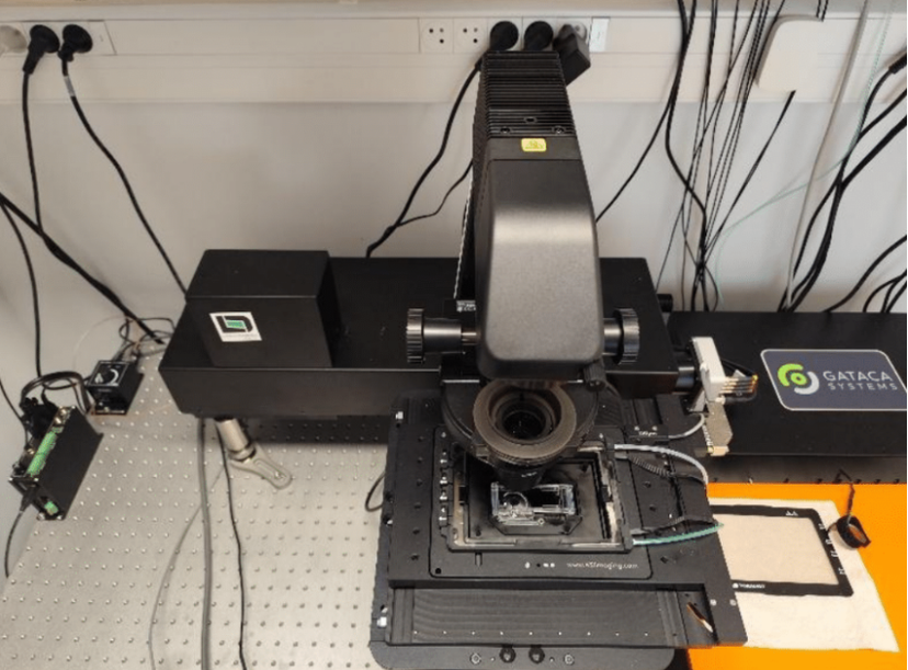
VertX® imaging module integrated with a microscope for label‑free sample analysis
Lutèce Dynamics has completed its first commercial sale and installation of its VertX® label-free live microscope module at Centre Hospitalier Chalon-sur-Saône William Morey in Chalon-sur-Saône, France. This installation is especially meaningful for Lutèce Dynamics as it takes place in Chalon-sur-Saône, the city where photography was invented by Joseph Niépce.
Announced at SLAS Europe 2026 in Vienna, Austria, the installation took place in collaboration with Dr. Thomas Maldiney and his research team from CTM DIJON INSERM and Université Bourgogne Europe. The VertX module, combined with a Nikon microscope and a confocal module from Confocal NL, will be used to transform high-resolution microscopy imaging of human biopsies and to better understand the pathophysiology of disease.
By enabling clinicians and researchers to visualize complex tissues with unprecedented clarity and precision, the system aims to support a deeper understanding of disease mechanisms and to enhance clinical and translational research capabilities at the hospital and its academic partners.
Supporting research on bacterial infections in perfusion catheters
Dr Thomas Maldiney’s main research focus is to understand the key factors driving bacterial infections through perfusion catheters, which represent a major public health risk.
The high-resolution dynamic imaging provided by the VertX module enables researchers to:
- Quantify bacterial growth in catheters
- Visualize and analyze the formation of bacterial biofilms
- Quantify the amount of metabolically active bacteria
These capabilities will play a pivotal role in identifying patient-specific perfusion formulas to reduce the risk of bacterial infections associated with catheter use.
A new vision for label-free 3D imaging of living tissues
At Lutèce Dynamics, the long-term vision is to transform diagnostics and biological research by enabling the detection of all living cells in complex tissues without the need for labeling or fixation.
By allowing life in 3D at microscopic scale to be observed in its natural state, the VertX module aims to open new possibilities for studying tissue organization, cell behavior, and disease processes in a way that preserves physiological conditions.
A collaborative achievement with key partners
The successful deployment of the VertX module at Centre Hospitalier Chalon-sur-Saône William Morey is the result of close collaboration between Lutèce Dynamics, its partners, and the clinical team.
On the system image, the fully deployed setup can be seen alongside a first image of a fixed kidney sample, as well as a view of the William Morey General Hospital. Although the fixed kidney image does not display the vivid colors associated with live imaging, it demonstrates the system’s capability and paves the way for future live-sample studies.
Want the latest science news straight to your inbox? Become a SelectScience member for free today>>
Frequently asked questions
How is the VertX label-free live microscope module transforming high-resolution imaging of human biopsies at Centre Hospitalier Chalon-sur-Saône William Morey?
The VertX® label-free live microscope module from Lutèce Dynamics has been integrated with a Nikon microscope and a confocal module from Confocal NL at Centre Hospitalier Chalon-sur-Saône William Morey in Chalon-sur-Saône, France. This combined system delivers high-resolution, label-free, live imaging of human biopsies.
By enabling clinicians and researchers to visualize complex tissues with unprecedented clarity and precision, it supports a deeper understanding of disease mechanisms and enhances clinical and translational research capabilities for the hospital and its academic partners, including CTM DIJON INSERM and Université Bourgogne Europe.
How does the VertX imaging module support research on bacterial infections in perfusion catheters led by Dr. Thomas Maldiney?
Dr. Thomas Maldiney’s research focuses on understanding the key factors driving bacterial infections through perfusion catheters, which are a major public health risk. The high-resolution dynamic imaging provided by the VertX module allows researchers to quantify bacterial growth in catheters, visualize and analyze the formation of bacterial biofilms, and quantify the amount of metabolically active bacteria. These capabilities are pivotal for identifying patient-specific perfusion formulas to reduce the risk of bacterial infections associated with catheter use.