Discover the Latest 3D Cell Culture News and Methods
Don't miss these new products and methods
3 Feb 2015
Editorial article
These new products and methods will improve your 3D cell culture
Discover the latest methods and technologies for 3D Cell Culture. Learn how you can automate your cell culture and find out about new technologies for imaging 3D cell cultures.
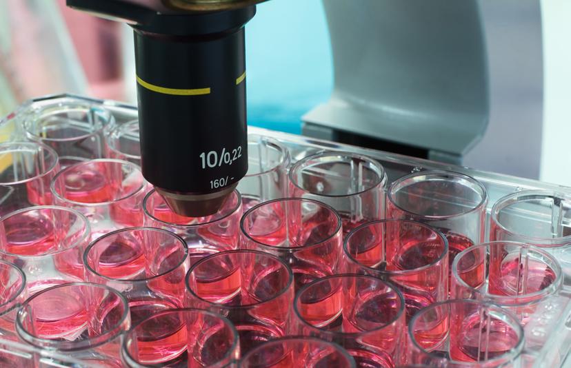
1. Application Note: Z-Stacking and Z-Projection using a Scaffold-based 3D Cell Culture Model
This application note demonstrates a technique using BioTek’s Gen5™ Image+ Data Analysis Software to perform z-stacking and z-projection of scaffold-based 3D cellular tumoroid structure images . Download application note
2. NEW Product: InSphero Launches Enhanced Ultra-low Attachment Spheroid Plate for Scaffold-free 3D Cell Culture
The new GravityTRAP™ ULA Plate design simplifies medium exchange and improves imaging during spheroid production and culture. The automation-compatible 96-well ULA format expands InSphero's portfolio of tissue culture platforms for scaffold-free 3D cell culture, offering a low-cost, enhanced ULA plate. More information
3. Application Note: Seeding, Culture and Cell-Based Assays of upcyte® and vericyte® Cells in the Mimetix® 3D Scaffold
This application note describes how to seed and culture cells, exemplified using Medicyte’s upcyte® Hepatocytes, as 3D cultures in the Mimetix® electrospun scaffold for subsequent endpoint measurements. This protocol can be adapted to all upcyte® and vericyte® cells. Download application note
4. Video: Easy Automation of Corning® 96- and 384-well Spheroid Microplates
Learn how Corning® 96- and 384-well Spheroid Microplates are designed to enable uniform, central, single spheroid formation across wells and allow culture and assay/imaging of spheroids on the same microplate. Watch the video
5. Application Note: 3D Cell Culture on Hamilton/GCS BioLevitator
This application note describes the general technology and a typical experiment with HEK293 cells cultured in the Hamilton BioLevitator™. This provides a more convenient cell culture process and delivers consistent results through continuous monitoring of cells on the Global Eukaryotic Microcarrier™ substrate. Download application note
6. NEW Product: FluoView FVMPE-RS Multiphoton System - Two New Microscopes for Live Cell and In-Vivo Imaging
With two new configurations of its FluoView FVMPE-RS multiphoton laser scanning microscope series, Olympus continues to expand the possibilities of multiphoton microscopy. The new Gantry microscope frame is designed to accommodate large samples and setups, and the inverted microscope frame is ideal for 3D cell culture. More information
7. NEW Webinar: Light Sheet Imaging for Fast 3D Live Cell and Tissue Imaging
Join Dr Planchon, Biophotonic Imaging Laboratory, Delaware State University and Dr Truong, Translational Imaging Centre, University of Southern California, in this fascinating discussion on light sheet microscopy. Case studies are presented in fast and dynamic 3D imaging. Watch the webinar on-demand
8. Application Note: Simplifying High Throughput 3D Spheroid Growth and Shrinkage Assays Using Live Content Imaging
This poster shows the assembly and validation of fully-kinetic, spheroid growth and shrinkage assays through the use of fluorescently labeled cells and Essen BioScience IncuCyte ZOOM™. IncuCyte ZOOM™ metrics and fluorescence intensity measurements are informative, and can be readily gathered to monitor spheroid growth and shrinkage over time. Download application note
Related products
Request Quote for All Products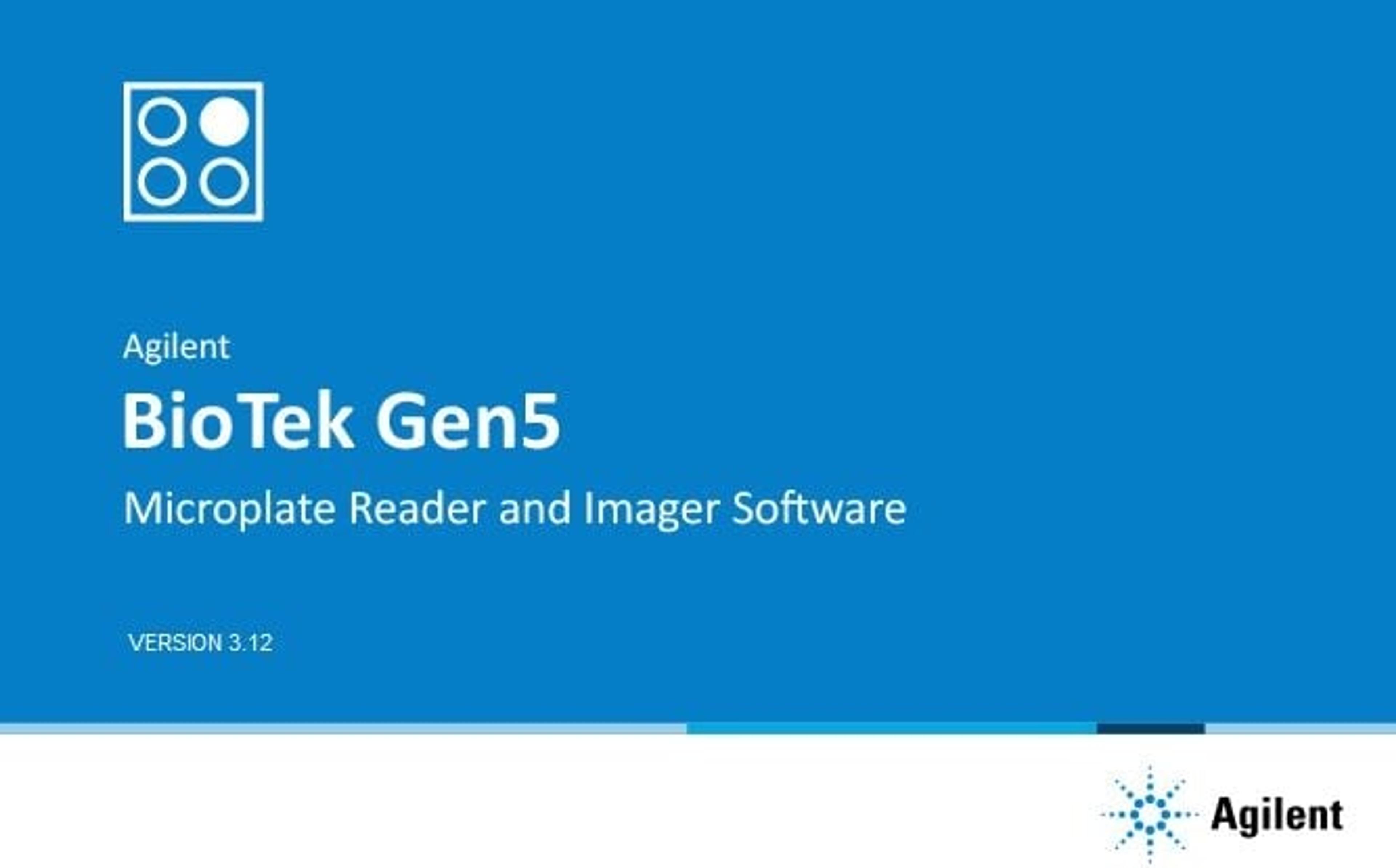
Agilent BioTek Gen5 Software for Detection
Agilent TechnologiesAgilent BioTek Gen5 software for Agilent BioTek multimode and single-mode microplate readers is an integrated tool for endpoint, kinetic, spectral scanning, and well area scanning. It controls all the functions of the plate reader and has powerful data analysis capabilities for a broad range of applications. Gen5 microplate reader software is also used to integrate BioTek plate readers to BioTek BioStack and other automated systems.
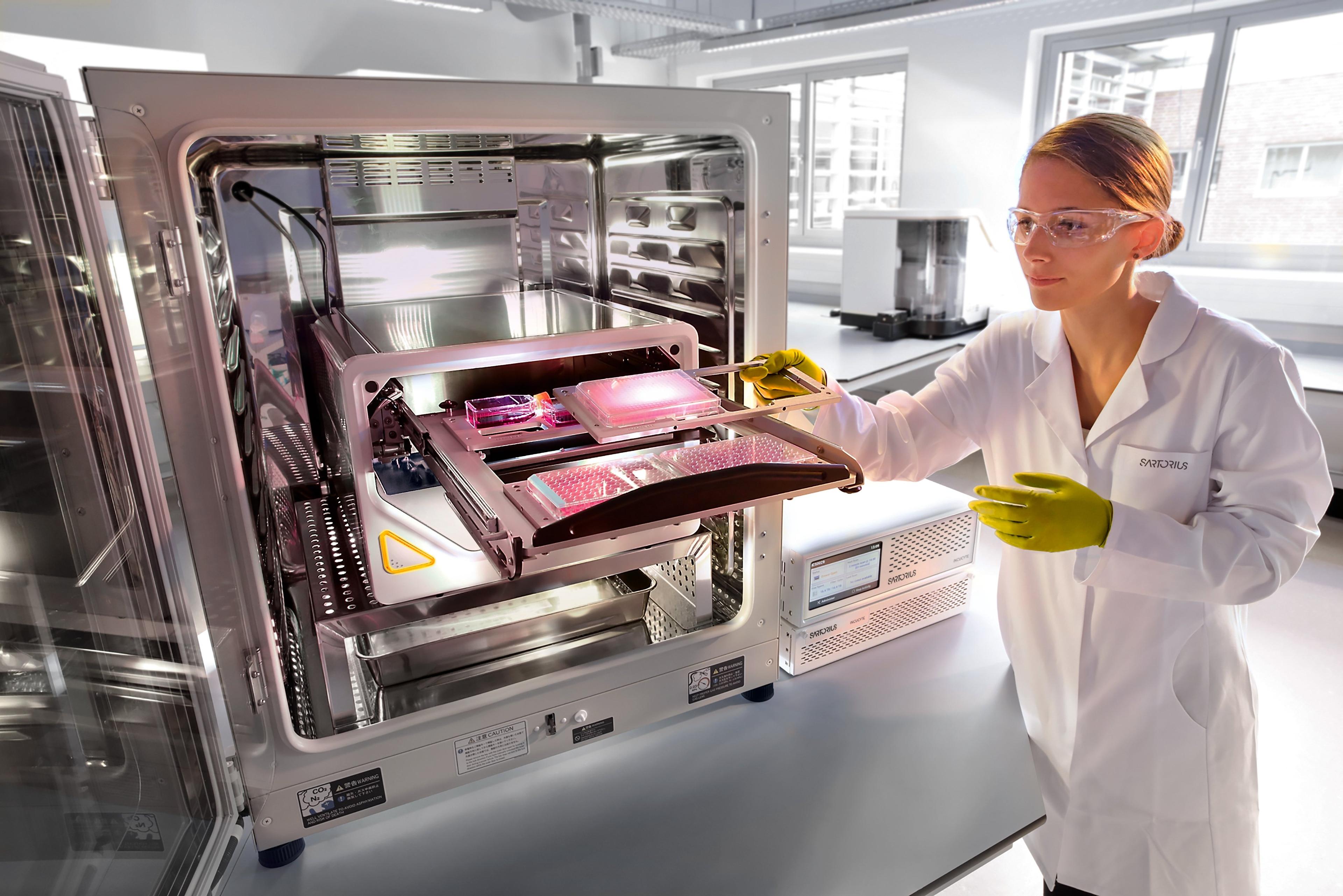
Incucyte® Live-Cell Analysis Systems
Sartorius GroupIncucyte ® , Empower Live-Cell Analysis Inside Your Incubator
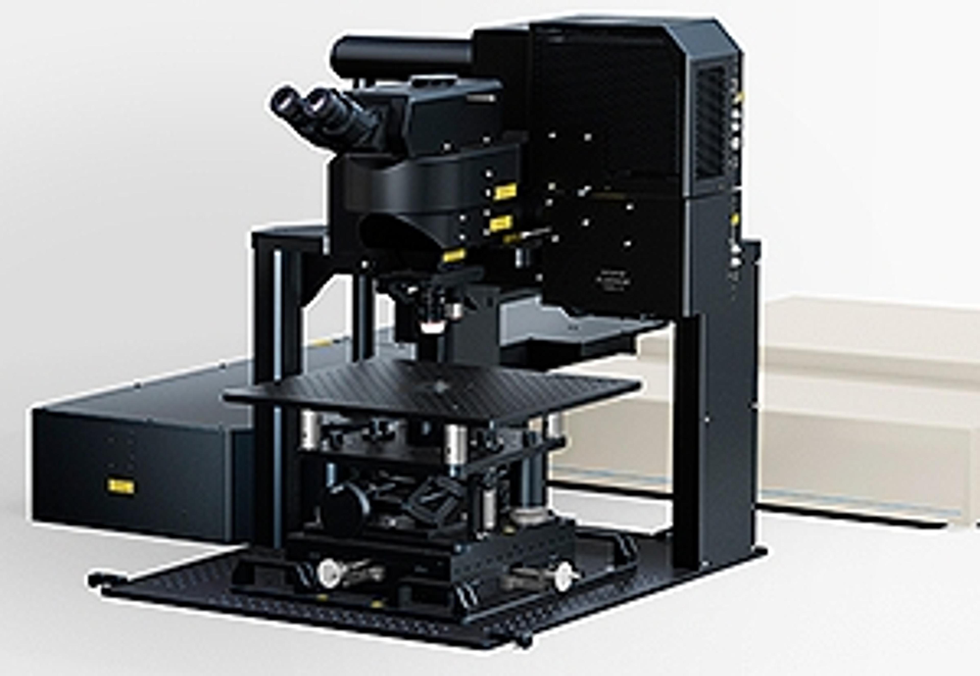
FVMPE-RS Multiphoton System - Two New Frames for Live Cell and In-Vivo Imaging
EVIDENTExpanding the possibilities of multiphoton microscopy, Olympus presents two new configurations of its FluoView FVMPE-RS multiphoton laser scanning microscope series: the Gantry microscope frame to accommodate large samples and setups, and the inverted microscope frame for 3D cell culture. The Olympus FluoView FVMPE-RS multiphoton laser scanning microscope series is optimised for life science research. Its high-speed scanner allows observation of ultra-rapid biological responses, and the system can obtain vivid images from as deep as 8 mm below the tissue surface. As a direct result of researchers requesting support for a wider range of applications and specimens, Olympus has now expanded the range beyond the original upright frame version. The new Gantry microscope and inverted microscope frames offer even greater scope and flexibility for research, opening up a wide range of observation possibilities suited to imaging a greater variety of biological specimens. The Gantry microscope frame features an ultra-stable arch-like structure that allows considerable space beneath the objective to accommodate experiments of varied sizes. A volume of 640 mm wide, 355 mm high and 520 mm deep is available if the stage is removed, providing sufficient space for the researcher’s own experimental apparatus and providing flexibility to suit different observation purposes. The inverted microscope frame is ideal for observation of cells in 3D cultures, where multiple layers of cells are cultured in a petri dish or similar vessel. Because inverted microscopes allow the researcher to observe the sample from below, this configuration is optimised for 3D cell culture imaging and allows cells that have adhered to the base of the petri dish to be observed without culture fluid touching the objective. Such capabilities meet the needs of researchers, enabling them to capture rapid biological responses deep within samples. Features: Gantry frame accommodates large samples and setups Three-dimensional Adjustment - A removable manual XYZ stage enables height adjustments. Changing between thin sample and whole animal imaging can be easily accomplished. Large Workspace Applications: Multiphoton confocal laser scanning microscopy Optogenetics Electrophysiology In vivo imaging with large samples Live cell imaging including 3D cell culture