Digital holographic microscopy redefines label-free live-cell imaging
What if you could watch living cells for days, or follow them at 100,000 frames per second, without adding a single marker?
7 Oct 2026

Figure 1. Benjamin Rappaz, Head of Life Sciences, Lyncée Tec Division, Park Systems
For decades, fluorescence microscopy has been the workhorse of live-cell imaging, lighting up specific structures and molecules that would otherwise stay hidden. That visibility comes at a price, however. Labels can change how cells behave, the light needed to excite them can damage the samples under study, and every staining step adds time, reagent costs and labor. For scientists who need to follow living cells over several days, or capture events that unfold in milliseconds, those trade-offs can limit which questions are even possible to ask.
Digital holographic microscopy (DHM) takes a radically different route. Rather than relying on chemical dyes, it takes advantage of the laws of optics. DHM captures the delay of light passing through a cell and encodes it into a hologram, instantly generating quantitative data on cell morphology, volume, and intracellular density.
In this interview, SelectScience speaks with Benjamin Rappaz, Head of Life Sciences at Lyncée Tec, the Swiss DHM pioneer that became part of Korean nanometrology company Park Systems in 2025. Rappaz is a biologist who has worked with DHM since his early research at EPFL, and he now leads efforts to demonstrate the technology's unique label-free advantages to academic and industrial partners. He explained how DHM works and where it is making the greatest difference in life sciences research.
Turning transparency into data
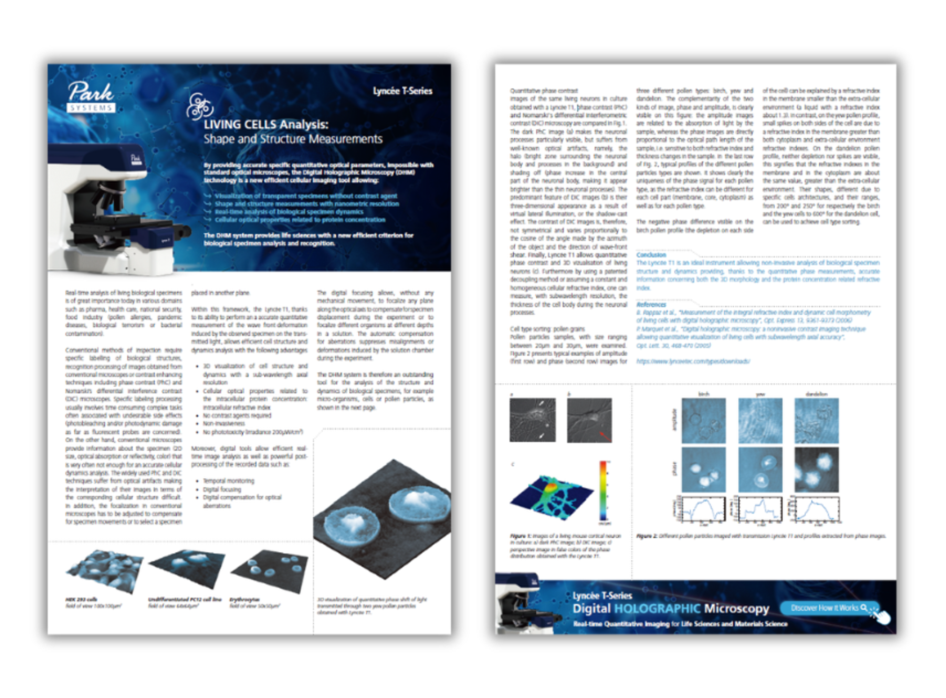
" Under a standard microscope, living cells are essentially invisible and lack the contrast needed to actually measure them," Rappaz explains. In his view, what really sets DHM apart is "the ability to extract quantitative data from otherwise transparent biological cells, noting that as the light goes through the cell, it slows down depending on cell thickness and intracellular density," he says. By capturing this phase delay, DHM simultaneously quantifies cell topography, revealing shape and volume, and refractive index, which reflects what is happening inside the cell in real time.
This turns a simple picture into a diagnostic tool, transforming subtle visual shifts into concrete data points that researchers can track in real time.
Why label-free matters for living cells
"The gold standard in microscopy is fluorescence imaging, but it comes with a few challenges," Rappaz notes, pointing out that dyes can interfere with native cell behavior, for example by causing phototoxicity. DHM needs no labels and no sample preparation. "We can just directly image the cell with minimal light intensity, and thus we're not perturbing or stressing the cells as we measure them," he says.
The payoff is time. "This opens a huge temporal window for the researcher," Rappaz observes. "You can leave your sensitive cells under the microscope for continuous multi-days monitoring to observe the slow processes like proliferation and differentiation."
DHM is also built for speed. "We can go up to nearly 200 frames per second with our standard camera or up to 100,000 frames with high-speed equipment," he says. That enables applications such as sperm cell tracking for fertility assessment and monitoring the rapid beat rate of cardiomyocytes during cardiotoxicity screening. No other methods can capture both the days-long arc of differentiation and the split-second rhythm of a beating heart cell on one platform.
From cell therapy to drug screening
One of the most compelling examples comes from cell and gene therapy. Assessing T cell viability, Rappaz explains, traditionally requires chemicals and individual sampling. "With DHM, we can do that continuously without touching the cell and immediately observe a potential drop in viability," he says.
Because modified T cells are ultimately reinfused into the patient, a label-free approach is essential. "Here it's really important to have a label-free technique so that you don't put anything back in the patient beside their own cells," Rappaz emphasizes.
Drug discovery is another natural fit. "Researchers drastically save on reagent cost and eliminate all the labor-intensive pipetting steps," he points out, adding that "cytotoxicity can be robustly quantified based entirely on the automated morphological measurements obtained with DHM." For core facilities running large plate-based assays, that simplifies workflows considerably.
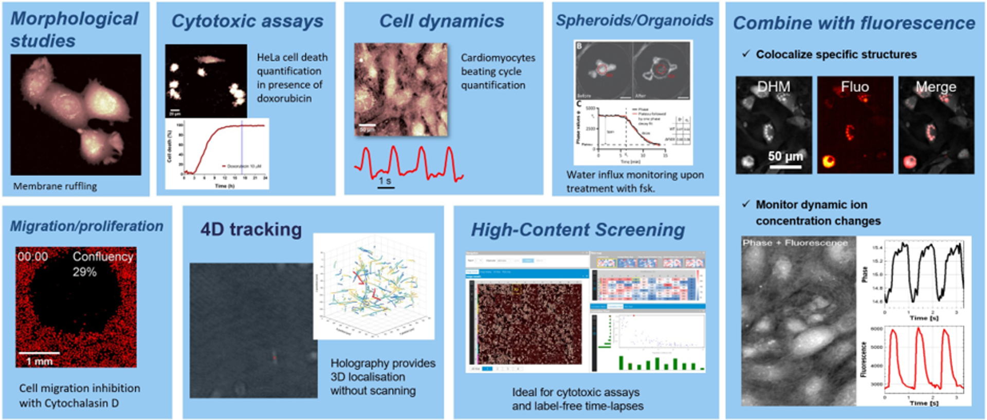
Figure 2. DHM has multiple applications in life sciences, ranging from Cytotoxicity, and high-speed cell dynamics to drug screening.
Tracking cells in four dimensions
DHM also shines in 4D tracking, following objects in three dimensions over time. Rappaz describes it as "currently the only technology capable of tracking the 3D position and precise trajectory of every single particle in the field of view at up to 200 frames per second." Because a hologram records the full optical field, the Lyncée T-Series can refocus images after acquisition. "This has led to many publications in microbiology for bacterial tracking and in reproductive medicine for human sperm viability analysis," Rappaz adds.
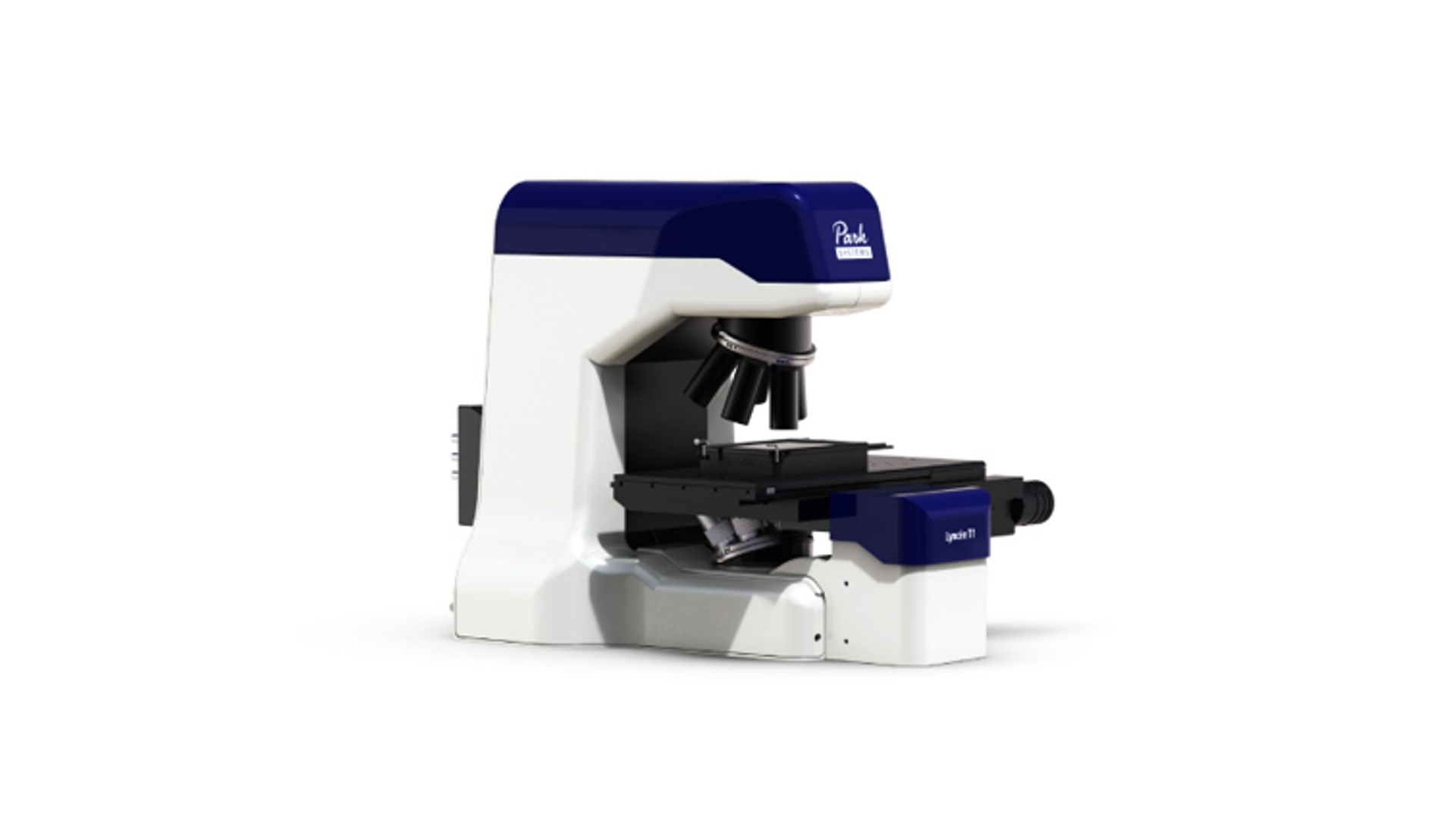
Figure 3. Lyncée T Series combines Digital Holographic Microscopy (DHM) with powerful analysis software to deliver real-time, label-free quantitative imaging of transparent and semi-transparent samples.
A partner to fluorescence, not a replacement
Rappaz sees the two approaches as complementary. DHM is ideal for continuous, noninvasive structural monitoring, while "fluorescence remains essential for specific molecular identification," he continues. Lyncée Tec therefore integrates a fluorescence module into its microscope "to take advantage of both modalities to maximize insight while minimizing cell stress."
A researcher might "use fluorescence to establish ground truth," confirming a structure with a specific marker or validating a subpopulation. "Once this validation step is done, then you can use the DHM for the rest of the experiment," Rappaz explains. The two can also run side by side: in cardiomyocytes, DHM can measure morphological changes during contraction while a fluorescent marker tracks intracellular calcium release. "Combining those two modalities provides a complete picture of the biological phenomenon," he says.
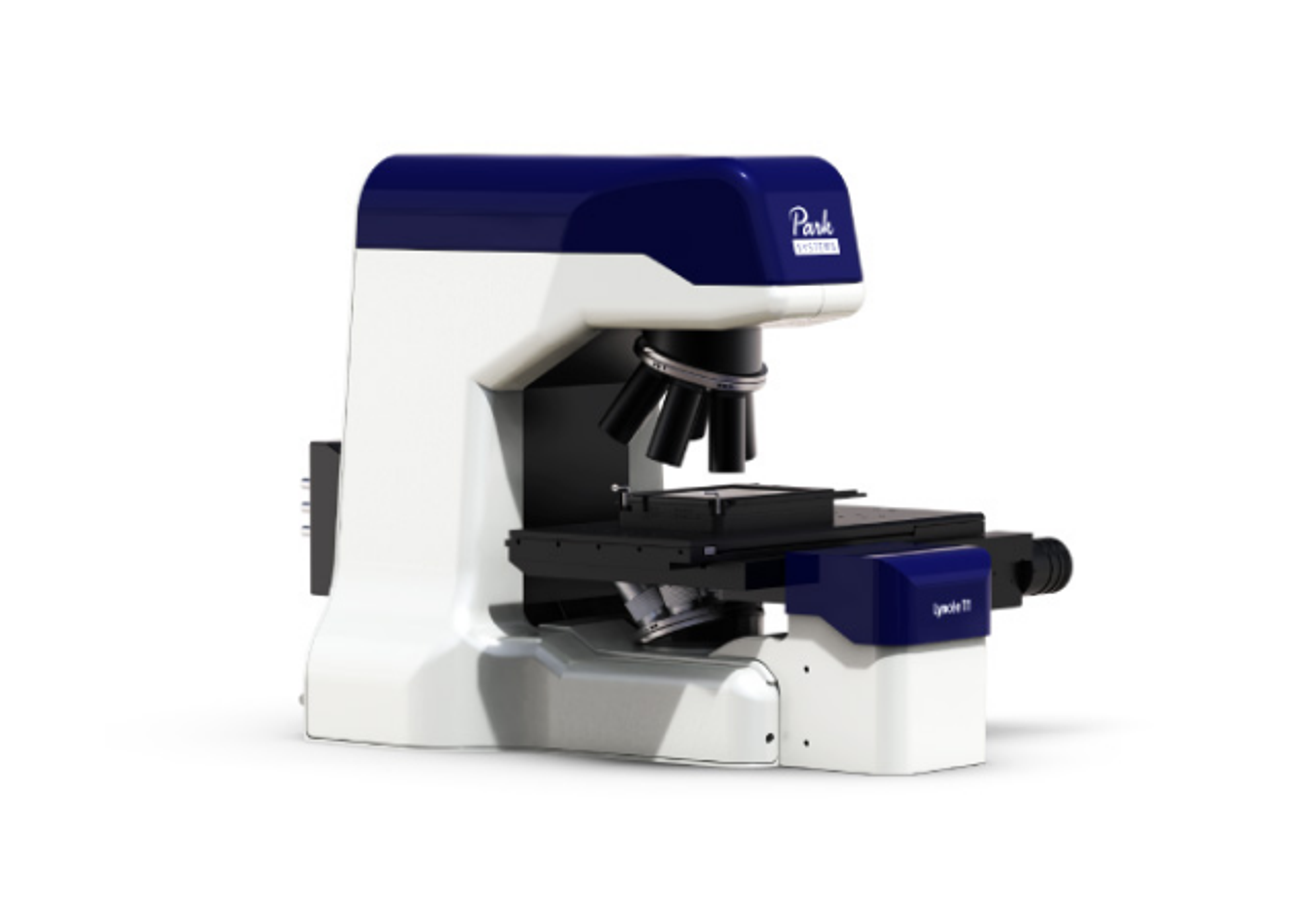
The Lyncée T-Series is built for that flexibility. Available in single-laser T1 and dual-laser T2 configurations, it offers a modular, upgradeable design with options including motorized stages, fluorescence modules and environmental chambers.
The future of quantitative live-cell imaging
Rappaz describes DHM as "a mature, robust technology backed by decades of refinement and publication," and believes its next chapter will be written with its users. "We started in biophysics, then scaled up to high-content screening, and now we're developing new applications in the cell and gene therapy and fast 4D tracking," he says. By delivering quantitative, label-free measurements that respect the biology of living cells, whether over days or in fractions of a second, DHM lets researchers see more while disturbing less. For Rappaz, the most exciting question is one he puts back to the scientific community: what new applications will researchers develop with DHM next?
Interested in exploring how digital holographic microscopy could support your live-cell imaging, cell therapy, drug screening, or high-speed tracking applications? Contact the Lyncée Tec team at lynceetec.info@parksystems.com to discuss your research needs and learn more about the T-Series platform.