Cellular Pathology Community Round-Up
Latest SelectScience® news and downloads related to cellular pathology
28 Mar 2016
Editorial article
Latest cellular pathology news and application article downloads from the SelectScience® Library
In the news recently, learn why ’de novo’ FDA clearance of whole slide imaging devices is significant, find out how a new device could diagnose cancer rapidly at the cellular level, and discover more about scanning near-field optical microscopy.
1. Article: Elephants in the Room: Whole Slide Imaging Devices
In mid-January the Digital Pathology Association (“DPA”) announced that the ‘de novo’ application process may be acceptable by the FDA for clearance of whole slide imaging devices (“WSI” devices) for primary diagnostic uses. Why is this significant? The de novo process lowers the primary hurdle for marketing approval from that of a class III device to essentially a class II device.
2. News: Tews Laboratory use LaVision BioTec Technology for Tumor Study
LaVision BioTec, developers of advanced microscopy solutions for the life sciences, report on the work in the Tews Laboratory which is studying the molecular mechanisms of tumor invasion using an UltraMicroscope for enhanced imaging of cells.
A University of Texas at Arlington electrical engineer has developed a novel cancer cell detection method that will improve early diagnosis through a tool that tracks cellular behavior in real time using nanotextured walls that mimic layers of body tissue.
Dako, an Agilent Technologies company and a worldwide provider of cancer diagnostics, has announced that the FDA has approved the expansion of the intended use of Dako PD-L1 IHC 28-8 pharmDx to include patients with melanoma.
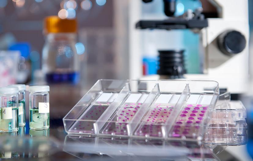
In this case study Lumenera demonstrate the high image quality of its microscopy cameras, image quality, especially clarity of color, is essential in dermatopathology as the diagnosis relies of microscopy photographs of stained tissue. Lumenera’s INFINITY 2-5 CCD camera was suited for the teaching side of the dermatopathology clinic as its ImageJ application allowed for easy editing and viewing on the Mac system.
6. Application Download: Scanning Near-Field Optical Microscopy - Histological Microtome Cuts
Scanning Near-field Optical microscopy allows tissue to be viewed in air or liquid at high resolution without taking up time with sample preparation. This application note from WITec GmbH uses a variety of rat organs to show the results of Scanning Near-field Optical microscopy, showing the different structures that can be clearly seen in the tissue after staining.
This application from Waters Corporation demonstrates that desorption electrospray ionization (DESI) imaging can be utilized as a non-destructive imaging technique. This allows multiple analyses on the same tissue section at different spatial resolution.
Image: anyaivanova/Shutterstock
Related products
Request Quote for All Products
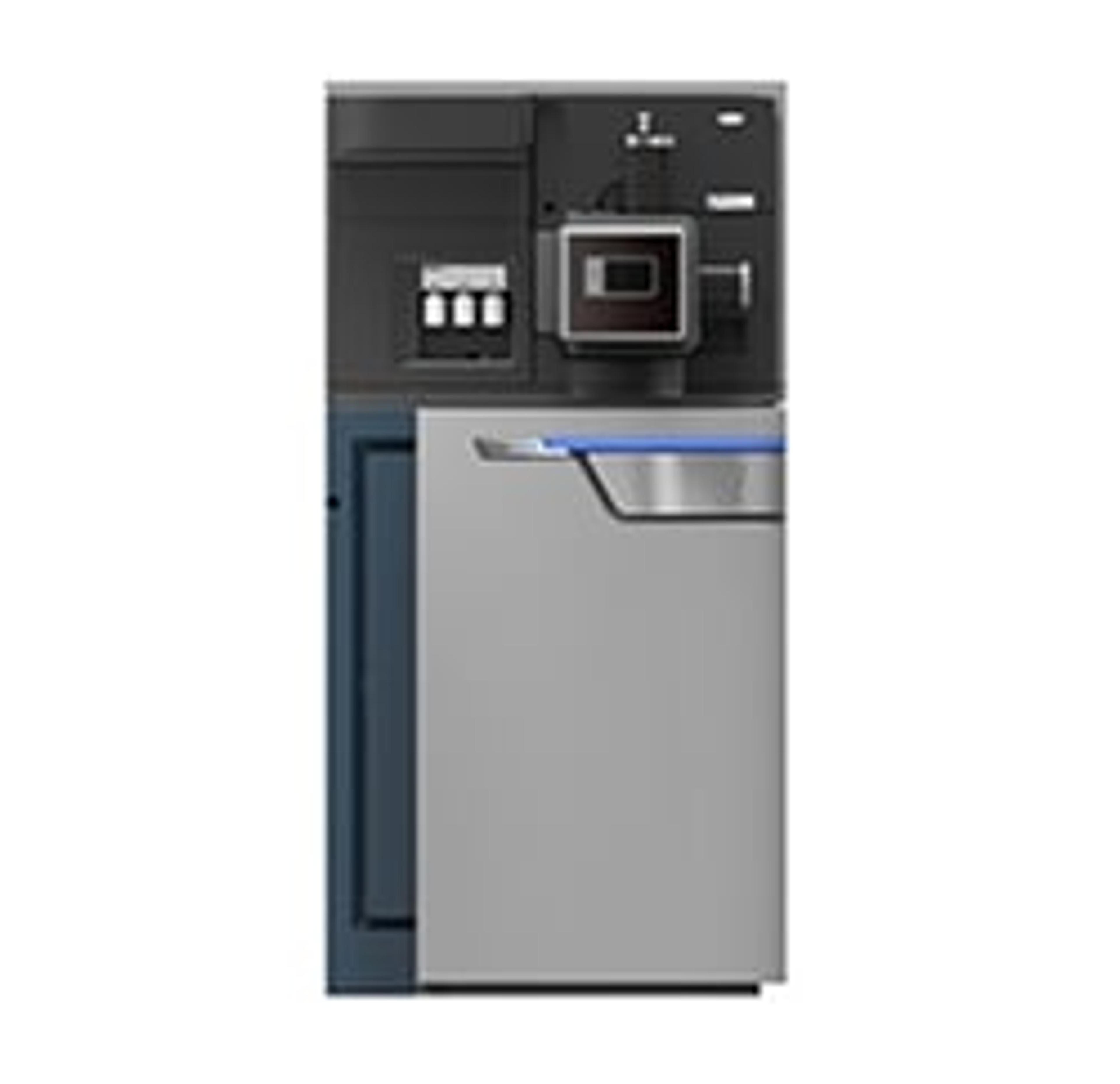
SYNAPT G2-Si MS
WatersTransform your lab's discovery capability with the SYNAPT G2-Si MS. Information. Informatics. Impact. SYNAPT enables extensive characterization of complex mixtures and molecules with uncompromising qualitative and quantitative performance, streamlined workflows and unparalleled platform versatility. With the SYNAPT G2-Si you get ultimate UPLC/MS/MS performance, powerful data independent & data dependant solutions, CID and ETD fragmentation capabilities, and a wide range of experimental options.
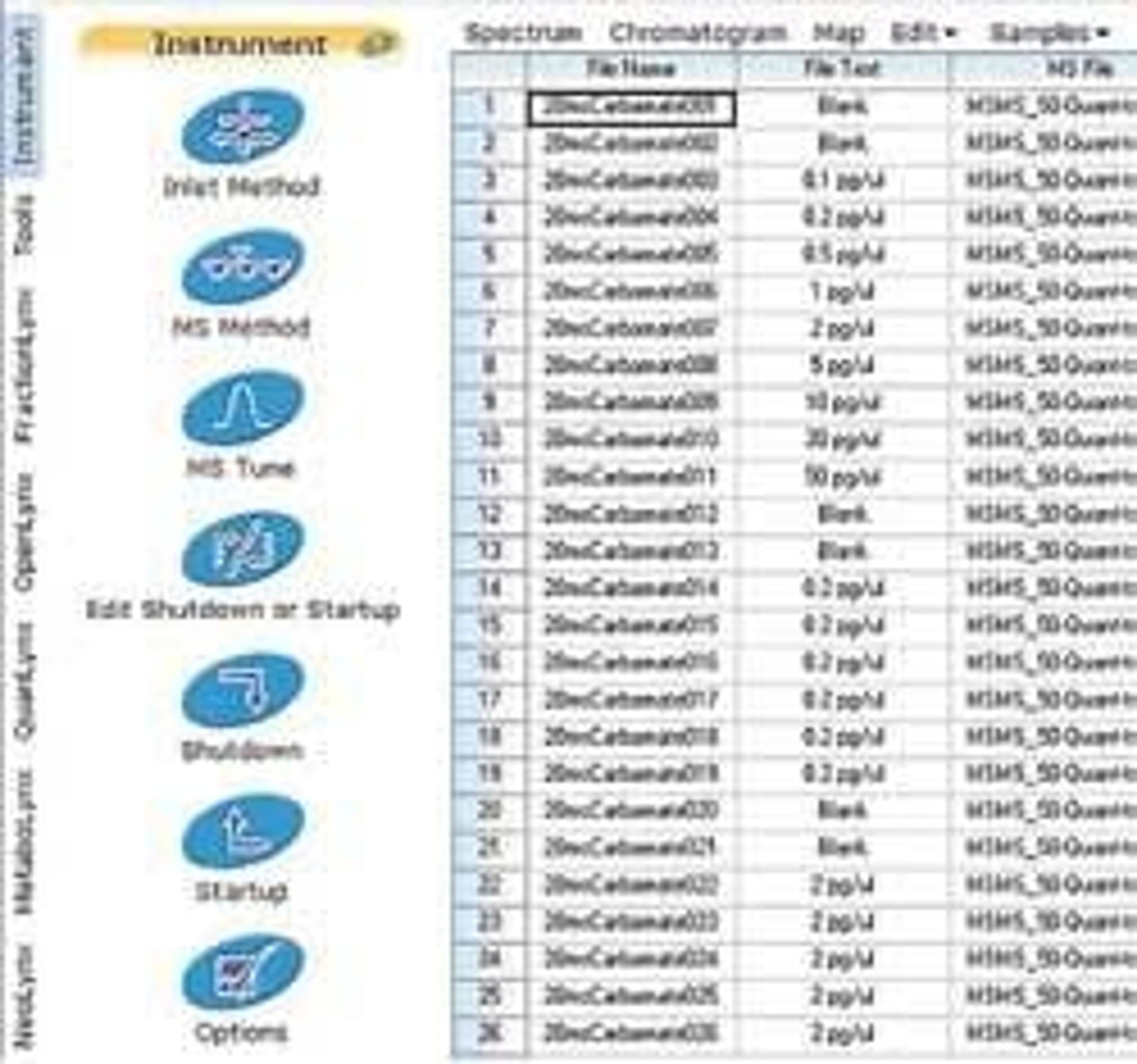
MassLynx MS Software
WatersAcquire, Analyze, Manage, and Share Mass Spectrometry Information MassLynx™ Software increases the speed at which you can convert your sample data into valuable knowledge. It provides you with the fundamental platform to acquire, analyze, manage, and share your mass spectrometry information. MassLynx intelligently controls any Waters mass spectrometry system, from sample and solvent management components to mass spectrometer and auxiliary detectors. MassLynx Software may acquire nominal mass, exact mass, MS/MS and exact mass MS/MS data. MassLynx’s Sample List is the core of the system. It maintains and consolidates the data on all of your samples. You also initiate any activities related to your sample from the Sample List. This "sample centric" approach simplifies the interaction with your LC/MS or GC/MS system, acquired data and processed results. Process Application-specific Data MassLynx Software features general purpose and specialized Application Managers that provide information for your specific MS analyses and data. Two come standard with MassLynx: QuanLynx™ for automated quantification included as standard with MassLynx OpenLynx™ for qualitative screening and identification Optional Application Managers can perform: Targeted quantitative analysis Mass-directed purification Metabolite identification Deconvolution of complex chromatograms Metabonomics & metabolomics Protein identification and protein characterization