
Automating Cell-Based Microplate Assays
6 Sept 2012One of the most important uses for fluorescent proteins like GFP, mCherry, and mOranage is real-time monitoring of cellular processes in live cells. Many applications use fluorescent proteins including intracellular transport, receptor desensitization, cell movement, cell division, apoptosis, differentiation, chemotaxis, transcription, and translation.
All of these applications, in turn, need an analytical instrument that can monitor these cell-based environments. The confocal microscope is the primary analytical instrument used to investigate cell-based fluorescent protein assays. Confocal microscopes and fluorescent proteins have allowed researchers to obtain critical snapshots of live biological cellular processes. However, a major drawback to confocal microscopy is that only one cell or cell cluster is viewed at a time. Therefore, it can take hours or even days to capture statistically significant data and the cell-based assay conditions may not be uniform across the entire test.
To increase throughput of fluorescent protein cell-based assays, researchers will turn to a fluorescent microplate reader. With the ability to measure down to nanoliter volumes and thousands of samples in one plate, microplate readers allow reproducible, cell-based assays to be performed in a matter of minutes.
Until recently, however, there was a limitation to cell-based experiments performed on a microplate reader – the sensitivity seen in a confocal microscope could not be obtained on a microplate reader. Therefore, translating the assay to a microplate reader for higher throughput from the confocal microscope (or other instruments like a FACS flow cytometer) did not always work.
To improve the sensitivity in microplate cell-based assays, BMG LABTECH has engineered a Direct Optic Bottom Reading system in a microplate reader that is analogous to the microscope. The PHERAstar FS microplate reader incorporates a series of software controlled, motor-driven mirrors to focus light through a free-air optical path directly onto the top or bottom of the microplate. In cell-based fluorescent protein assays tested, Direct Optic Bottom Reading produces at least a three-fold higher signal-to-blank ratio than an instrument without this feature. This will allow for the miniaturization and detection of fluorescent protein cell-based assays down to volumes not possible on other readers. In addition, with some cellular fluorescent protein assays providing only a 25% change in signal upon assay activation, a 300% higher overall signal will enable experiments that were previously impossible on a microplate reader.
With their discovery of the Green Fluorescent Protein, Osamu Shimomura, Martin Chalfie and Roger Y. Tsien gave laboratories around the world a fantastic new tool to investigate biological and chemical processes in cells. Correspondingly, BMG LABTECH has engineered a microplate reader that can now fully utilize the endless potential of this Nobel Prize winning discovery.
Related products
Request Quote for All Products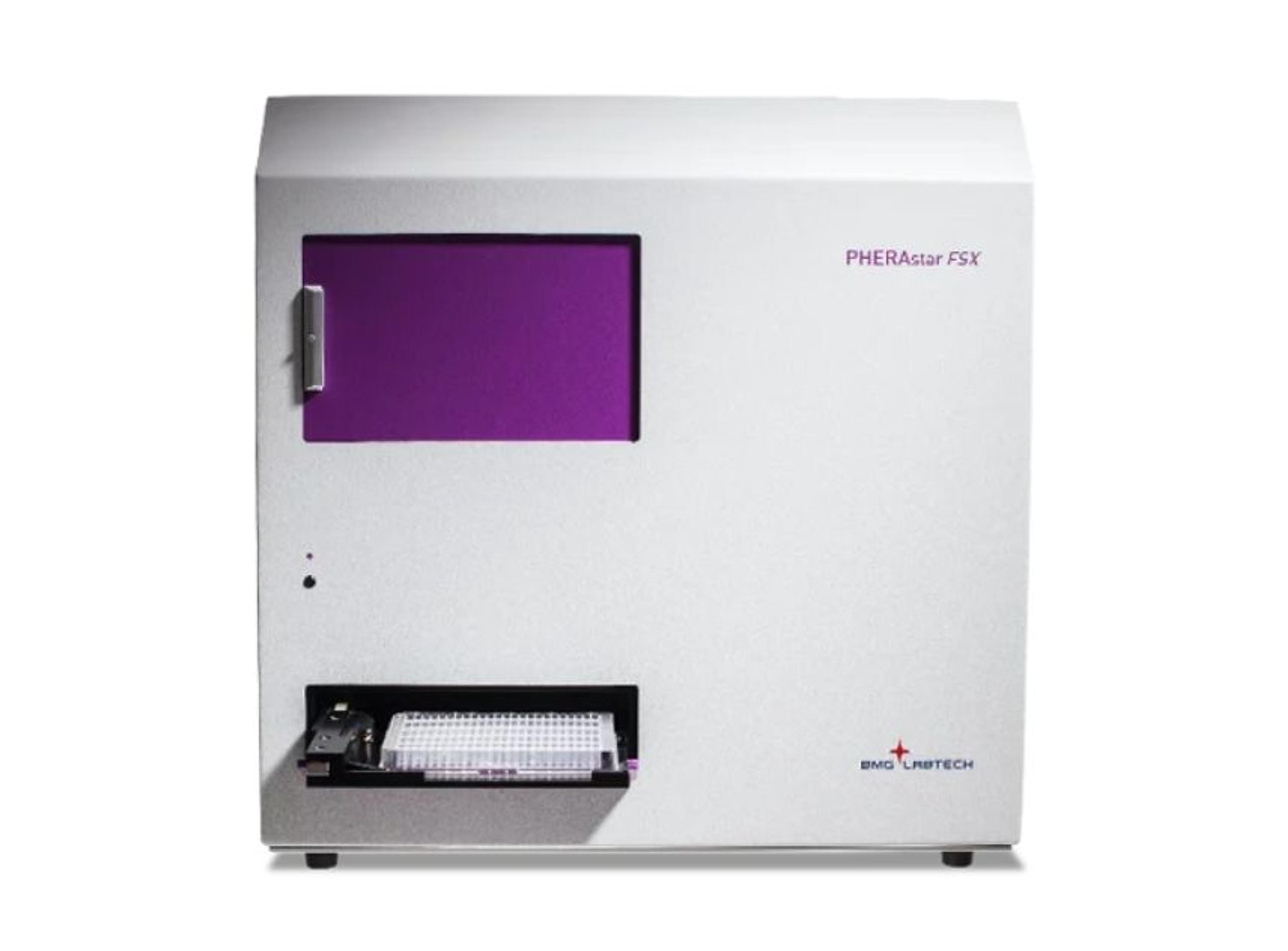
PHERAstar® FSX HTS Microplate Reader
BMG LABTECHThe most sensitive microplate reader for high-throughput screening