Transforming cancer research: Advanced imaging solutions for early detection and monitoring
6 May 2025Editorial article
Early cancer detection and accurate monitoring are critical in improving patient outcomes and advancing cancer research. Despite significant progress, challenges remain in identifying cancer at its earliest stages, tracking its progression, and predicting recurrence. Imaging technologies are revolutionizing precision oncology, offering new hope in the fight against cancer. Bruker preclinical imaging companies, MOLECUBES and Spectral Instruments Imaging are at the forefront of these innovations, providing cutting-edge solutions to address these challenges.
The evolving landscape of cancer detection and monitoring
The field of cancer care is shifting from reactive to proactive approaches, emphasizing early detection, monitoring metastasis, and tracking minimal residual disease (MRD). Non-invasive screening, whole-body molecular imaging, and AI-assisted analysis are key trends driving this transformation. These advancements enable more precise and timely interventions, ultimately improving patient outcomes.
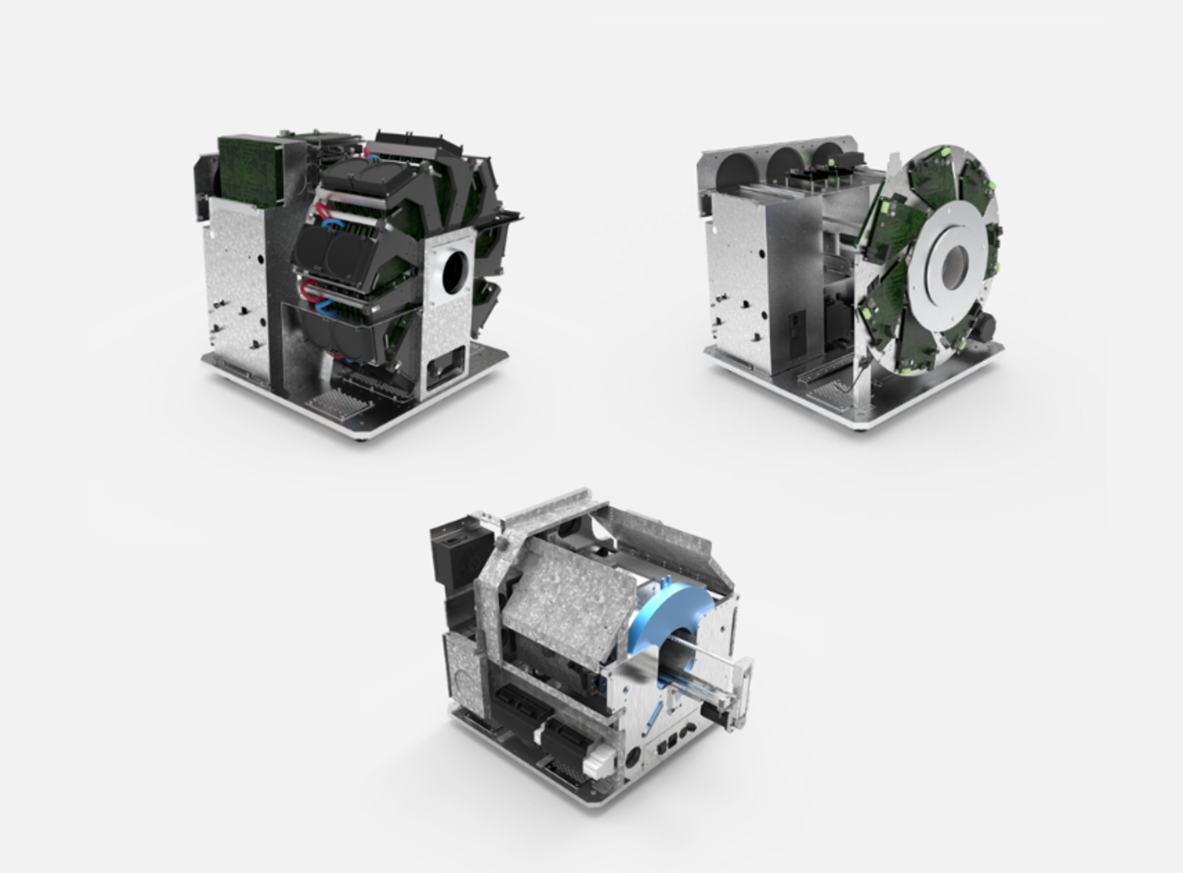
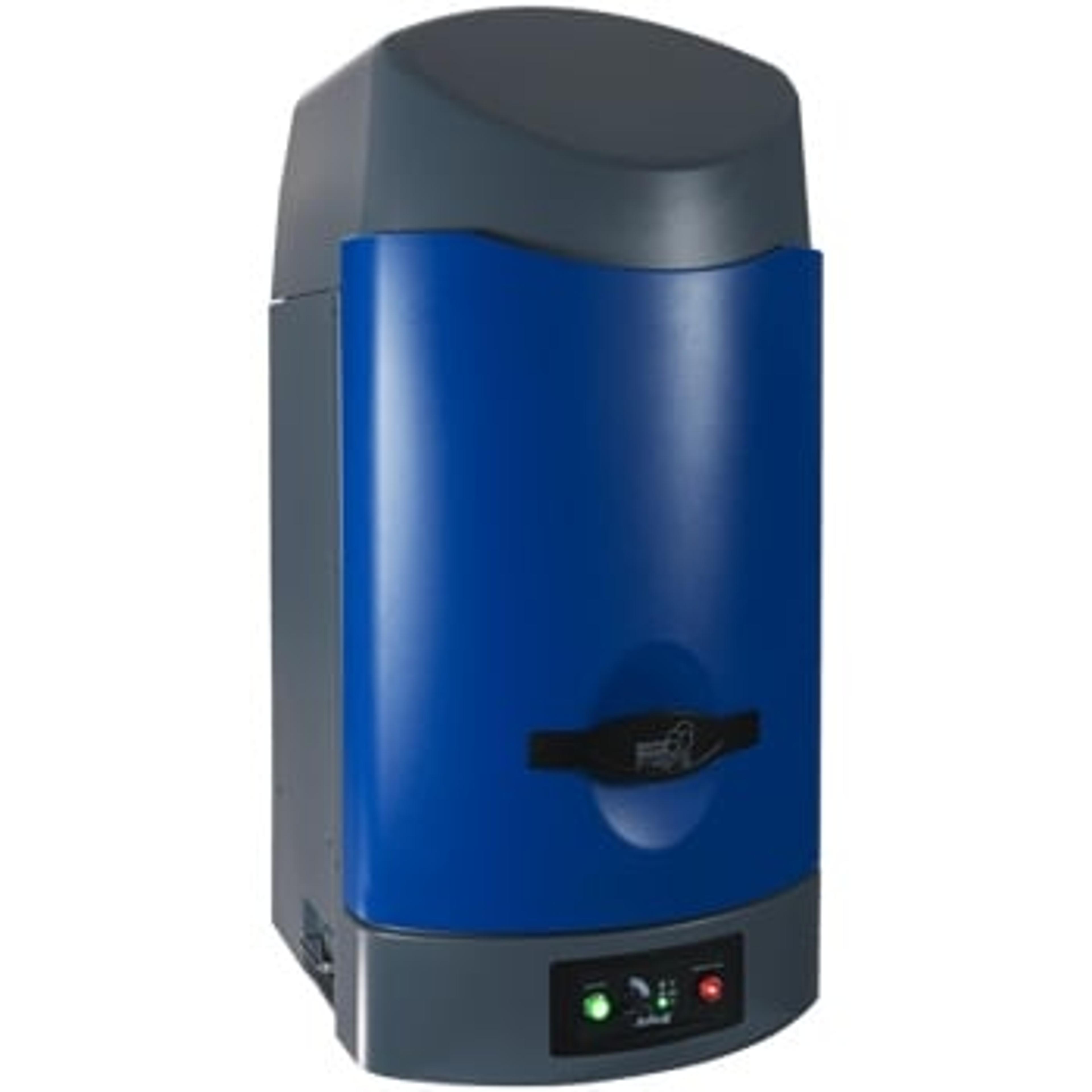
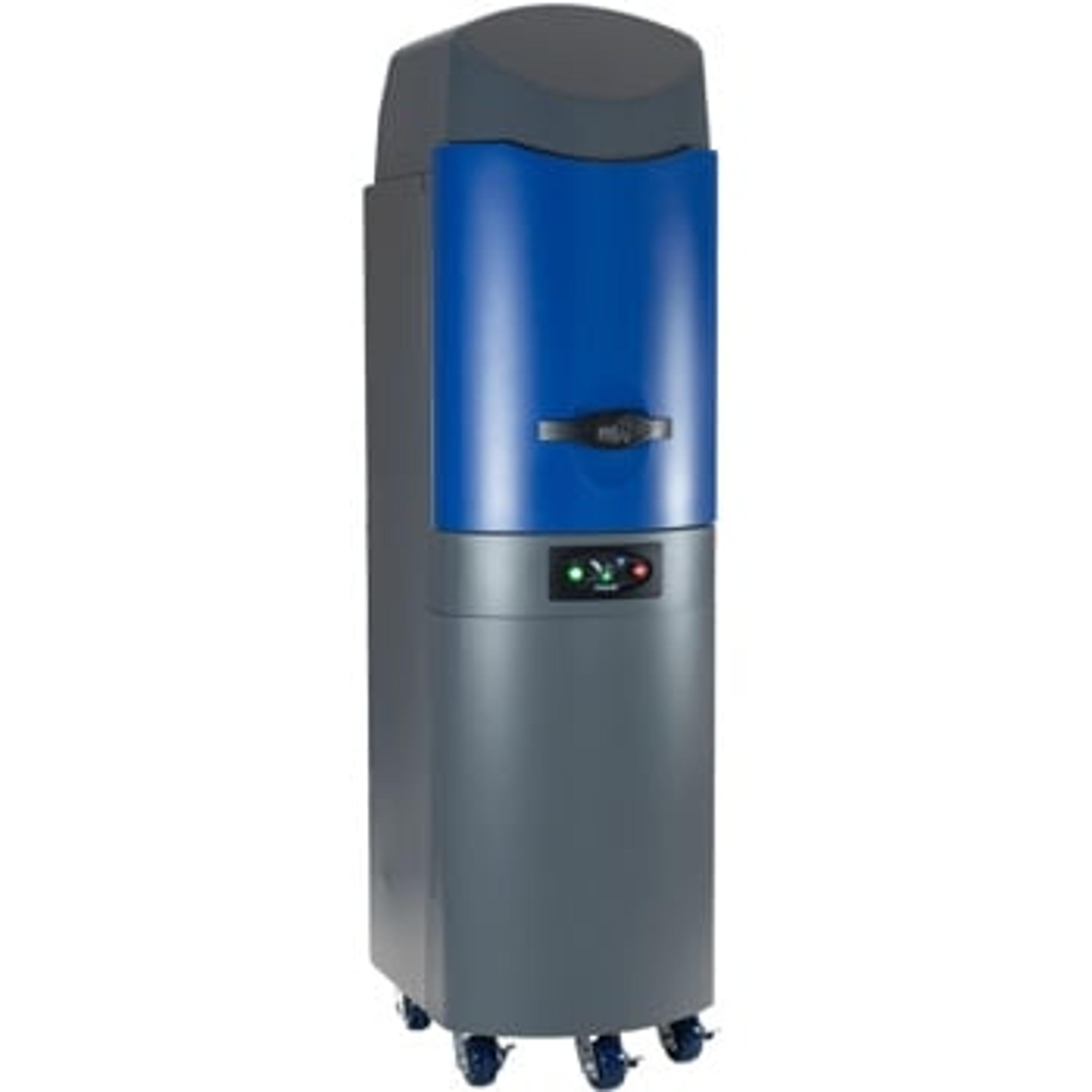
MOLECUBES: Compact high-performance preclinical imaging for oncology research
MOLECUBES offers high-performance PET, SPECT, and CT systems designed for preclinical imaging. These compact, bench top systems are ideal for longitudinal and noninvasive monitoring of pathologies and treatment response, studying molecular changes with high sensitivity, and evaluating the biodistribution of novel compounds. For example, MOLECUBES systems have been used to track metastatic spread in mouse models, providing valuable insights into cancer progression and treatment efficacy.
Learn more about how CT imaging can help provide detailed, high-resolution three-dimensional images of tumors at various timepoints within the same animal, in a humane manner. Request the download
Preclinical imaging with MOLECUBES allows researchers to visualize pathologies comprehensively and quickly. Widely available tracers for PET-CT and SPECT-CT imaging, such as 18F-FDG, 68Ga-PSMA, 99mTc-MDP, 131I, and 99mTc-sestamibi, can evaluate tumor metabolism, proliferation, hypoxia, and metastasis. This imaging provides unique and quantitative results, such as tumor uptake and receptor binding, which are crucial for developing targeted therapies and molecular imaging agents.
Learn more about how molecular imaging techniques can help accelerate translation from bench to bedside. Request the download
Case study: PET-CT imaging for non-invasive longitudinal therapy evaluation in a glioblastoma mouse model
In a study by Valtorta et al., preclinical PET-MR imaging was used to evaluate the efficacy of a new treatment strategy in glioblastoma mouse models. The study utilized two radiotracers, 18F-FLT and [18F]VC701, to predict tumor response to a combination therapy of metformin (MET) and temozolomide (TMZ). The results demonstrated that MET addition improved TMZ efficacy, increasing survival time and reducing tumor relapse rates. This case highlights the power of MOLECUBES' imaging systems in providing valuable insights into treatment responses and advancing precision oncology.
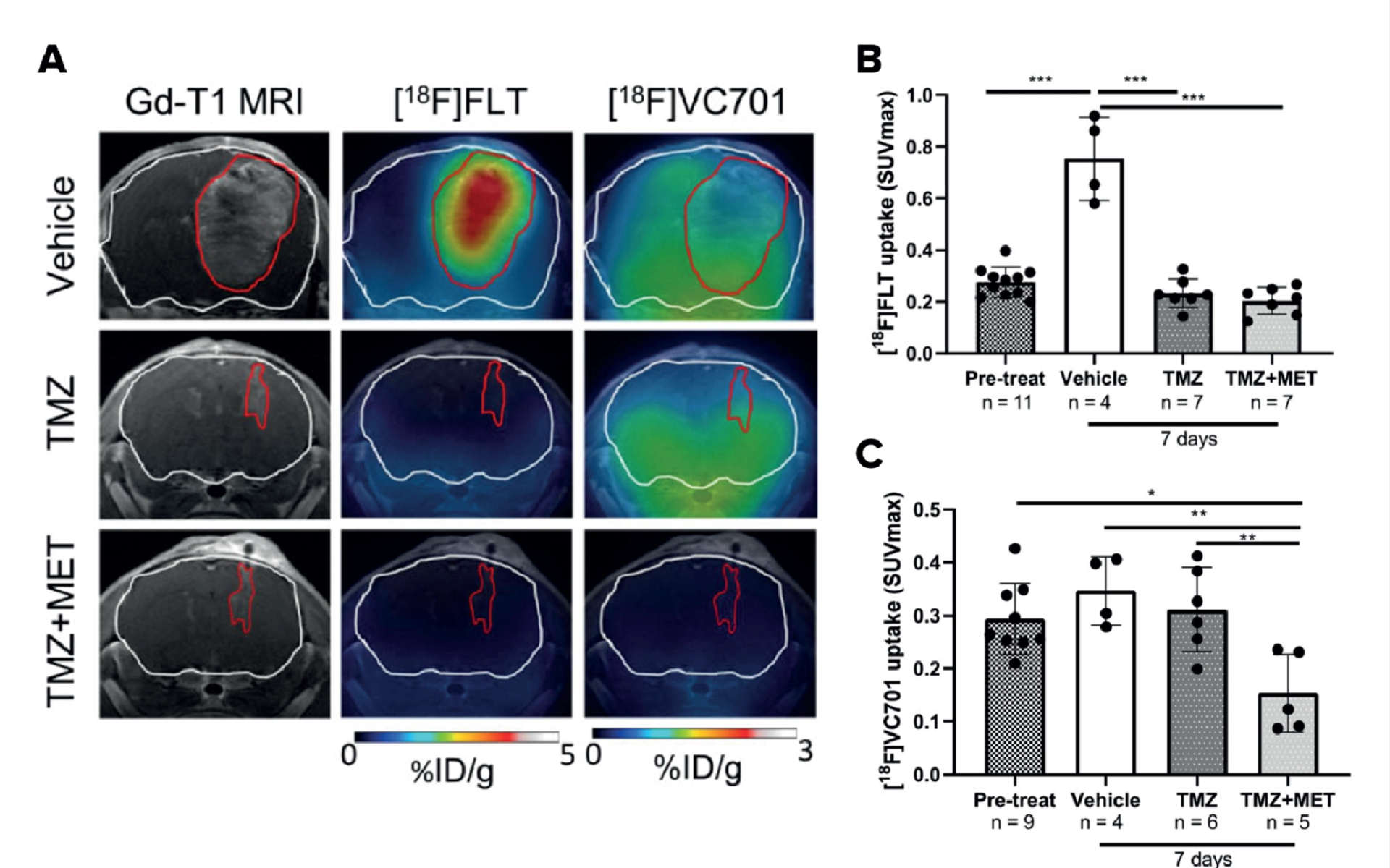
Figure 1: Adapted from Valtorta et al.
A: PET-MR images of a TMZ sensitive GBM model (Gli36_EGFR-1 bearing mice). The white line represents the brain and the red line represents the tumor delineation as defined on the gadolinium T1w MR image.
B: 18F-FLT uptake expressed as SUV max in tumor bearing mice after 7 days of vehicle, TMZ and TMZ+MET treatment. C: 18F-VC701 uptake expressed as SUVmax in tumor bearing mice.
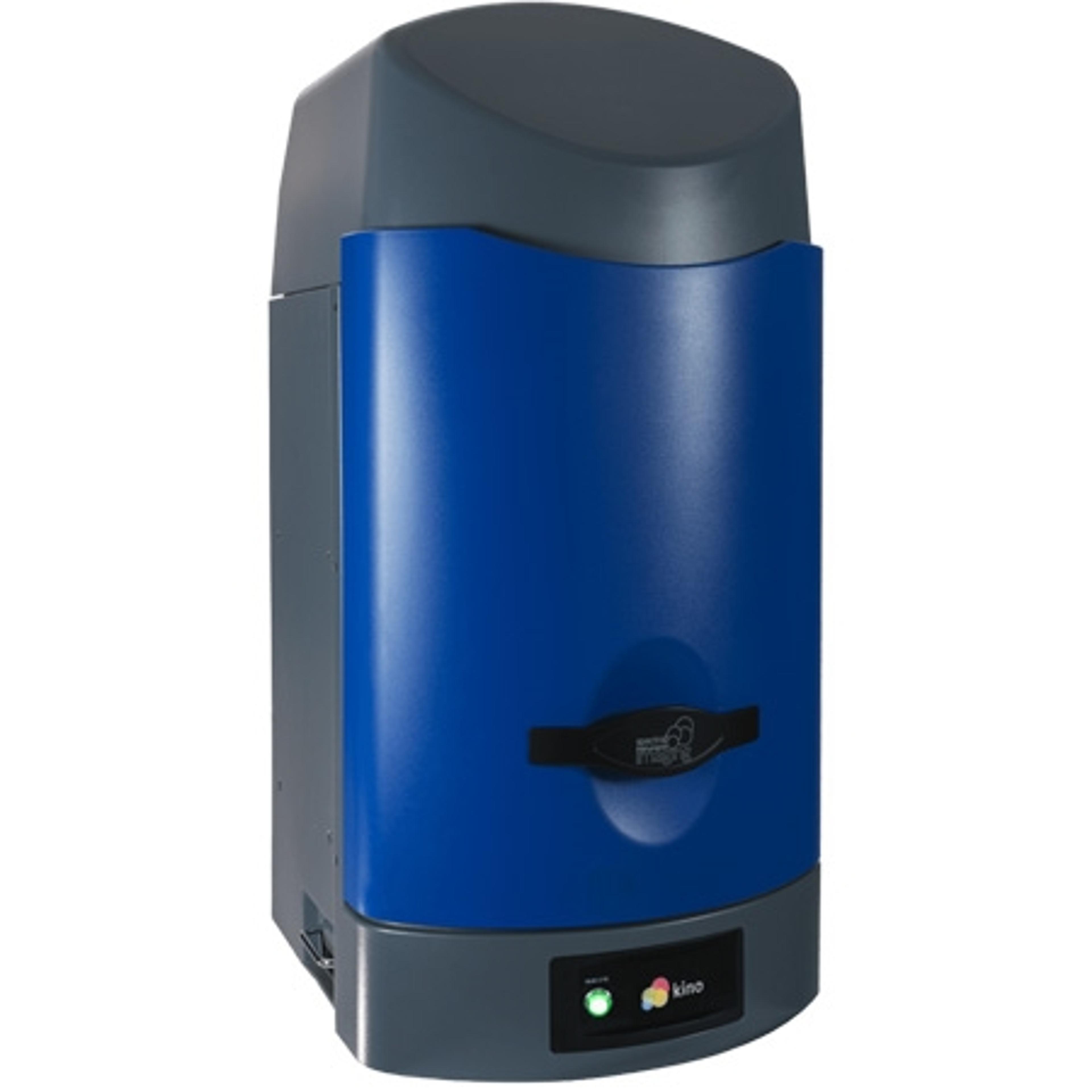
Spectral Instruments Imaging: Illuminating cancer progression with optical imaging
Spectral Instruments Imaging specializes in bioluminescence (BLI) and fluorescence (FLI) imaging, with capabilities for multi-modal integration. These technologies are particularly effective in MRD and metastasis research, allowing for the detection of deep tissue bioluminescence with high sensitivity and longitudinal tracking of tumor burden and spread. Spectral's systems have been used to monitor cancer therapy response and recurrence, providing critical data for optimizing treatment strategies.
Guidelines for establishing a cancer tumor mouse model for in vivo bioluminescence and fluorescence imaging
Creating a cancer tumor mouse model for in vivo BLI or FLI imaging involves careful planning and consideration of various factors, such as cell line choice, inoculation route, and reporter gene selection. Regular monitoring of tumor growth and progression through BLI and FLIprovides valuable insights into tumor behavior and treatment responses. These guidelines ensure that the imaging results are accurate and reliable, contributing to the advancement of cancer research.
Case study: Automated, real-time acquisition and quantification of peak, plateau-phase in vivo bioluminescent data in multiple oncology models
Kinetics software from Spectral Instruments Imaging
Spectral Instruments Imaging has developed a new software feature, Kinetics, which automates the acquisition and real-time presentation of bioluminescent (BL) kinetic data. This feature allows researchers to identify the peak, plateau-phase BL kinetics for any given BL mouse model easily and efficiently. By providing access to camera settings and enabling predictive use in larger studies, the Kinetics feature enhances the quality and reproducibility of in vivo BL data, further advancing cancer research.
The Kinetics software operates within Spectral's established Aura software platform, automatically acquiring and analyzing BL signal data flux (total photons/second) from up to 10 mice concurrently. It presents both individual and group mean BL signal data in live, auto-generated graphs and summary data tables. This enables investigators to identify peak BL data values in real-time, ensuring optimal sensitivity and reproducibility.
Kinetics was tested in hematologic oncology models, including B-cell leukemia and myeloma, demonstrating distinct BL peak times and plateau-phase kinetics. The software also evaluated BL kinetic curves in solid-tumor models, such as lung adenocarcinoma, pancreatic ductal adenocarcinoma, and orthotopic breast cancer, highlighting its versatility and effectiveness in various cancer research applications. Notably, it was observed that each cancer model demonstrated different kinetics, individual mice in a cohort often had unique kinetics and that kinetics evolved dramatically over the course of a study.
Combining modalities: A comprehensive imaging approach
Integrating PET/SPECT/CT with BLI/FLI offers comprehensive insights at both molecular and anatomical levels. This multi-modal approach enhances detection sensitivity and improves the prediction of treatment response and relapse. Bruker's multimodal image processing and quantification software pmod provides a toolkit for flexible image analysis, with a focus on accurate reproducible quantification.
Preclinical imaging enables longitudinal and non-invasive monitoring of pathologies and treatment responses. Bruker’s multimodal imaging approach allows researchers to obtain multiparametric information on tumor properties in animal models regarding interrelated aspects of metastasis and therapeutic response including early phase metastasis, metabolic activity, receptors, and tumor boundaries. Longitudinal cross-modal imaging also facilitates earlier detection and characterization of disease, extending the detection and monitoring from early-stage disease engraftment to late-stage disease progression.
Real-world impact and future directions
Bruker's imaging solutions are making a significant impact on preclinical research, bridging the gap between early-stage discoveries and clinical applications. These tools play a crucial role in identifying biomarkers and tailoring experimental therapies, advancing the field of precision oncology. Looking ahead, the integration of AI, machine learning, and multi-omics will further enhance the capabilities of these imaging platforms, driving future innovations in cancer research.
Preclinical imaging adheres to the 3R principle in animal research and reduces and refines the number of animals required for studies. It allows each animal to serve as its own control, increasing statistical power and reducing the need for euthanizing multiple animals at several time points.
Conclusion
Cutting-edge imaging solutions from Bruker, MOLECUBES, and Spectral Instruments Imaging are enabling earlier and more accurate cancer tracking, empowering researchers in the global fight against cancer. By exploring these advanced platforms, researchers can accelerate their discoveries and contribute to the development of more effective cancer treatments.