Automation and Image-Based Analysis of a Hanging Drop MicroHole Plate Model to Create and Differentiate 3D Mesenchymal Stem Cell Spheroids for Downstream Tissue Formation
4 Apr 2017Human mesenchymal stem cells (hMSCs) have recently emerged as a leading candidate in cellular therapies due to their unique properties. This application note demonstrates the ability to automate the steps necessary to place hMSCs into microplate inserts, track spheroid formation, and monitor spheroids during differentiation into chondrocyte lineages.
Related Products
Request Quote for All Products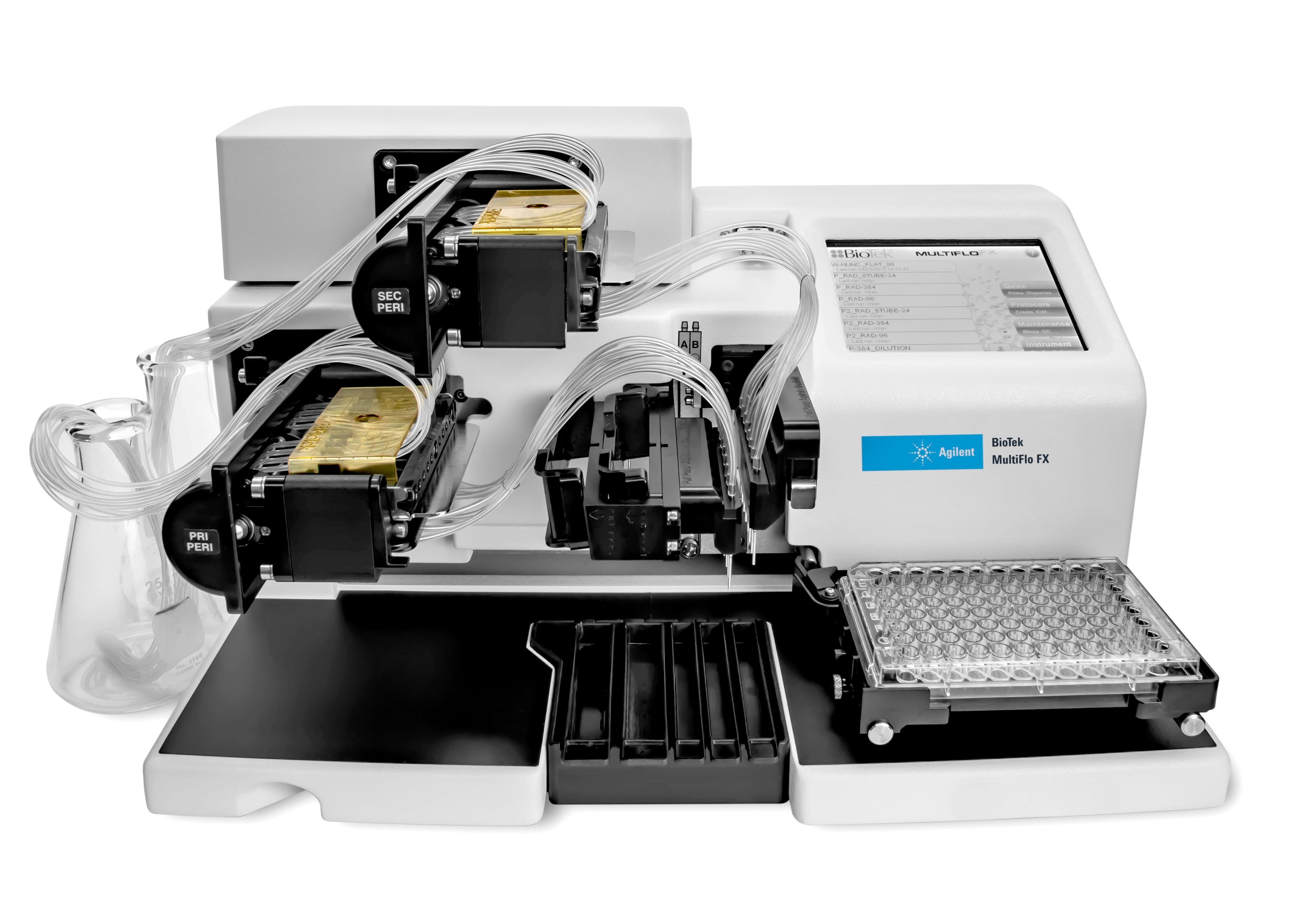
Agilent BioTek MultiFlo FX Multimode Dispenser
Agilent TechnologiesMultiFlo FX can dispense up to 4 independent reagents into 6- to 1536-well microplates.
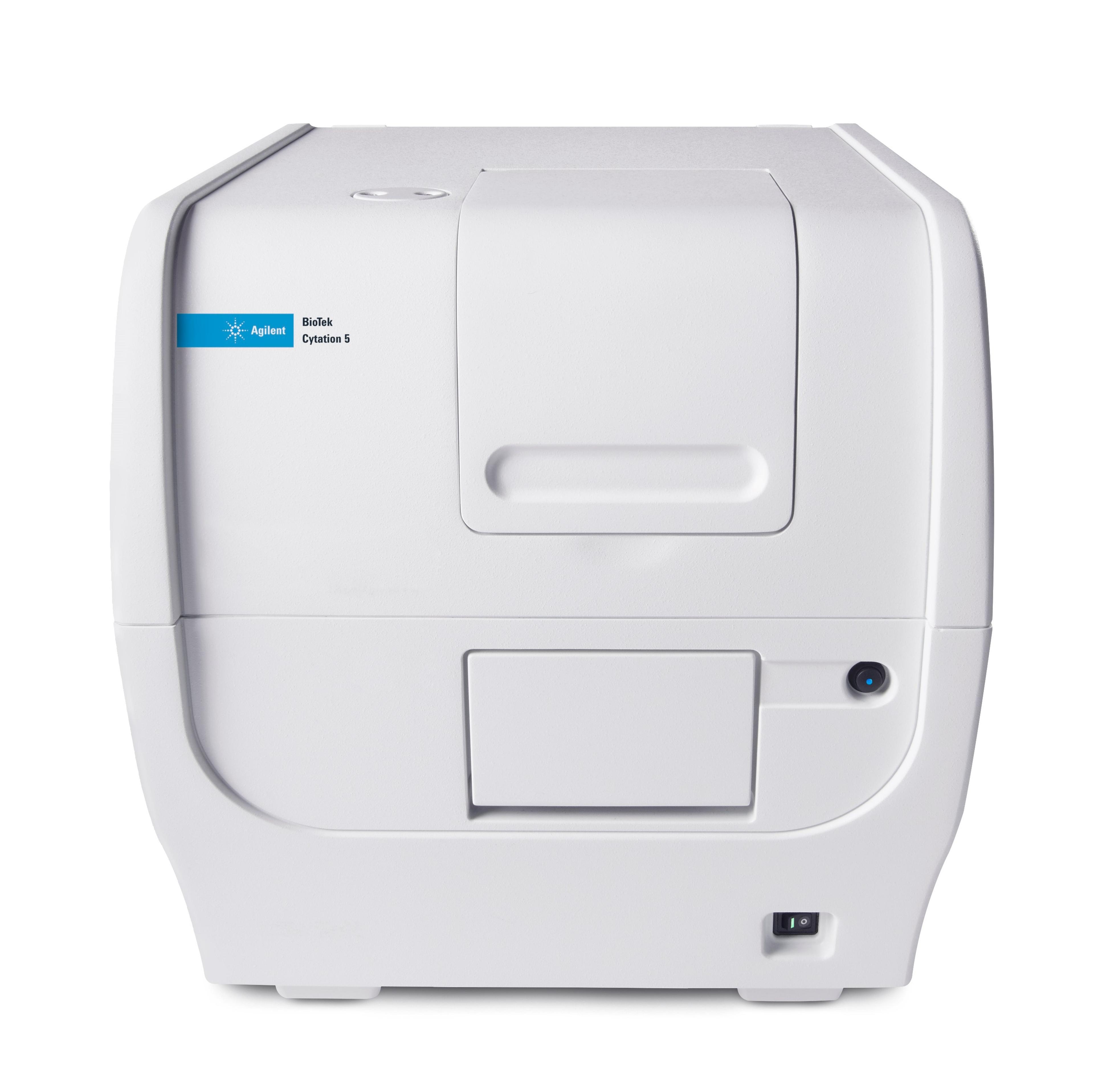
Agilent BioTek Cytation 5 Cell Imaging Multimode Reader
Agilent TechnologiesAgilent BioTek Cytation 5 is a uniquely integrated, configurable system that combines automated digital widefield microscopy with conventional multi-mode microplate detection to provide phenotypic cellular information and well-based quantitative data.