Getting to Know CellCelector
7 Jul 2022CellCelector is a unique and fully automated cell imaging and picking system developed for detection, selection and isolation of single cells, clusters, spheroids and organoids as well as single cell clones and adherent colonies. It is widely used in a multitude of research areas such as circulating tumor cell/CTC screening, stem cell research as well as cell line development and antibody discovery.
The platform combines an automated inverted fluorescence microscope with a high-speed scanning stage, a high-precision microfluidic cell picking robot and a PC workstation running powerful image processing software typing all system components together. The unique versatility is achieved by three exchangeable picking modules utilizing either high-precision glass capillaries, reusable (autoclavable) stainless steel scraping tips or disposable plastic tips. The operation principle can be summarized as following:
- Image the source plates
- Detect/Select cells, clusters or colonies of interest
- Pick target cells and transfer them into destination plates
The whole workflow is fully automated and documented by providing a live image during picking, cell tracking data from source plate to destination plate as well as before and after picking images.
Benefit by applying the CellCelector:
- Open platform allowing the development of individual imaging, detection and picking protocols
- Versatility: single cell picking, isolation of clusters, spheroids, organoids, adherent colonies, single cell clones from a variety of liquid and semi-solid medias
- Up to 100 % picking/transfer efficiency
- Extremely gentle cell transfer resulting in high cell integrity and outgrowth rates after the cell transfer
- Isolation of living or fixed cells
- Fully automated or interactive picking modes with live on-screen visualization of the picking process
- High scanning and picking throughput resulting in fast retrieval of cells
- Optionally deck tray with cooled destination positions available to prevent degradation of RNA of picked cells
- Automated adjustment of picking position and height
- Validated protocols for detection and isolation of single cells in suspension, adherent cells or colonies, 3D clones in liquid or semi-solid media
- Compatibility with standard and custom cell culture labware
- Complete documentation and traceability from source plate to destination including cell tracking data as well as before and after picking images
- The high-content imaging subsystem can also be used for imaging applications without picking
- Easy to operate with no routine maintenance necessary
Related Products
Request Quote for All Products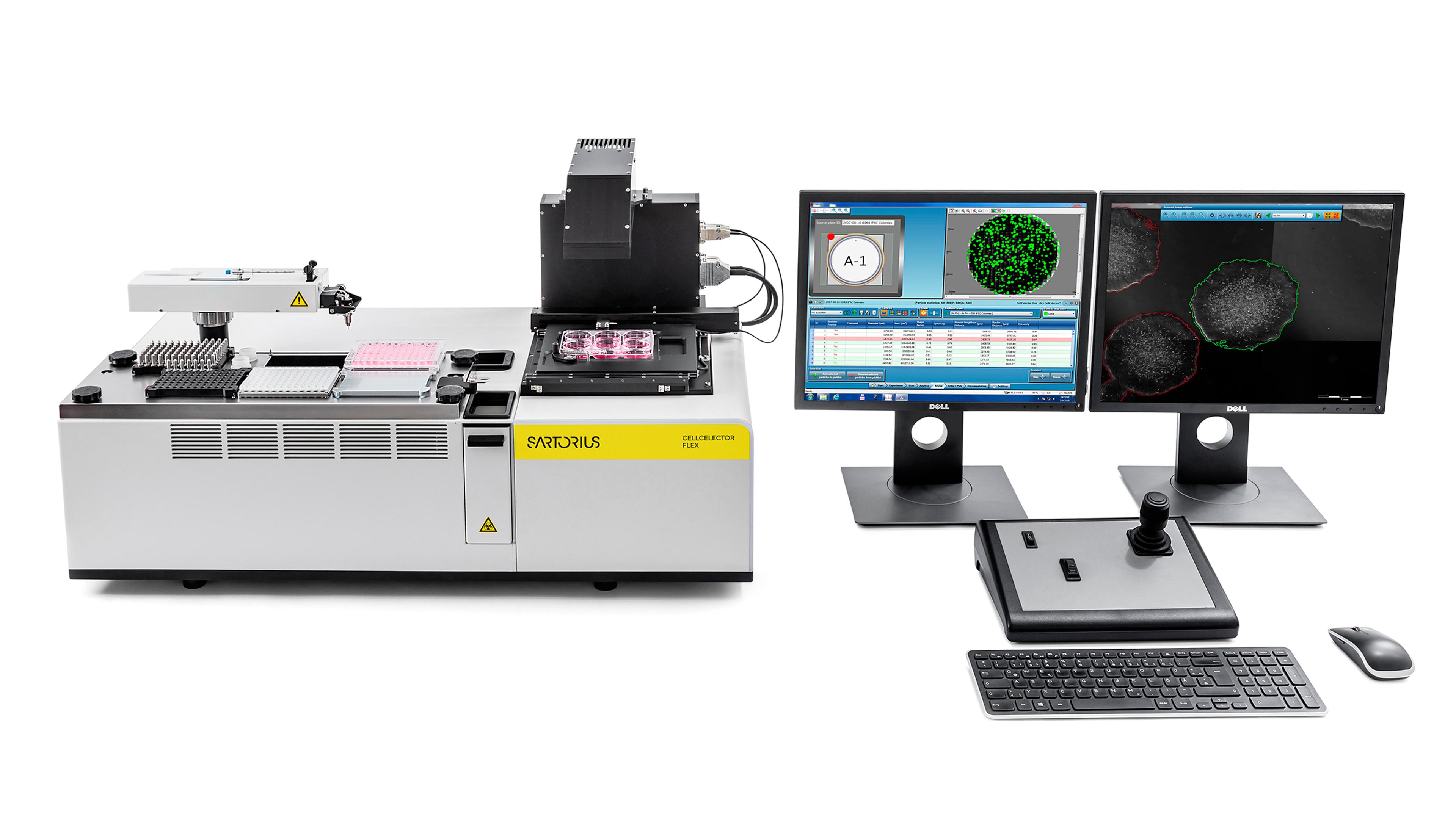
CellCelector Flex
Sartorius GroupFully automated cell imaging and picking system developed for the screening, detection, selection and isolation of single cells, clusters, spheroids and organoids, as well as single cell clones and adherent colonies.