Bio-Rex™ 70 Cation Exchange Resin, analytical grade, 100–200 mesh, sodium form, 500 g
Bio-Rad500 g, cation exchange resin, sodium form, macroreticular, 100–200 dry mesh size, 75–150 µm wet bead size, >1,000,000 MW limit
500 g, cation exchange resin, sodium form, macroreticular, 100–200 dry mesh size, 75–150 µm wet bead size, >1,000,000 MW limit
500 g, anion exchange resin, chloride form, 4% crosslinkage, 100–200 dry mesh size, 75–150 µm wet bead size, ~1,400 MW limit
500 g, anion exchange resin, chloride form, 4% crosslinkage, 200–400 dry mesh size, 45–75 µm wet bead size, ~1,400 MW limit
500 g, analytical grade anion exchange resin, chloride form, macroporous, 100–200 dry mesh size, 75–150 µm wet bead size, >1,000,000 MW limit
500 g, analytical grade anion exchange resin, chloride form, macroporous, 200–400 dry mesh size, 38–75 µm wet bead size, >1,000,000 MW limit
500 g, analytical grade cation exchange resin, hydrogen form, 2% crosslinkage, 50–100 dry mesh size, 300–1,180 µm wet bead size, ~2,700 MW limit
500 g, analytical grade cation exchange resin, hydrogen form, 2% crosslinkage, 200–400 dry mesh size, 75–180 µm wet bead size, ~2,700 MW limit
500 g, analytical grade cation exchange resin, hydrogen form, 12% crosslinkage, 200–400 dry mesh size, 53–106 µm wet bead size, ~400 MW limit
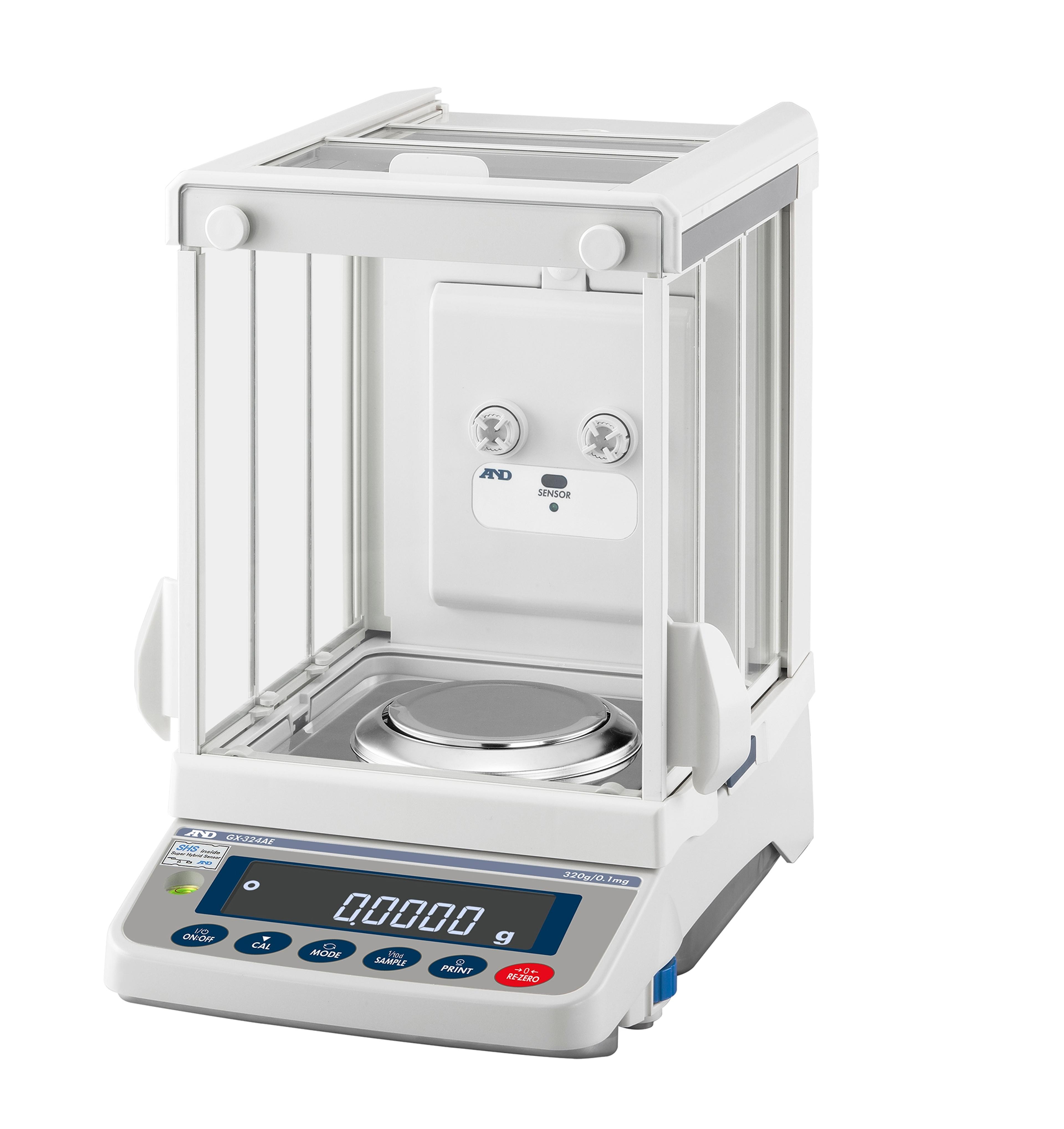
Our patented Smart Super Hybrid Sensor (SHS) technology improves response speed and minimizes maintenance costs. The new Apollo Analytical comes with a built in Ioniser, keeping the robust design of the precision version.
Our patented Smart Super Hybrid Sensor (SHS) technology improves response speed and minimizes maintenance costs. The new Apollo Analytical comes with a built in Ioniser, keeping the robust design of the precision version.
New CRISPR screening services substantially broaden range of possible genetic studies
GenScript provides lipid nanoparticle (LNP) formulation services for efficient delivery of nucleic acids and other therapeutic payloads across a wide range of research applications, including gene editing, vaccines, immunotherapy, and cell engineering. Leveraging expertise in payload synthesis and LNP formulation, GenScript supports the development of customized delivery solutions, including advanced targeted and novel LNP sys…
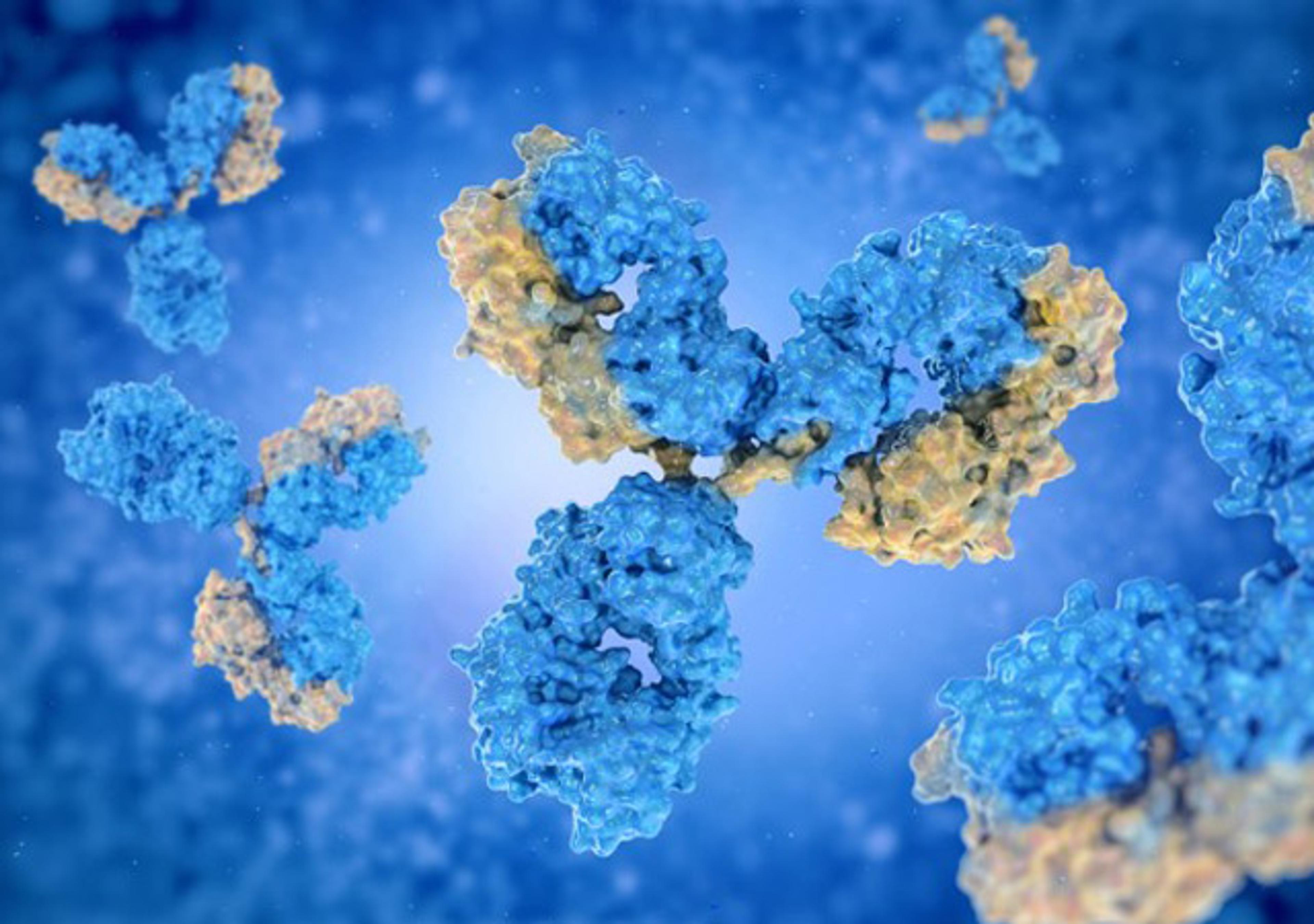
Creative Biolabs has proudly developed the unique Native™ Antibody Discovery Platform to develop native monoclonal antibodies using antigen-specific B lymphocytes cytometry technology.
GenScript provides research-grade lentiviral vector (LVV) packaging services for gene delivery, cell engineering, and therapeutic research applications. Leveraging optimized packaging systems and integrated workflows, GenScript delivers high functional titer lentiviral vectors with fast turnaround times, including reliable packaging support for large insert constructs.
Get the best performance from your water purification system from the people who designed and built it. We provide best-in-class service plans and maintenance options to meet your individual needs. Plus, MyMilli-Q™ Digital Services simplifies system management and allows remote monitoring and support.