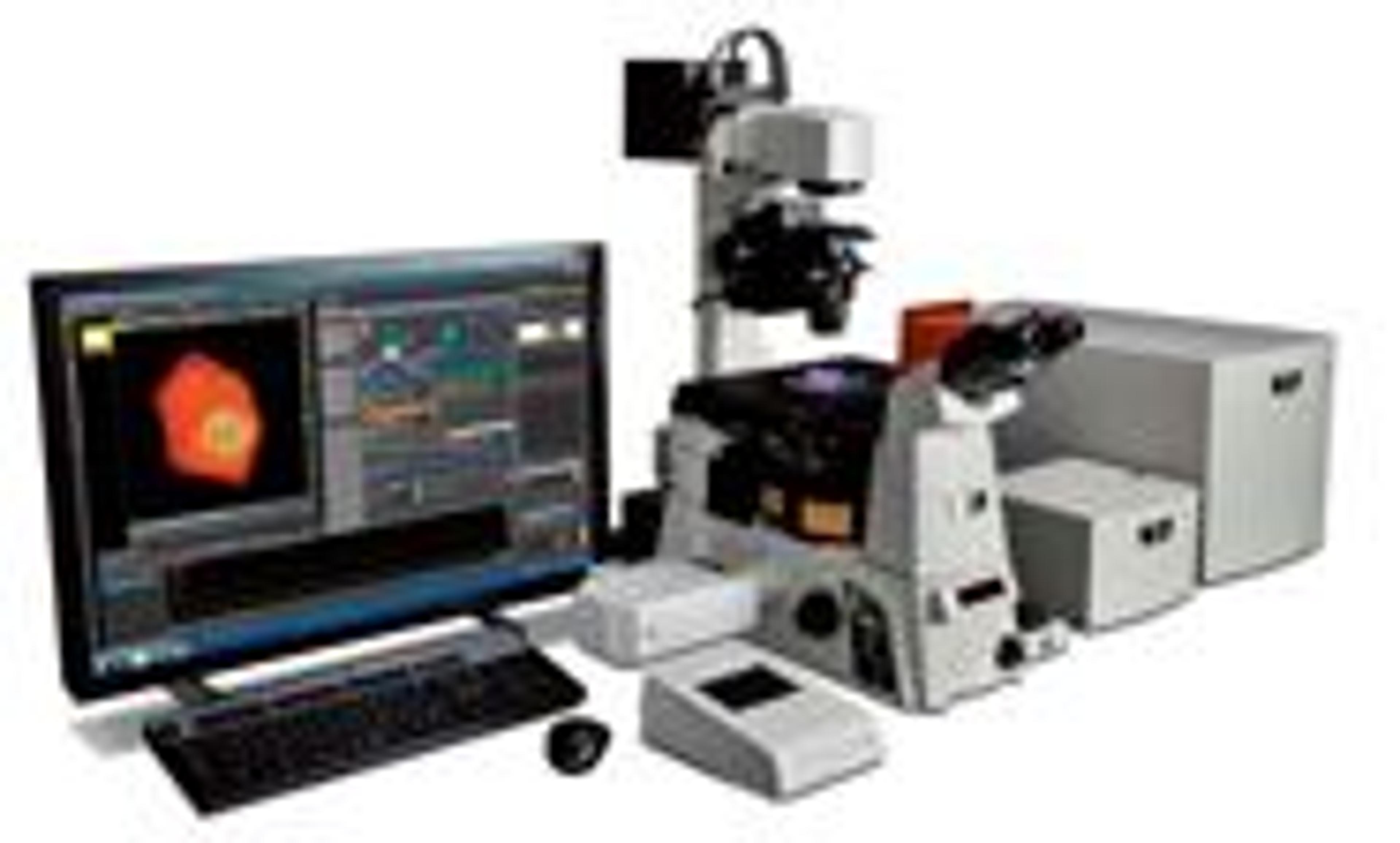
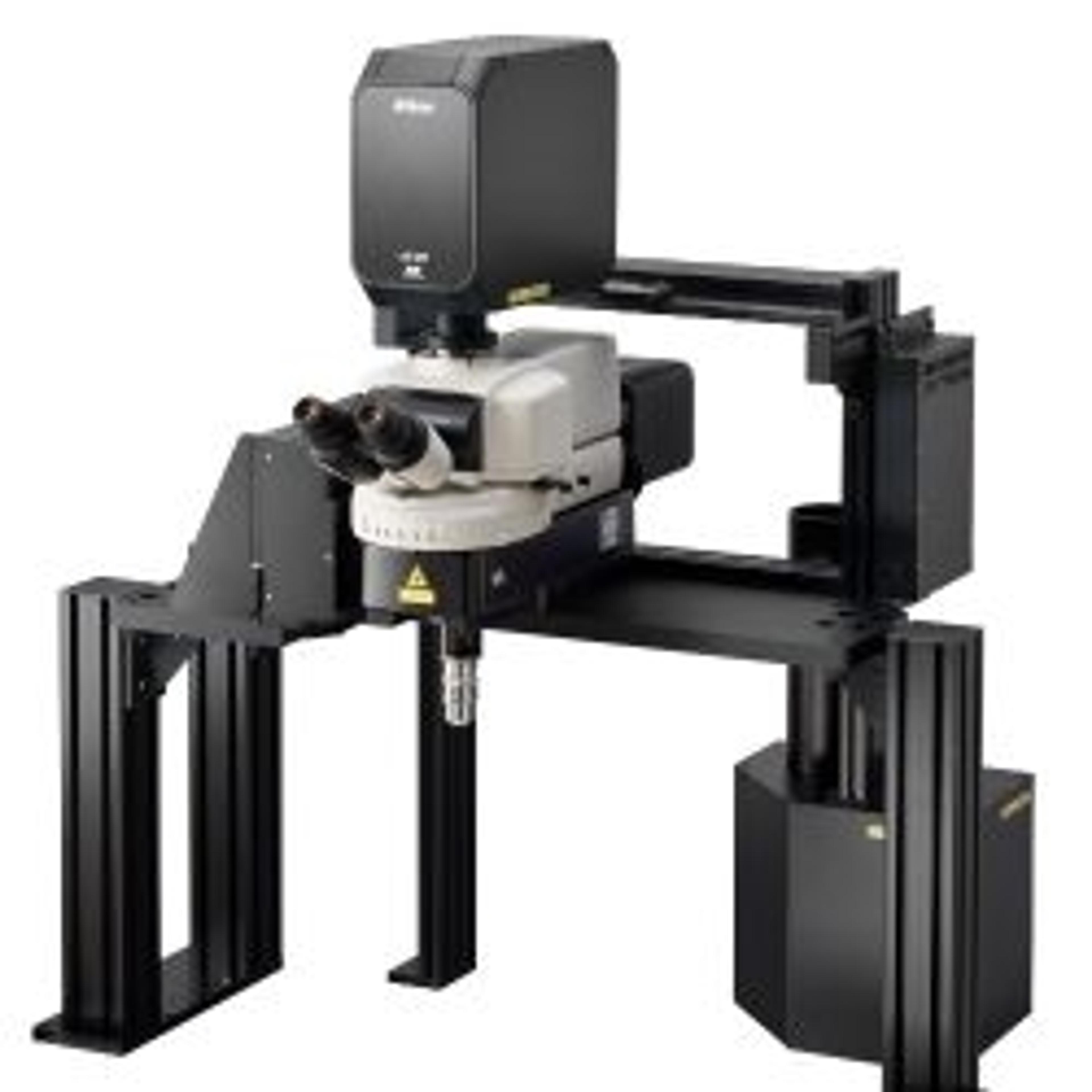
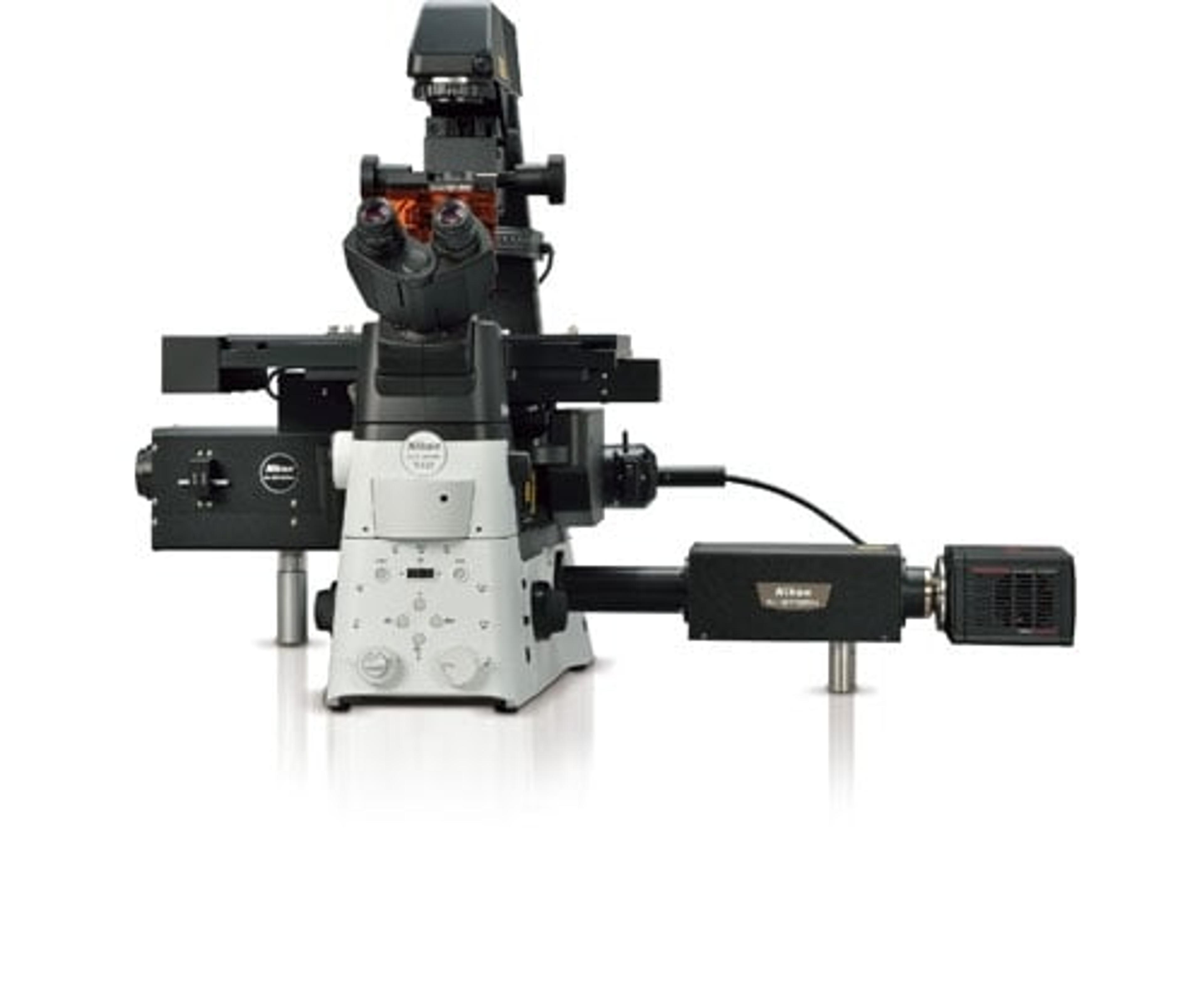
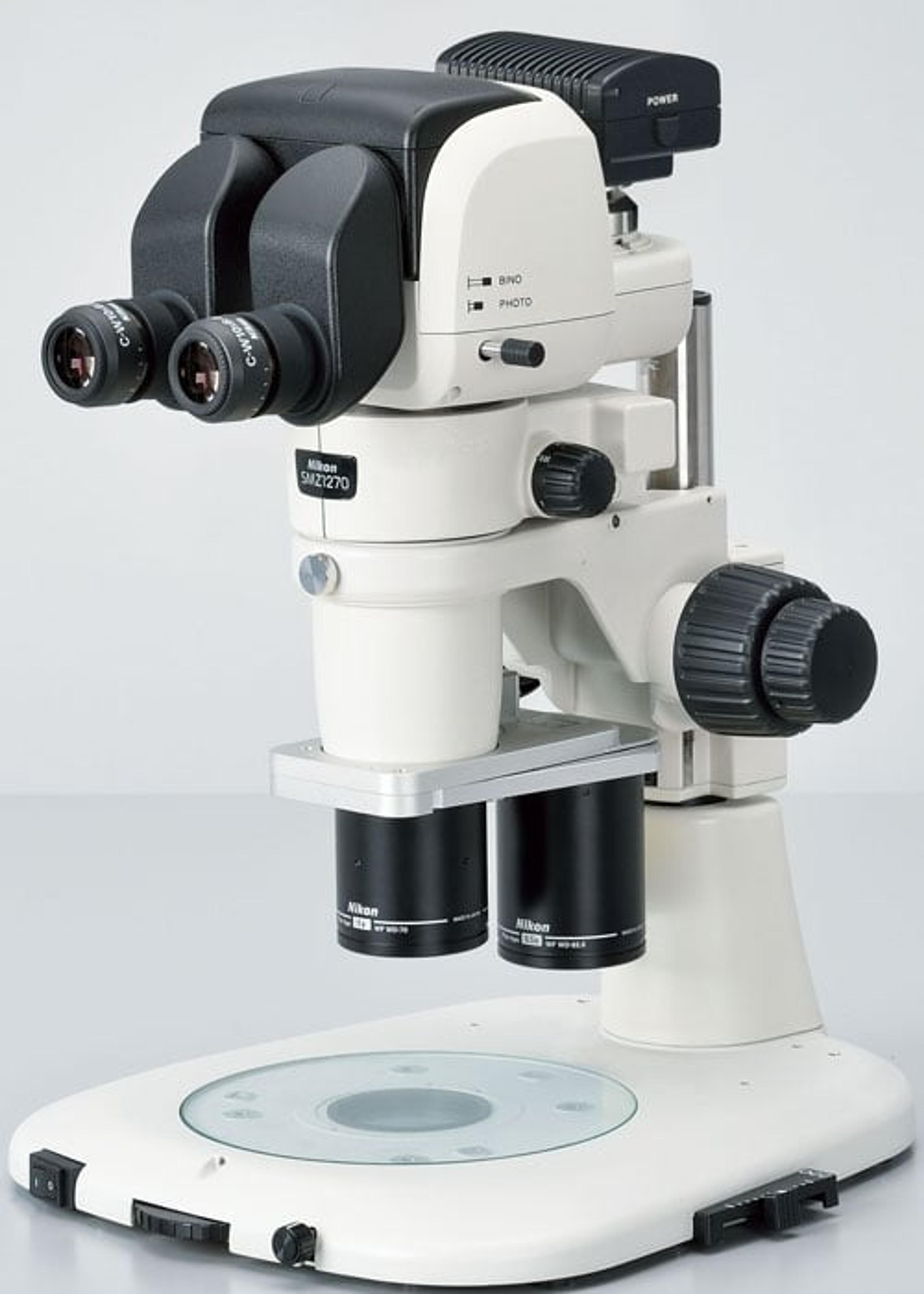
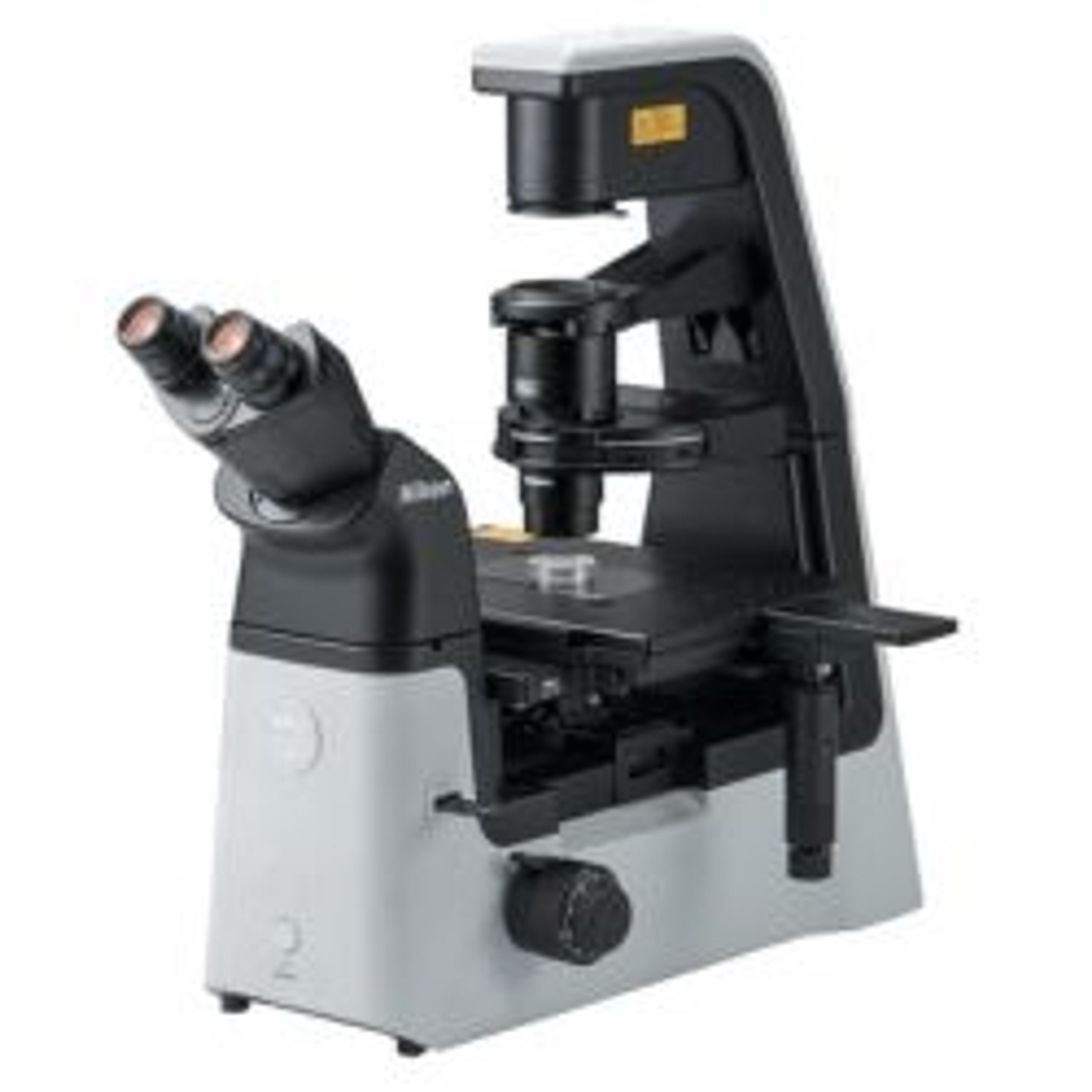
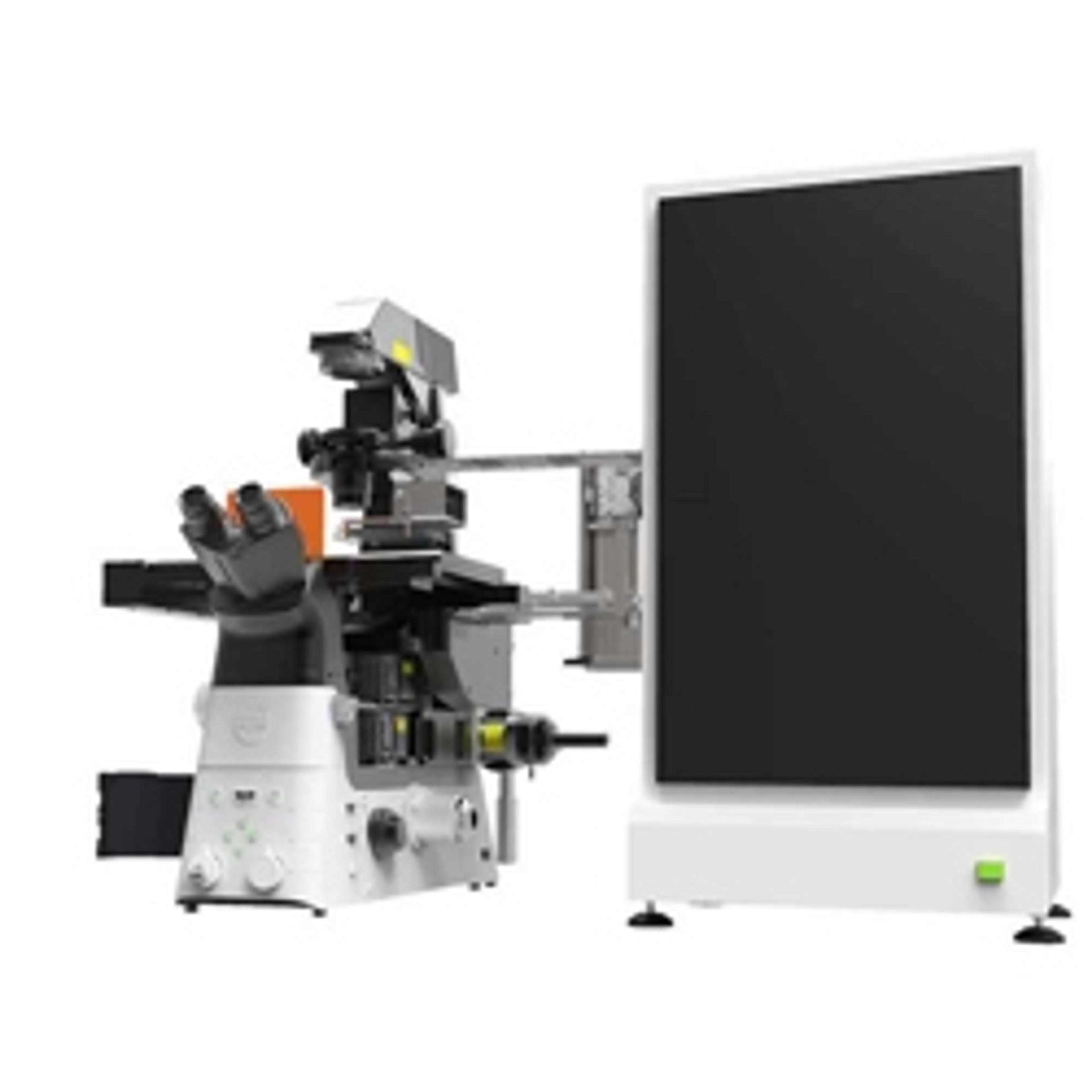
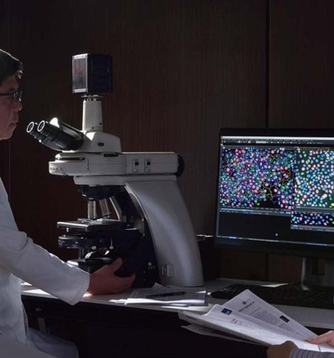
NIS-Elements Microscopy Software
Flexible software platform for controlling Nikon microscopes and 3rd-party components, with powerful custom programming tools for image acquisition and analysis.


Receive your quote directly from the manufacturer.
Ok but not the best
Shahjahanpur
I think this is oke
Review Date: 27 Jul 2022 | Nikon Healthcare Business – Microscope Solutions
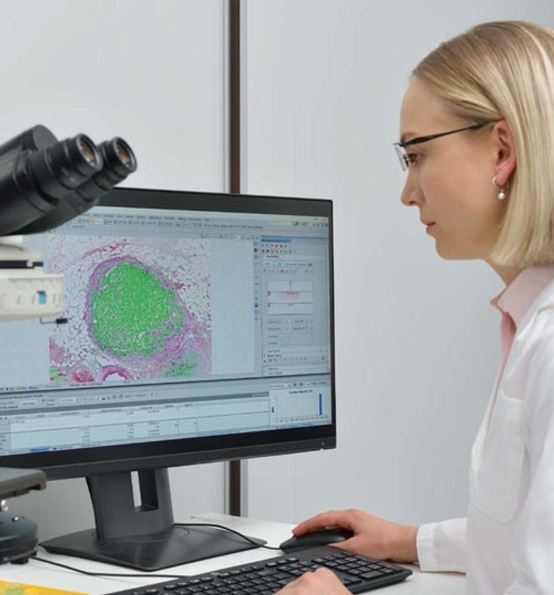
Powerful analysis software
Analysis use
This fluorescence microscope lets us capture high-quality images and detects fluorescence in the protein expression. Easy to use once learnt under supervision but requires careful handling. It is worth spending money on it.
Review Date: 22 Jul 2022 | Nikon Healthcare Business – Microscope Solutions
For my research work, the protein expression analysis wouldn't be completed without this microscope
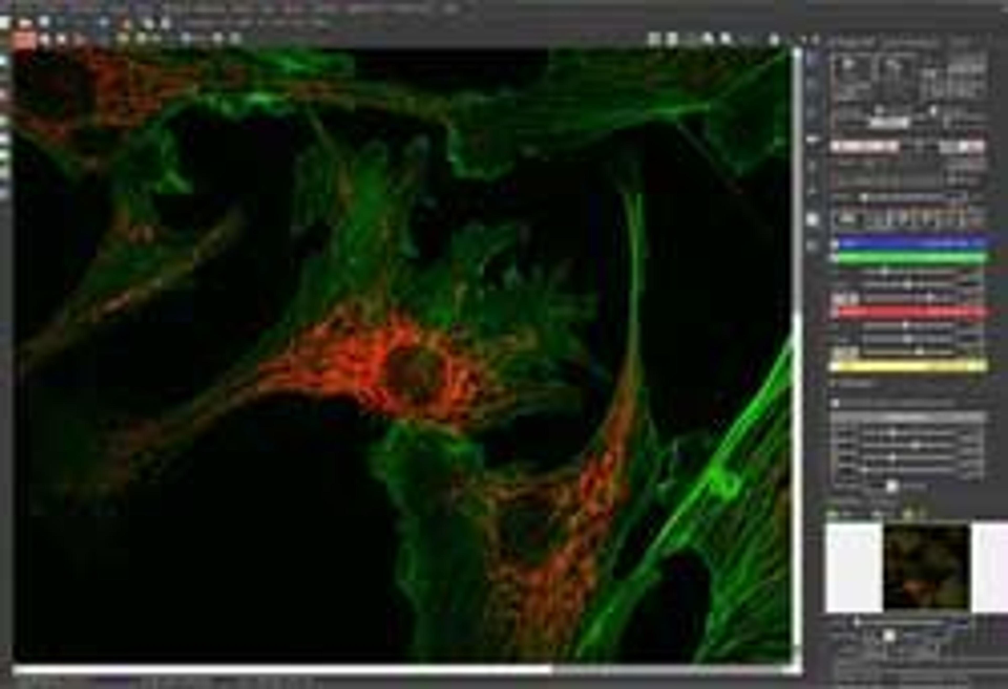
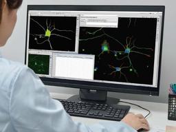
Fluorescence detection in immunocytochemistry
This fluorescence microscope lets us capture high-quality images and detects fluorescence in the protein expression. Easy to use once learnt under supervision but requires careful handling. It is worth spending money on it.
Review Date: 24 Aug 2021 | Nikon Healthcare Business – Microscope Solutions
User friendly.
Immunohistochemistry quantification
Easy to quantify a variety of histology applications, with multiple targets per image. Very user friendly.
Review Date: 13 Nov 2020 | Nikon Healthcare Business – Microscope Solutions
Great at analysis, not ideal for image acquisition.
Image acquisition and analysis process
It is a very powerful piece of software with comprehensive analysis functions. However, the image acquisition process on a microscope is slow. Sometimes it has communication problems between the software and the hardware.
Review Date: 25 Apr 2020 | Nikon Healthcare Business – Microscope Solutions
Quantification excellence
Neurotoxicology
I love the ability to customize quantification within the program. It's very user-friendly and has a wide application base for cellular or tissue imaging. The reps are knowledgeable and will quickly assist you if a solution is needed.
Review Date: 27 Feb 2020 | Nikon Healthcare Business – Microscope Solutions
I can never go back.
Software for running microscope
This software is very intuitive and easy to work with.
Review Date: 15 Mar 2019 | Nikon Healthcare Business – Microscope Solutions
Imaging
I've used several imaging software programs and instruments and Nikon has an excellent optics reputation. The software is easy to use and the customer service is phenomenal, the reps are always there for support and tutorials. The resolution is improved over other brightfield and fluorescence microscopy.
Review Date: 28 Nov 2012 | Nikon Healthcare Business – Microscope Solutions
Bio-imaging
The real-time measurement from the Elements doesn't work for me and the output is sometimes corrupted.
Review Date: 30 Apr 2012 | Nikon Healthcare Business – Microscope Solutions
This program crashes a lot.
Review Date: 2 Feb 2010 | Nikon Healthcare Business – Microscope Solutions
The Nikon NIS-Elements platform is an investment that addresses ever-changing protocols, new technology and system components. With NIS-Elements’ upgradability and ease in training and navigation, you create a resource that can be passed on through generations of your laboratory and research transitions.
Nikon also believes that having a single software platform for all imaging modalities is vital. NIS-Elements provides the same interface, control, workflow, and terminology whether it’s used for widefield, confocal, or super resolution imaging. With one platform to learn, users can easily switch between microscope systems when their applications require different imaging modalities. Imaging results from different Nikon systems can also be easily combined and analyzed to expand your research direction.
The NIS.ai module expands the NIS-Elements platform by building in tailor-made solutions for acquisition, visualization and analysis. Artificial Intelligence (AI) and deep learning methods are making seemingly impossible tasks now possible. Results only managed by challenging acquisition parameters or by painfully long or manual segmentation methods can now be automated thanks to AI.
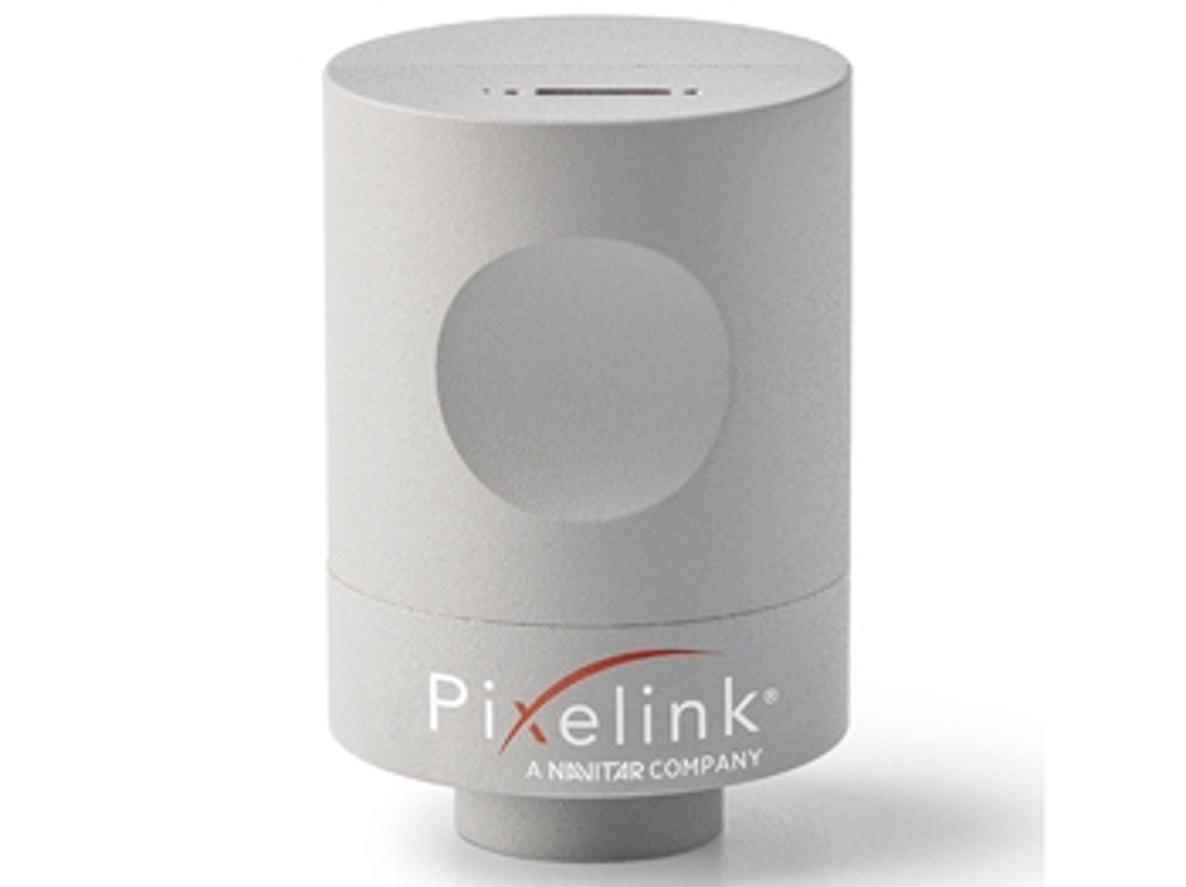
New Partnership to Deliver High-Quality Industrial Microscopy Cameras
Nikon Metrology NV and Pixelink announce new strategic partnership to distribute both standalone and integrated solutions to mid-range customers