Spatial biology techniques in tumor microenvironment analysis
Researchers in the US are using multiplex immunofluorescence to unravel the complexities of the spatial biology within the microenvironment of brain tumors
28 Mar 2024Editorial article
Gaining greater insight into the complex immune processes within the tumor microenvironment is technology-dependent and requires advances in resolution and reproducibility. While immunotherapies have shown great promise in preclinical studies, clinical trials have yielded more heterogeneous responses. One reason for this complexity is the interplay between molecular and genetic elements influencing T-cell antigen recognition and immune responses within the tumor microenvironment. The latest spatial biology techniques offer unparalleled insight into brain tumor microenvironments at a single cell resolution and prospects for identifying more effective treatments.

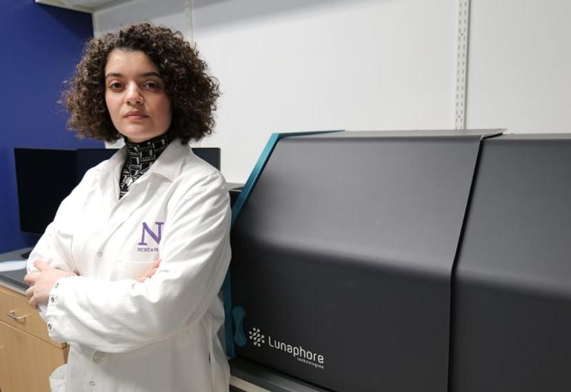
In this editorial, we speak with Hinda Najem, a Postdoctoral Research Fellow at Northwestern University in Chicago, USA. Working in the Department of Neurological Surgery at Northwestern, Dr. Najem spends a significant amount of her time using spatial proteomics techniques to study both primary and metastatic cancers of the central nervous system. She acknowledges the importance of having the latest cutting-edge tissue profiling technologies that are nimble for achieving her translational research goals.
Generating reproducible data from a single sample
The heterogeneity of brain tumors is one of the main challenges of their treatment. Tumors can exhibit diverse genetic backgrounds, molecular organization, cell, and molecular compositions. The development of innovative spatial biology techniques, such as multiplex immunofluorescence, has enabled researchers, by bringing spatial context to multi parameter studies, to gain a deeper understanding of the spatial organization of cells and molecules at the proteomic level with high resolution imaging. The COMET™ platform, developed by Lunaphore, is a leading example of a fully automated, hyperplex immunofluorescence platform for the spatial analysis of tissue samples and is the equipment of choice in the Malanti Brain Tumor Institute where Dr. Najem works.
Dr. Najem explains, “The COMET™ platform is a powerful tool for both proteomic validation and spatial discovery. Proteomic analysis is usually needed to validate any type of transcriptomic or genomic data, especially for brain tumors with a high degree of epigenetic regulation. The proteome represents the end of the cell manufacturing chain”.
Dr. Najem highlights that the COMET™ platform uses off-the-shelf antibodies and that each analysis is easy to customize to a specific research hypothesis. Additionally, the platform preserves the specimen, allowing for other types of “omic” interrogation of the same slide.
Choosing the right tool and provider
Dr. Najem added that single-cell resolution is crucial to conduct studies such as cell-cell interactions and is required for getting meaningful insights from the TME . “We specifically invested in this platform as benchtop equipment, but it can also be acquired within a core facility,” she describes. Adding, “The data quality and acquisition are easy, rapid, and highly satisfactory at the single cell level.”
She notes that different labs have different needs and hypotheses, and an increase in yield per run achieved on COMET™ would be well received. Nonetheless, she remains impressed with the throughput. She has been able to validate over 200 targets and address over 50 distinct research queries spanning many different central nervous system pathologies in less than a year.
As with any cutting-edge technology, the long-term support of a technology provider can be key to adopting the equipment. Najem speaks highly of Lunaphore, attesting that “the appropriate training was received upon the acquisition of the machine, and that all support and help necessary was also provided when needed in a timely fashion.”
A neuroscientist’s best friend
Looking more broadly at the future of spatial proteomics in biology, Dr. Najem believes that this technology will be needed for the foreseeable future. “The information gathered through this technique is so vast and powerful that, as translational scientists, we see its importance on the clinical side as well, especially in supplementing standard techniques such as H&E staining and immunohistochemistry,” she predicts.
Dr. Najem concludes, “With emerging AI technologies, the future of proteomic spatial analysis is very promising at all levels.”
Learn how Lunaphore Technologies’ COMET™ hyperplex immunofluorescence platform can accelerate your research.
Related products
Request Quote for All Products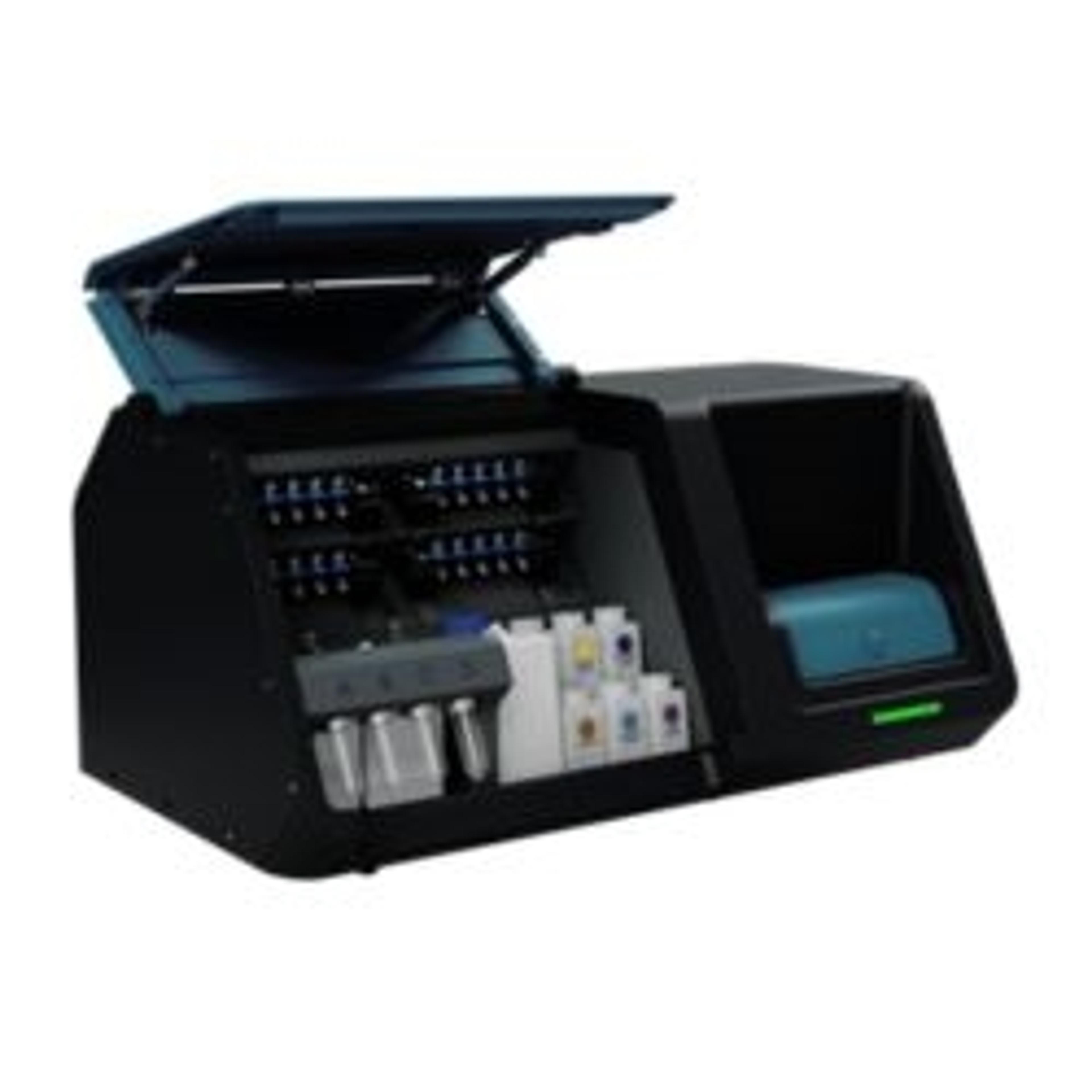
COMET
Bio-Techne SpatialCOMET™ is a fully automated, high-throughput, hyperplex platform for the analysis of Formalin-Fixed, Paraffin-Embedded (FFPE), and frozen tissues.