Safeguarding public health against antimicrobial-resistant bacteria
Infectious disease expert shares how robust LC columns are facilitating the analysis of complex pathogen samples
4 May 2021

The Robert Koch Institute’s aim is to protect the population against disease and improve its state of health through research into non-communicable diseases, such as diabetes and cancer, and infectious diseases, most recently, COVID-19.
In this exclusive interview, we speak with Dr. Jörg Döllinger, head of the proteomics service in the core facility of the RKI, about his collaborative work developing proteomic methods to facilitate infectious disease research.
What project are you working on currently?
JD: My research focuses on the development of proteomic methods to facilitate infectious disease research including the identification and characterization of human pathogens. At the moment, I am working on method development to turn mass spectrometry-based proteomics into a high-throughput technology for clinical microbiology. We hope that this will translate into personalized patient treatment and help control the spread of antimicrobial resistance, which is currently a major issue for global health.
In the last year, we have published three technical papers and a proof-of-concept study that shows that species identification, as well as antimicrobial resistance detection, is possible using proteomics with high sensitivity and specificity1. The first paper outlines our sample preparation method, named SPEED, which is based on acid extraction2. The second deals with the optimization of data-independent acquisition (DIA), especially using in silico predicted libraries3. The final technical paper introduces a new concept for bacteria identification using peptide mass barcodes instead of using sequences4.
What are the intended impacts of your work on research, healthcare, and patients?
JD: I would like to be able to analyze whole bacterial proteomes in a very short time and then translate the output directly into patient treatment. In measuring whole proteomes of bacterial infections, we can get much deeper insights into the expression of virulence factors and how antimicrobial resistance develops. We hope to use this information to enable personalized patient treatment and improve our view of the progress of infections within patients.
How do you use µPAC technology in this work?
JD: We have been using the micropillar array columns for about one and a half years now. We use the long, 200-centimeter column routinely for complex samples like eukaryotic cells and tissue samples. We use the smaller, 50-centimeter column for less complex samples like bacteria, fungi, or in interaction studies with shorter gradients (30-60 minutes).
What does this technology enable you to achieve?
JD: These columns provide us with a new level of reproducibility to peptide separation. In our DIA paper, we showed that we were able to develop a method that uses only 1.5 data points per peak, greatly improving proteome coverage a lot - up to more than 8,000 human proteins in a single-shot experiment - and we can still maintain very high quantitative reproducibility.
In addition, I think the most overlooked aspect of the columns is the low back pressure, because you can run samples at pressures below 350 bar, far below the limit of current LC systems. For me, this translates into fewer LC problems. We are still using nano-LC even for our routine diagnostic measurements because we have this robustness and can then keep the sensitivity of the low flow rate.
Why are robustness and reproducibility especially important for clinical proteomics?
JD: In routine diagnostics, reliability is very important and so proteomics can't enter the clinic without improved robustness. LC-MS is a complex technology and still has some big limitations with sensitivity, throughput, and ease of use. For example, this is obvious when you compare LC-MS for SARS-CoV-2 diagnostics to the gold standard PCR.
Currently, when you look at diagnostic LC-MS papers, sample cohorts and the awareness of quality control are both increasing. We are moving in the right direction, but there's still room for improvement.
How have you applied your methods to COVID-19?
JD: In my experience, LC-MS still has severe limitations for routine virus diagnostics, namely sensitivity and throughput, in comparison to PCR. In my opinion, it is currently better suited for untargeted virus screening5. We conducted an extensive study along with the virology unit in our department (ZBS1) using deep proteome profiling of SARS-CoV-2-infected human lung cells, which provided new insights into the differences between SARS-CoV and SARS-CoV-2 with respect to immune modulation6.
Are there any exciting opportunities or challenges that still need to be addressed in the future?
JD: I think that one day LC-MS could replace MALDI-ToF MS in clinical microbiology. At the moment, laboratories will use several different methods to address different questions. For example, MALDI-TOF MS is used for species identification and phenotypic methods for antimicrobial resistance detection. We hope to do all this investigation within one LC-MS-based method that also provides additional data, such as the expression of bacterial toxins and other virulence factors. Today, this technology can already analyze near-complete bacterial proteomes and this could translate into new insights into the biology of the cells.
We work hard on method development and I think it will soon be possible to analyze hundreds to thousands of bacterial proteomes, which should hopefully translate into personalized patient treatments. However, I think the cost and the technical complexity of LC-MS instrumentation are still major challenges for the distribution of the technology, because at the moment LC-MS is still limited to experienced and specialized laboratories.
References
1. Blumenscheit C, Pfeifer Y, Werner G, John C, Schneider A, Lasch P, et al. Unbiased antimicrobial resistance detection from clinical bacterial isolates using proteomics. bioRxiv. 2020:2020.11.17.386540.
2. Doellinger J, Schneider A, Hoeller M, Lasch P. Sample Preparation by Easy Extraction and Digestion (SPEED) - A Universal, Rapid, and Detergent-free Protocol for Proteomics Based on Acid Extraction. Mol Cell Proteomics. 2020;19(1):209-22.
3. Doellinger J, Blumenscheit C, Schneider A, Lasch P. Isolation Window Optimization of Data-Independent Acquisition Using Predicted Libraries for Deep and Accurate Proteome Profiling. Anal Chem. 2020;92(18):12185-92.
4. Lasch P, Schneider A, Blumenscheit C, Doellinger J. Identification of Microorganisms by Liquid Chromatography-Mass Spectrometry (LC-MS(1)) and in Silico Peptide Mass Libraries. Mol Cell Proteomics. 2020;19(12):2125-39.
5. Grossegesse M, Hartkopf F, Nitsche A, Schaade L, Doellinger J, Muth T. Perspective on Proteomics for Virus Detection in Clinical Samples. J Proteome Res. 2020;19(11):4380-8.
6. Grossegesse M, Bourquain D, Neumann M, Schaade L, Nitsche A, Doellinger J. Deep time course proteomics of SARS-CoV and SARS-CoV-2-infected human lung epithelial cells (Calu-3) reveals strong induction of interferon-stimulated gene (ISG) expression by SARS-CoV-2 in contrast to SARS-CoV. bioRxiv. 2021:2021.04.21.440783.
Related Products
Request Quote for All Products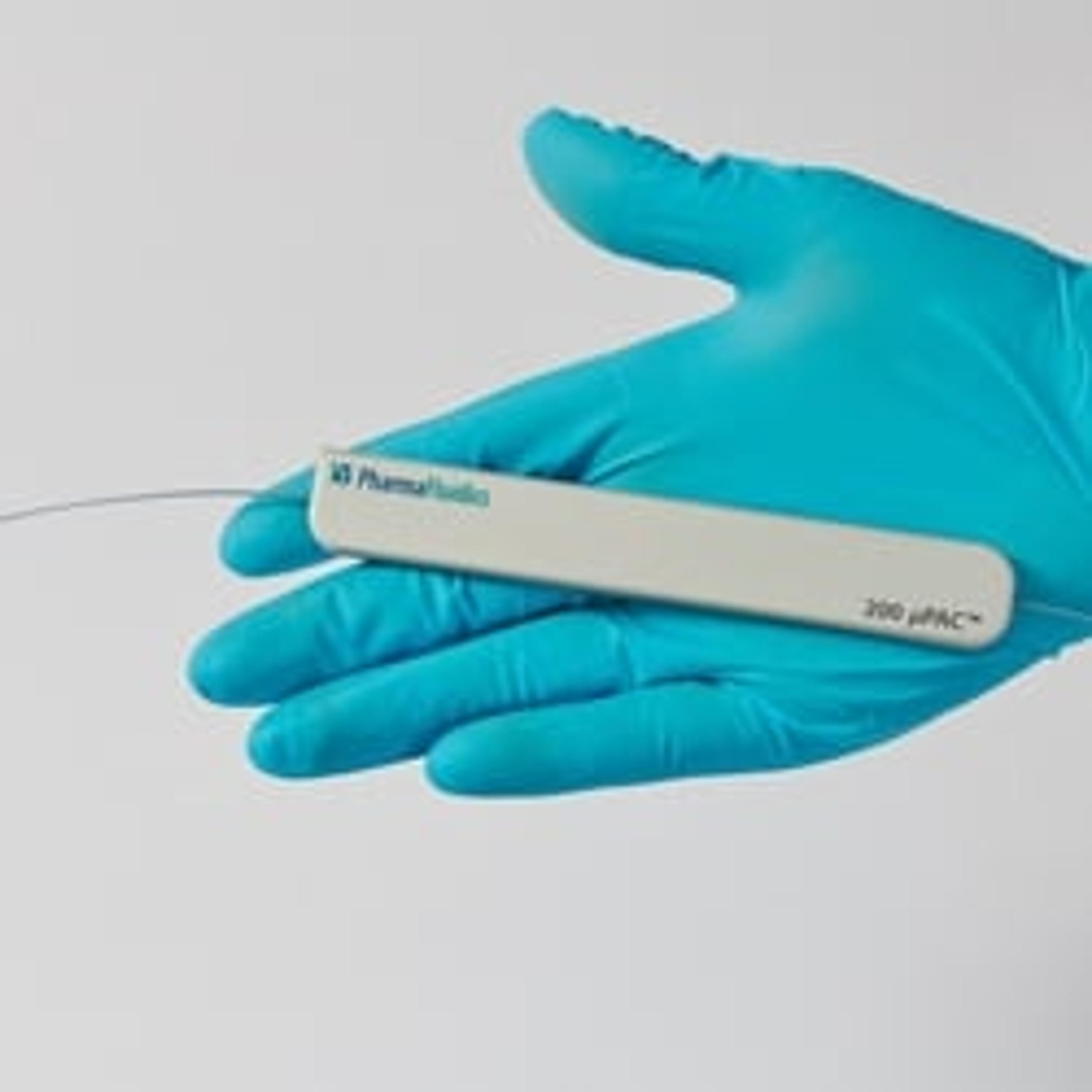
200 cm µPAC™ column
PharmaFluidicsAs an alternative to conventional columns, PharmaFluidics offers micromachined nano-LC columns or micro Pillar Array Columns (µPAC™). These µPAC™ columns display exceptional separation properties with unprecedented peak capacity and narrow peak shapes at low operating back-pressure. µPAC™ column, 200 cm length, with pillar array backbone at interpillar distance of 2.5µm, C18.
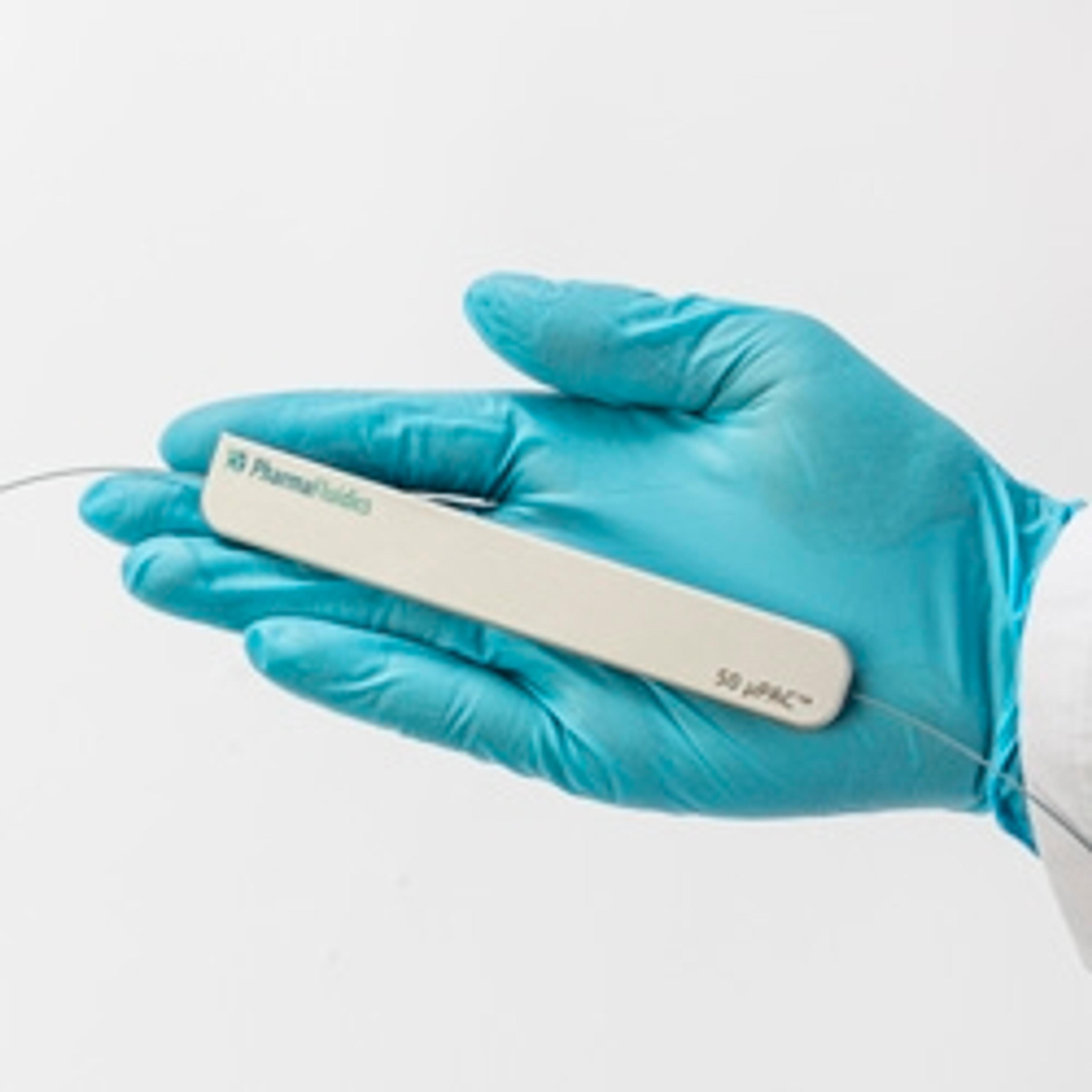
50 cm µPAC™ column
PharmaFluidicsWith an internal volume of 3µL, this 50 cm long RP-C18 µPAC™ column is perfectly suited to perform high throughput analyses with shorter gradient solvent times (30, 60 and 90-minute gradients) and it can be used over a wide range of flow rates, between 100 and 2000 nL/min. µPAC™ column, 50 cm length, with pillar array backbone at interpillar distance of 2.5µm, C18.