From Phenotype to Genotype: Unlocking the Power of Correlative Microscopy for Cell Biology
SelectScience® spoke to Dr Peter O'Toole, Head of Imaging and Cytometry, Department of Biology at the University of York, about the technology serving his department
6 Jun 2016

SelectScience® spoke to Dr Peter O'Toole, Head of Imaging and Cytometry, Department of Biology at the University of York, about the technology serving his department
European Molecular Biology Laboratory (EMBL) Founded in 1974, EMBL is Europe’s flagship laboratory for the life sciences – an intergovernmental organization with more than 80 independent research groups covering the spectrum of molecular biology.
The Bioscience Technology Facility at the Department of Biology provides the University of York with access to a range of technologies and expertise to enable cost effective, efficient, research. At the European Molecular Biology Laboratory’s (EMBL) workshop ‘From 3D Light to 3D Electron Microscopy’ (EMBL 3D), ran in conjunction with ZEISS Microscopy, we caught up with Dr Peter O’Toole, Director of the Bioscience Technology facility, about the innovation that has made it possible.
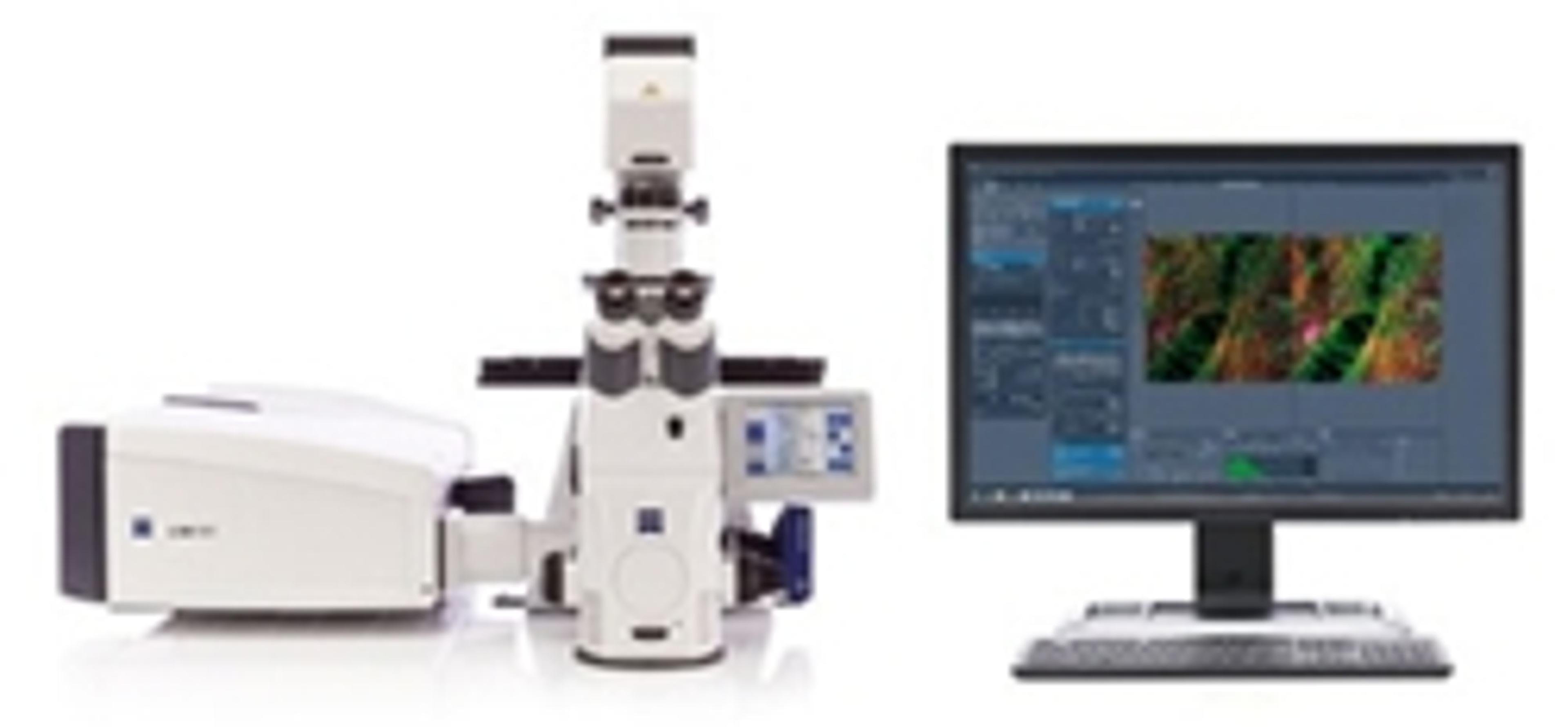
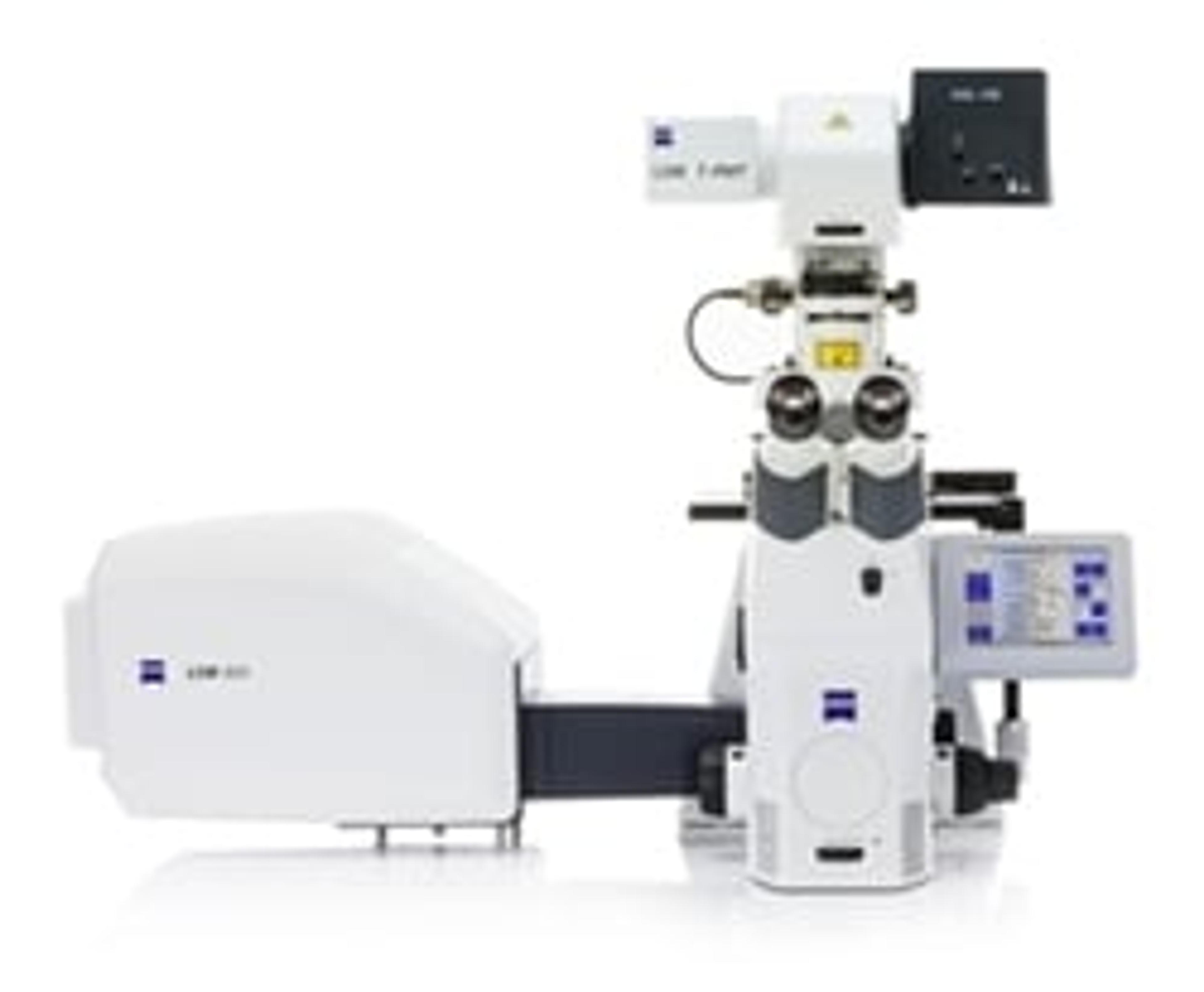
“The Bioscience Technology Facility encompasses lots of different core technologies, from genomics, proteomics and metabolomics, to molecular interactions to protein production bioinformatics”, Dr O’Toole explained. An important facility is the Imaging Cytometry lab, which has “an array of different microscopy techniques”. “We have the high end or electron microscope going all the way through to the life cell confocal microscopes, through to photon microscopes, through to more basic label free and developmental microscopes that are bridging those gaps”, Dr O’Toole revealed.
The facility aims to have the technology available to “best serve the department, which is a broad discipline department”, explained Dr O’Toole, providing services to “immunologists, cancer researchers and neurobiologists”. This means that the microscopes have to cope with a very diverse range of samples, from “plant samples, to your bacteria, to your yeast, to your tissue sections.”
Live cell imaging
One area where new technology is starting to provide insights is the field of live cell imaging. Previously, scientists have been able to “look at cell behaviors”, which doesn’t give information on “how many proteins there are or where the proteins actually reside in the cell”, Dr O’Toole explained.
That’s where correlative light and electron microscopy comes in. “Now, we can take proteins in cells and can label them with fluorescent dyes, and see where they are. By coupling that with electron microscopy, we can look at the protein diffusions right the way through to the fine ultrastructure where it is within that single cell.” The York facility first started exploring these different sample types using the ZEISS Airyscan module, “looking at protein distribution in bacteria or the ring structures of neuromuscular junctions and synapses”. The other side of correlative microscopies is looking at a bigger picture”, Dr O’Toole explained. Researchers can “take microscope images and correlate them back to the genomic information, to the proteomic information, to the metabolomics information”.
Sample preparation and integration
This technology is already providing “a better understanding and much greater precision” than was available before, Dr O’Toole revealed. “The future and the advancement of correlating light and electron microscopy is to make that whole workflow much easier, both through sample preparation and the integration of the systems themselves.” Dr O’Toole believes that this will allow “non-specialist biologists to have the courage to apply it to their work”.
Find out more about the other speakers and presentations from EMBL 3D, or learn more about the applications of Correlative Microscopy.
Watch our interview with Dr O'Toole here.