A New Streamlined Method for DNA Repair and Telomere Stability
Dr. Jaroslav Fulneček, CEITEC MU, tells SelectScience about a new high-throughput assay for analysis of DNA-protein interactions
28 Feb 2018
Many essential biological processes are regulated through nucleic acid-protein interactions, e.g. DNA replication and repair. Understanding of this intricate system holds great potential for elucidating the mechanisms of physiological processes, as well as human disease, and is essential for developing new treatments. Now, an exciting innovation may be about to change the way geneticists are working.
While an array of methods can be used to assess DNA-protein interactions, many are time consuming and labor intensive and there is much need for a simplified, sensitive alternative. Scientists at the Central European Institute of Technology (CEITEC MU) have addressed this issue with the development of a new assay for the high-throughput quantitative analysis of DNA-protein interactions that eliminates the need for protein labeling.
In this exclusive interview, the assay’s co-inventor, Dr. Jaroslav Fulneček, shares details of the method, which is based on protein-induced fluorescence enhancement (PIFE) using microwell plate fluorescence readers (mwPIFE), and explains how it was used to analyze DNA interactions with the Ku complex, a heterodimeric protein complex that has a role in both DNA repair and telomere stability.
Tell us a little about yourself and your background
JF: My name is Jaroslav Fulneček and currently I work as senior scientist in the lab of Dr. Karel Říha at Central European Institute of Technology, Masaryk University, in Brno, Czech Republic. I graduated in biochemistry and did my Ph.D. in biophysics in Brno. I worked at the Institute of Biophysics, Academy of Sciences of the Czech Republic, for 16 years where I studied epigenetics in plants, plant DNA methylation, polyploidization and the evolution of plant genomes. In 2014, I moved to the Masaryk University, where I helped to move and start the Dr. Karel Říha lab from the Gregor Mendel Institute in Vienna. In the Říha lab, I joined Dr. Soňa Valuchová, who is studying the function of Ku complex in DNA double-strand break repair and telomere maintenance.
Describe your current research into protein-nucleic acid interactions – what is your goal?
JF: We study the evolutionary conserved Ku complex which is important for DNA repair and telomere stability. While it is a crucial factor in the non-homologous DNA end joining pathway that mediates ligation of double-stranded breaks, it also protects the natural chromosome ends, telomeres, from DNA repair protein activity. We try to understand how Ku proteins implement these seemingly contradictory functions in genome maintenance. One of our approaches is to describe the mechanism of Ku-DNA interaction and to determine requirements for Ku-DNA binding in DNA repair and telomere maintenance. In our most recent study, we showed that Ku requirements for DNA binding differ in non-homologous end joining and telomere protection (Valuchova et al 2017).
We use several methods to study Ku protein-DNA interaction including fluorescence anisotropy, electrophoresis mobility shift assay and electron microscopy. Recently, we have developed a new assay based on protein-induced fluorescence enhancement (PIFE), a phenomenon that had previously been used to study DNA-protein interactions at single molecule level (Hwang and Myong 2014). We adopted the PIFE method for average, steady-state measurements of DNA – protein interactions in microwell plates. (Valuchova et al 2016).
Describe the novel mwPIFE method you have developed. How will this method benefit research?
JF: The principle of the method utilizes the PIFE phenomenon, whereby binding of a protein to cyanine-3 (Cy3) labeled DNA or RNA hinders isomerization of the Cy3 dye, resulting in a higher quantum yield upon fluorophore excitation. Usually, NeutrAvidin or Streptavidin coated black 96-well plates are used but it is also possible to use 384-well plates if high PIFE signal is expected. Cy3 labeled oligonucleotides, either at the end or internally, and single- or double- stranded DNA or RNA, with biotin at one end, are immobilized at the bottom of coated wells. Fluorescence of immobilized oligonucleotides is measured using a plate reader in well-scanning mode. The measurement is repeated after an addition of a protein. Higher fluorescence signal indicates binding of a protein in close proximity to a Cy3 label. Compared to FRET methods, where energy transfer works at some distance, PIFE requires binding of a protein directly very close to the dye, because the isomerization of the fluorophore has to be prevented by the bound protein. This is advantageous for searching for exact interaction sites or for monitoring movement of proteins along oligonucleotides. Furthermore, no protein labeling is required. Adopting the method to microplates makes the method high-throughput so we can quickly, for example, optimize buffer composition or determine the optimal sequence for binding by comparing various oligonucleotides.
Which microplate reader do you use in your research and why?
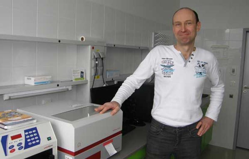
JF: We bought FLUOstar Omega plate reader and specific filters for Cy3, 540BP10 for excitation and 580BP10 for emission from BMG LABTECH. The reader can measure 96-well plates and also 384-well plates in scanning mode, which is essential to measure PIFE.
How will you use the mwPIFE method in your own research? What other areas of research do you see this method benefiting the most?
JF: Generally, the mwPIFE method is suitable for DNA, RNA – protein interaction studies where PIFE is observed. I also adopted the method for kinetic studies of Ku complex interactions with DNA using stop-flow system. Here, I could distinguish individual kinetic steps as binding, docking and translocation and compare it using mutated variants. The assay can also be adopted for a high-throughput format to screen chemical libraries or a wide range of binding conditions.
What are the future goals of your research and how will you achieve these?
JF: We want to describe in detail the mechanisms of Ku complex-DNA interaction and use this knowledge to identify chemical inhibitors that can specifically affect different modes of Ku-DNA binding. Such inhibitors will be useful for functional studies of the Ku complex in cells, as well as for biomedical applications, for example in the areas of genome editing or cancer treatment.
Learn more about research in the Říha lab.
Do you use the FLUOstar® Omega plate reader in your research? Write a review today for the chance to win a $400 Amazon voucher.
Literature:
Helen Hwang, Sua Myong
Protein induced fluorescence enhancement (PIFE) for probing protein–nucleic acid interactions
Chem Soc Rev. 2014 Feb 21; 43(4): 1221–1229.
Sona Valuchova, Jaroslav Fulnecek, Zbynek Prokop, Peggy Stolt-Bergner, Eliska Janouskova, Ctirad Hofr, Karel Riha
Protection of Arabidopsis Blunt-Ended Telomeres Is Mediated by a Physical Association with the Ku Heterodimer
Plant Cell. 2017 Jun; 29(6): 1533–1545
Sona Valuchova, Jaroslav Fulnecek, Alexander P. Petrov, Konstantinos Tripsianes, Karel Riha
A rapid method for detecting protein-nucleic acid interactions by protein induced fluorescence enhancement
Sci Rep. 2016; 6: 39653
Related Products
Request Quote for All Products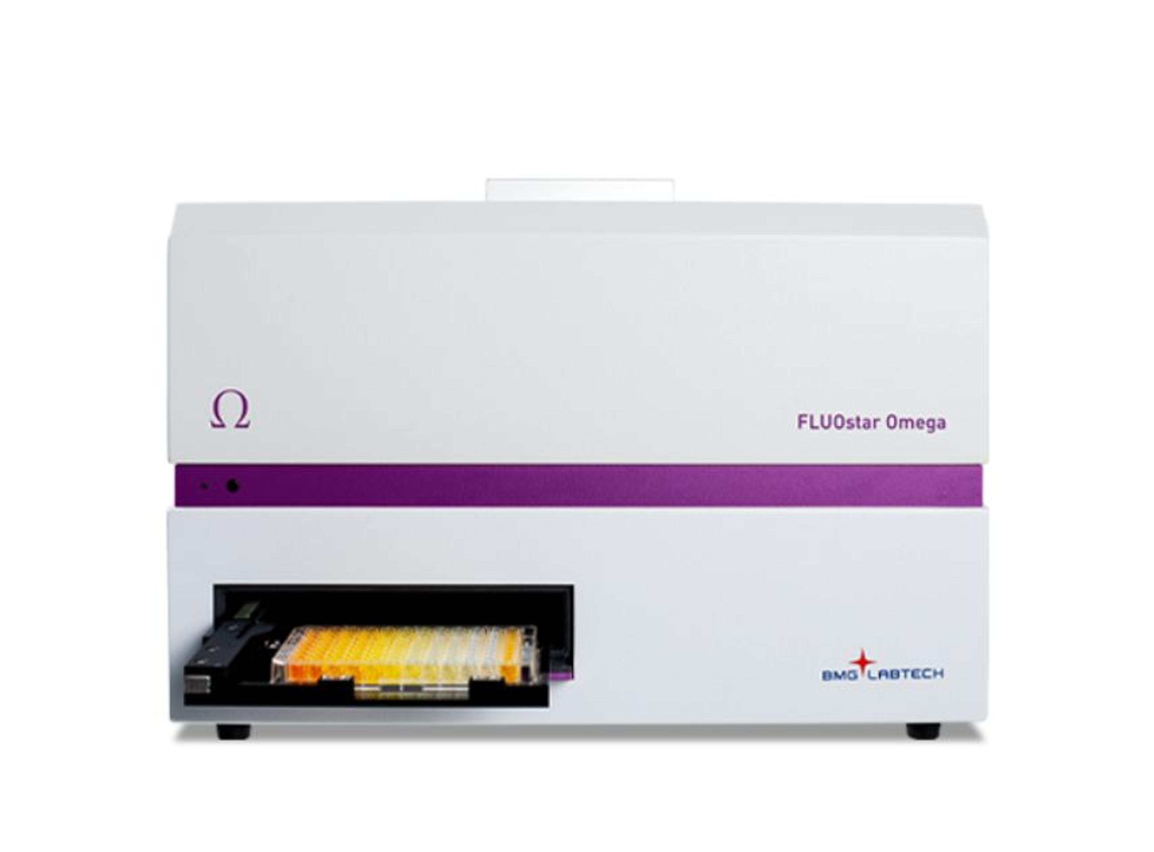
FLUOstar® Omega Plate Reader
BMG LABTECHThe FLUOstar Omega is a multimode microplate reader perfect for life science research laboratories.