Innovating Correlative Light Electron Microscopy and Volume Imaging Technology Through Discussion at EMBL 3D
SelectScience® spoke to Dr Yannick Schwab, Team Leader and Head of Electron Microscopy Core Facility at the European Molecular Biology Laboratory (EMBL), about a recent conference organized in conjunction with ZEISS Microscopy
24 May 2016
SelectScience® spoke to Dr Yannick Schwab, Team Leader and Head of Electron Microscopy Core Facility at the European Molecular Biology Laboratory (EMBL), about a recent conference organized in conjunction with ZEISS Microscopy
European Molecular Biology Laboratory (EMBL) Founded in 1974, EMBL is Europe’s flagship laboratory for the life sciences – an intergovernmental organization with more than 80 independent research groups covering the spectrum of molecular biology.
EMBL is an intergovernmental organization specializing in basic research in the life sciences, offering important services to scientists from its member states and developing new technologies and methods. In March 2016, at its main site in Heidelberg, Germany, EMBL hosted a workshop on 3D Light and 3D Electron Microscopy (EMBL 3D), in conjunction with ZEISS Microscopy. We spoke to one of the organizers, Dr Yannick Schwab, about the exciting innovation discussed at the event.
“Our goal at this conference was to bring together as many leaders in the field as we could”, Dr Schwab explained. The conference enabled them “to share the latest developments in the field and also to take new people in the field and enable them to catch up”. “Even though this is a focused conference, we have many, many things to share, many different techniques and approaches, fostering new ideas.”
“Correlative microscopy is not a new technique, but it bridges two worlds - the study of functional dynamic events in biological systems, from cultured cells to organisms, together with high resolution anatomical data”, revealed Dr Schwab. “We are now witnessing an interesting time where correlative microscopy is becoming a measurement tool to answer questions about more and more complex biological systems”.
EMBL3D
EMBL3D offered presentations on “correlative light electron microscopy, volume imaging using mostly scanning electron microscopy and a bit of transmission electron microscopy”. “Volume imaging and electron microscopy are very interesting because, now, we can address cells, cell populations and tissues at high resolution and for large areas of volumes of cells”, Dr Schwab explained. “Together with correlative microscopy, this is getting very powerful.”
The great thing about the electron microscopy community, revealed Dr Schwab is “people are willing to share a lot. For example, during this conference, a few speakers were even sharing data that was not published”. He believes this sharing mentality is “fruitful for the whole community, and is moving the whole field forward”. Although this conference was “very technology oriented”, scientists are already seeing that “this advanced technology is enabling science” in a range of applications. “Answering biological questions with correlative volume microscopy is a recent advance, and that’s really exciting.”
Find out more about the other speakers and presentations from EMBL 3D, or learn more about the applications of Correlative Microscopy.
Watch our interview with Dr Schwab here.
Related products
Request Quote for All Products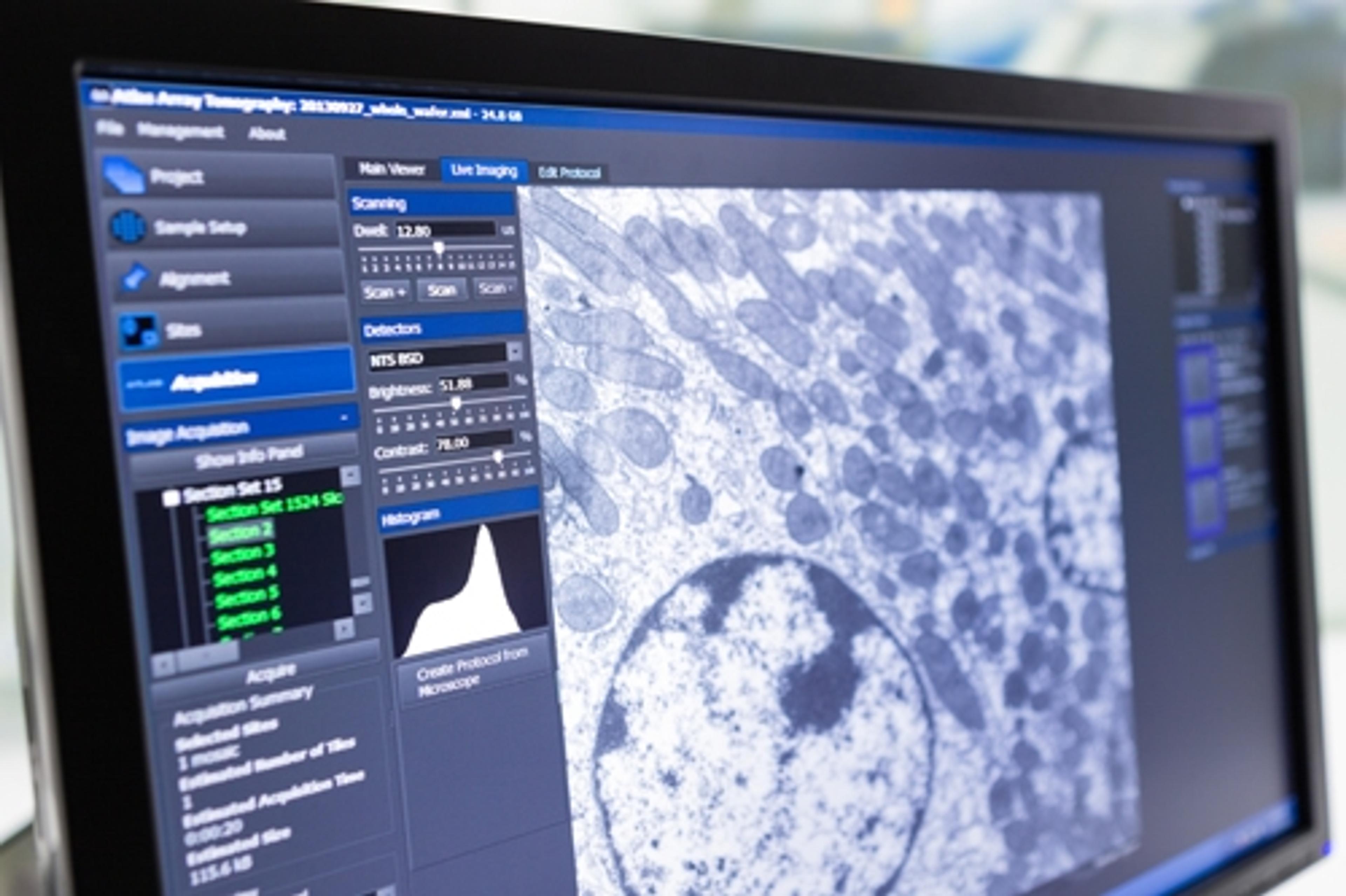
ZEISS Atlas 5
ZEISS Research Microscopy SolutionsLarge area imaging for SEM, FE-SEM & FIB-SEM ATLAS combines a 16 bit scan generator and dual super-sampling signal acquisition hardware with image processing and control software for your ZEISS electron microscope. Acquire images up to 32 k x 32 k pixels, with dwell times from 100 ns to > 100 s, adjustable in 100 ns increments. Save your images with eight or sixteen bits of intensity. With the ATLAS “Mosaic Tool” you create large image montages, automatically moving from image tile to tile, and mosaic site to site, resulting in an “Extreme Field of View” image, at SEM nanometer scale resolution. ATLAS provides • reduced number of tiles to acquire, reducing stage motion delay and areal fraction of each image “lost” to overlap • reduced number of overlap “seams”, leading to less beam damage and degradation of the sample • reduced computational complexity
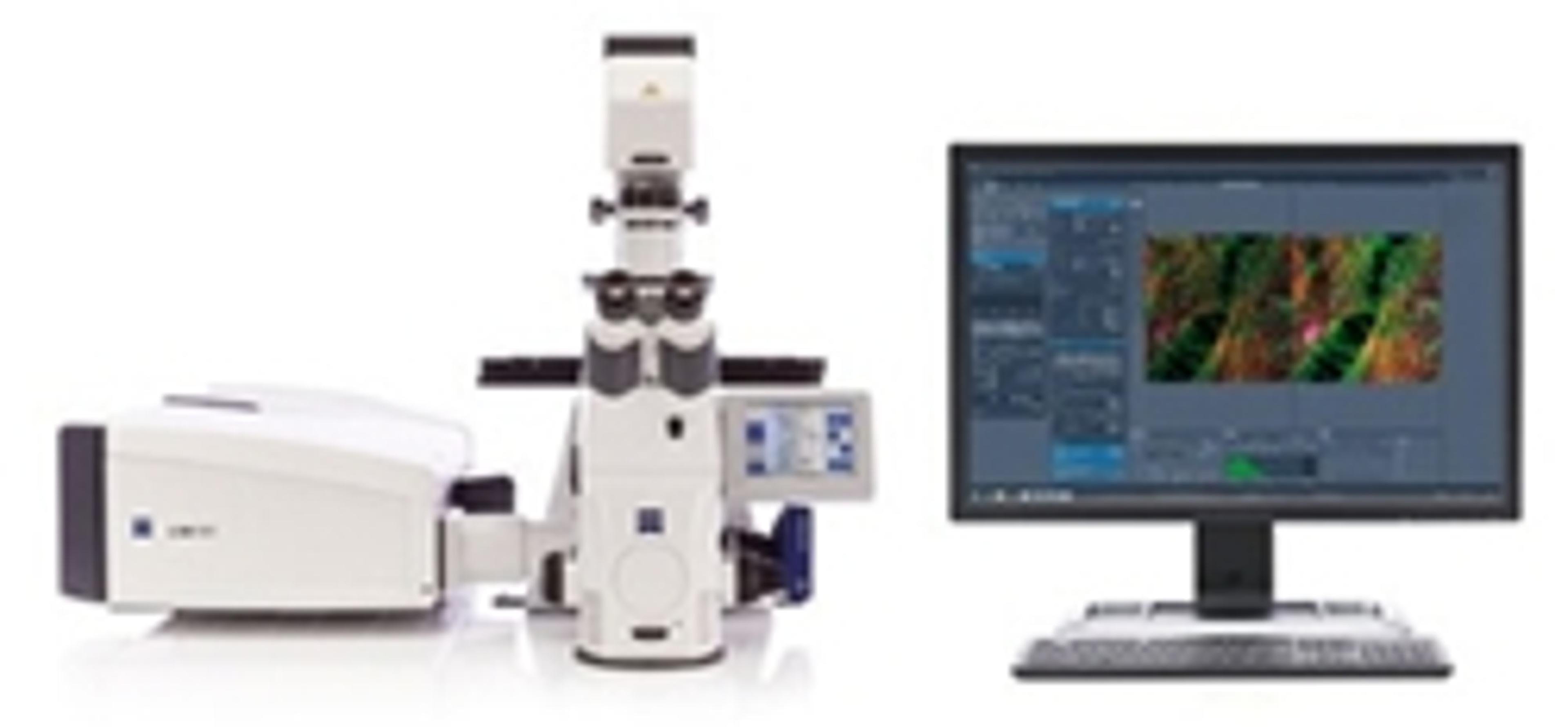
ZEISS LSM 880 with Airyscan
ZEISS Research Microscopy SolutionsYour New Standard for Fast and Gentle Confocal Imaging
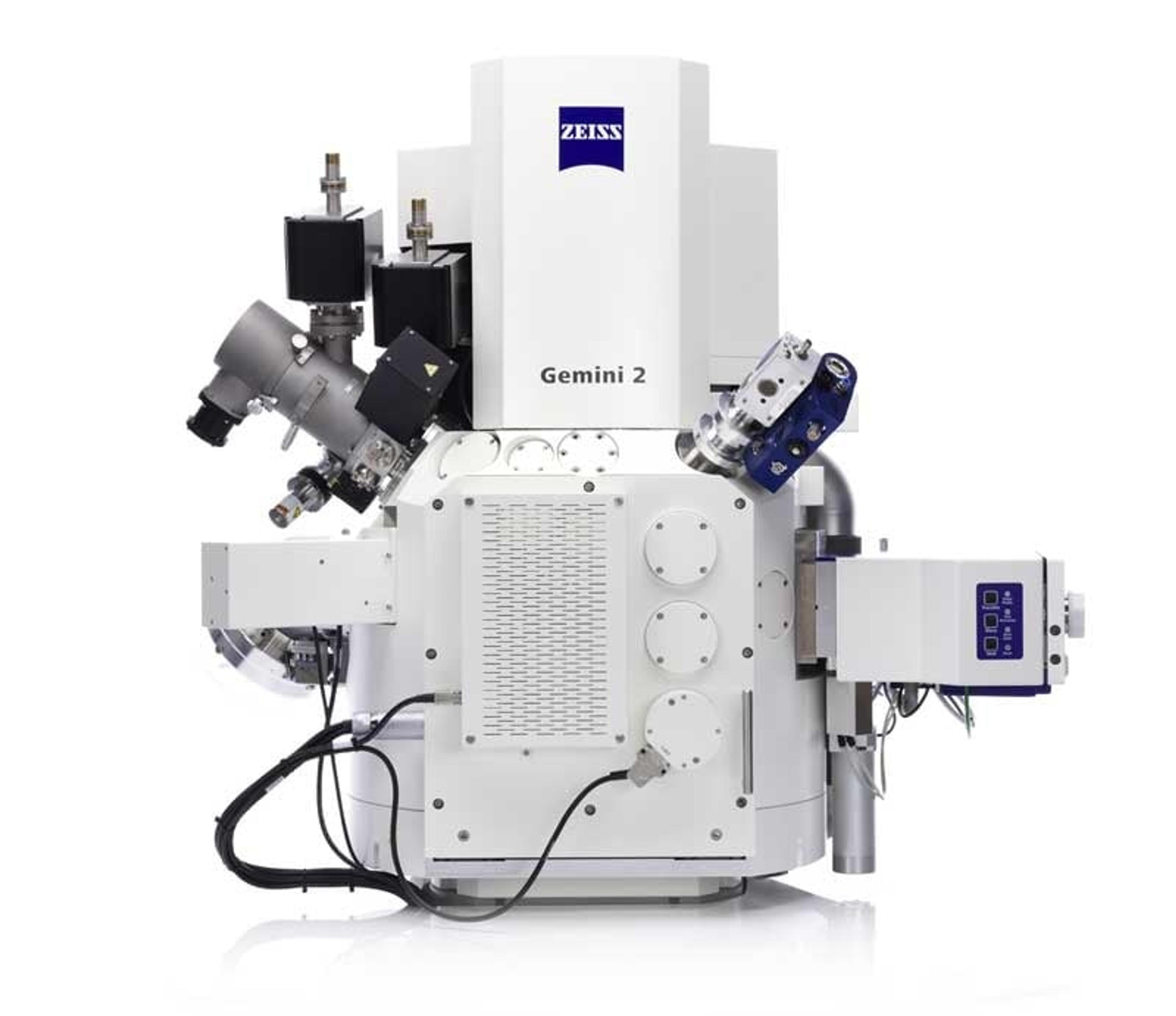
ZEISS Crossbeam Family
ZEISS Research Microscopy SolutionsWithin ZEISS Crossbeam Family you have the choice between Crossbeam 340 or Crossbeam 550. Exploit the variable pressure capabilities of Crossbeam 340. Or use Crossbeam 550 for your most demanding characterizations and choose the chamber size, standard or large, that best suits your samples.