The Role of Free Light Chains in the Diagnosis of Monoclonal Gammopathies
Improving monoclonal gammopathy diagnosis
2 Jul 2015
Editorial article
Professor Jerry Katzmann discusses the role of free light chains in the diagnosis of monoclonal gammopathies at Binding Site's 7th International Symposium
Jerry A. Katzmann, Laboratory Director, Mayo Clinic, Rochester, USA Jerry Katzmann is a Laboratory Director and Associate Professor of the Immunology Laboratory in the Department of Laboratory Medicine & Pathology at Mayo Clinic, Rochester, MN. Dr Katzmann has a specific interest in the laboratory detection of monoclonal gammopathies.
At the recent 7th International Symposium on Clinical Applications of Free Light Chain and Heavy/Light Chain Analysis, hosted by The Binding Site, Professor Jerry Katzmann gave a talk on the role of free light chains in the diagnosis of monoclonal gammopathies.
Traditional methods of diagnosing monoclonal gammopathies
Dr Katzmann began his presentation by reminding the audience that the monoclonal gammopathies are a diverse group of diseases. These plasma cell disorders secrete a wide range of monoclonal proteins, varying significantly both in molecular weight and in the unique variable region sequences. As such, there are three tests currently used to diagnose monoclonal gammopathies – protein electrophoresis (PEL), immunofixation electrophoresis (IFE) and free light chain (FLC) analysis.
Historically, PEL and IFE methods were used to identify abnormal plasma proteins. PEL, a well-established test allows for the detection and quantitation of monoclonal protein, but cannot identify the type. IFE is more sensitive than PEL and enables the monoclonal protein to be characterized into heavy or light chain subclasses, but cannot quantify the amount. Both PEL and IFE do not detect all instances of monoclonal gammopathy, mainly due to their lack of sensitivity.
A third test is developed
The Binding Site Freelite® assay, launched in 2000, offers a third assessment method; a highly sensitive and specific assay for measuring free light chains (FLCs) in serum. Freelite® antisera recognizes the epitope only on free light chains present and not those bound to the heavy chain. This enables the assay to quantify free kappa and lambda light chains in serum, and provide a diagnostic ratio.
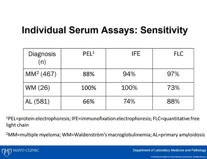
Table showing the sensitivity of individual serum assays
Professor Katzmann discussed the fact that all of these assays are good, but none of them are perfect. As a result, for the highest sensitivity, the assays are used in conjunction with each other to form a test panel that clinicians can use to detect monoclonal gammopathies.
In an analysis of 460 myeloma patients, using the traditional serum and urine analysis of PEL and IFE, 99% of myeloma patients were detected (see table below). When the FLC assay was added to the panel, 100% of abnormalities were detected. If you then remove the urine analysis tests, the number of abnormalities detected remains at 100%. This allows for the removal of urine tests from the panel, simplifying the test process and making it more cost-effective.
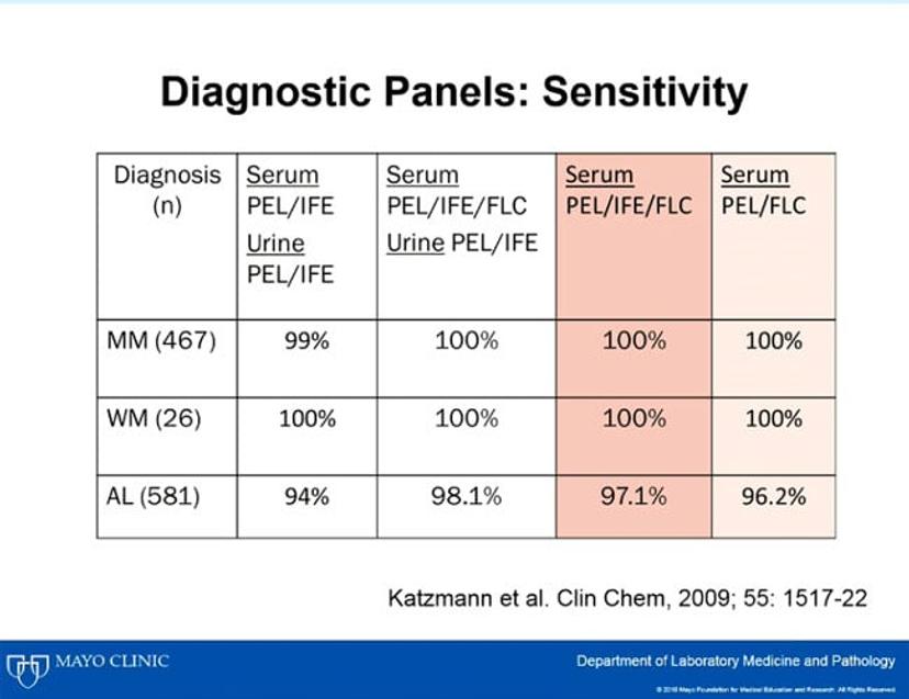
Diagnostic panels used to detect monoclonal gammopathies
The current gold standard panel that the International Myeloma Working Group recommends uses PEL, IFE and FLC (see table above). Professor Katzmann asserted that he hoped that the Working Group would consider the possibility of reducing this panel to just PEL and FLC (the last column in the above table). This panel would still detect all of the Multiple Myeloma and Waldeström's patients, but causes a drop of 1% in detection of Primary Amyloid cases.
In recommending this simplified panel, Professor Katzmann raised the point that even though the three test panel is currently recommended as the gold standard, not enough laboratories have implemented its use (in one example, a few hundred panels ordered out of a possible 24,000). It is Professor Katzmann’s view that a simplified panel would encourage more widespread adoption as a gold standard. Use of the two test panel would also improve turnaround times, and reduce the cost of diagnosis by removing the need to perform IFE, as well as reducing diagnostic and hospital staff time.
In the final part of his presentation, Professor Katzmann went on to discuss the use of FLC as a biomarker for predicting prognosis and the development of a risk stratification model. You can view Professor Katzmann's full presentation here.