Regenerative medicine technique development and the importance of cooperative research
Learn how a new imaging center in South Korea is working to overcome existing experimental limitations with hopes to improve quality of life for patients
13 Dec 2019
Editorial article
SelectScience talks with Eunsoo Lee, Director of Research Support at Ewha Womans University Fluorescence Core Imaging Center, about its work on fine-tuning methods to study the microenvironment of cancer cells as well as tissue-specific cell differentiation. Here, Lee highlights the team’s focus on designing and supporting experiments that require cooperative research and reveals a key technology underpinning its work.
Tell us about the Fluorescence Core Imaging Center and your role there.
Our center is still new, having opened just last month. It provides a variety of imaging systems to support the observation of cellular activity/the proteins and morphology of tissue samples. There is a research scientist in charge of each device to offer analytical services. I work on designing and supporting research experiments that require joint/cooperative research.
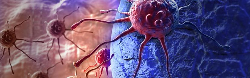
We are currently involved in research about the regulation of the microenvironment of cancer cells, as well as studying the regulation of reactive oxygen species (ROS) by organelles to determine the role and mechanisms of ROS. My personal research is to investigate the effects of tissue-specific extracellular matrix proteins on cell differentiation.
What are the current challenges you face?
We are developing new experimental methods to control the microenvironment or control the levels of ROS freely, with the goal of further subdividing and fine-tuning them into organelle- and tissue-specific methods. For cell differentiation experiments using extracellular matrix proteins, we have developed a new method to secure tissue-specific extracellular matrix proteins and are looking to determine whether culturing stem cells in these proteins cause them to differentiate into the respective tissue’s cells. In this way, we try to overcome the existing experimental limitations by developing new techniques optimized for our research.
What technologies do you use at the center?
We frequently use both the LUNA-FL Dual Fluorescence Cell Counter and X-CLARITY Tissue Clearing System from Logos Biosystems - we keep the LUNA-FL where we culture our stem cells, and it is used for the qualitative and quantitative analysis of cells at each passage.
We appreciate that Logos Biosystems is quick to respond and I have peace of mind as a customer as I can rely on their dependable customer service. We're also grateful for their support in providing better features through continuous software development.
Eunsoo Lee Ewha Woman's University Fluorescence Core Imaging Center
How has the LUNA-FL impacted your work?
When studying the differentiation of stem cells according to their location in the brain, we have to isolate six regions of the brain and extract stem cells from each region. When using the traditional method to count cells, it is not only time consuming, but cell quality also slowly deteriorates over time. Using the LUNA-FL makes it possible to get stem cell counts from all six regions quickly while checking cell quality at the same time. Because of this, we can use a similar standard of cells every time, making our experiments more reliable.
Would you recommend the LUNA-FL?
Yes, Logos Biosystems’ products are generally designed with customers in mind and have the advantage of being very easy to use and fast. When it comes to the LUNA-FL, it is especially convenient that, after it counts the cells, there is a dilution calculator for subsequent experiments, and live/dead ratios that need to be recorded can be reviewed directly on the LUNA-FL. Lastly, I think most consumers would choose a product with an attractive design when considering similarly priced devices.
Coming soon
Early next year, Logos Biosystems will launch the newest member of the LUNA™ family, designed to build on the success of its predecessors: the LUNA-FX7™ Automated Cell Counter.
What do you see or hope for your field in the future?
As my research is mainly focused on using tissue scaffolds to study recellularization and tissue-specific cell differentiation, I am hopeful about this ultimately leading to the development of implants that can be used for patients. The development of tailored, purpose-specific functional scaffolds would be a huge contribution to improving patient quality of life in the field of regenerative medicine.
Do you use Logos Biosystems products in your lab? Write a review today for your chance to win a $400 Amazon gift card>>
Related products
Request Quote for All Products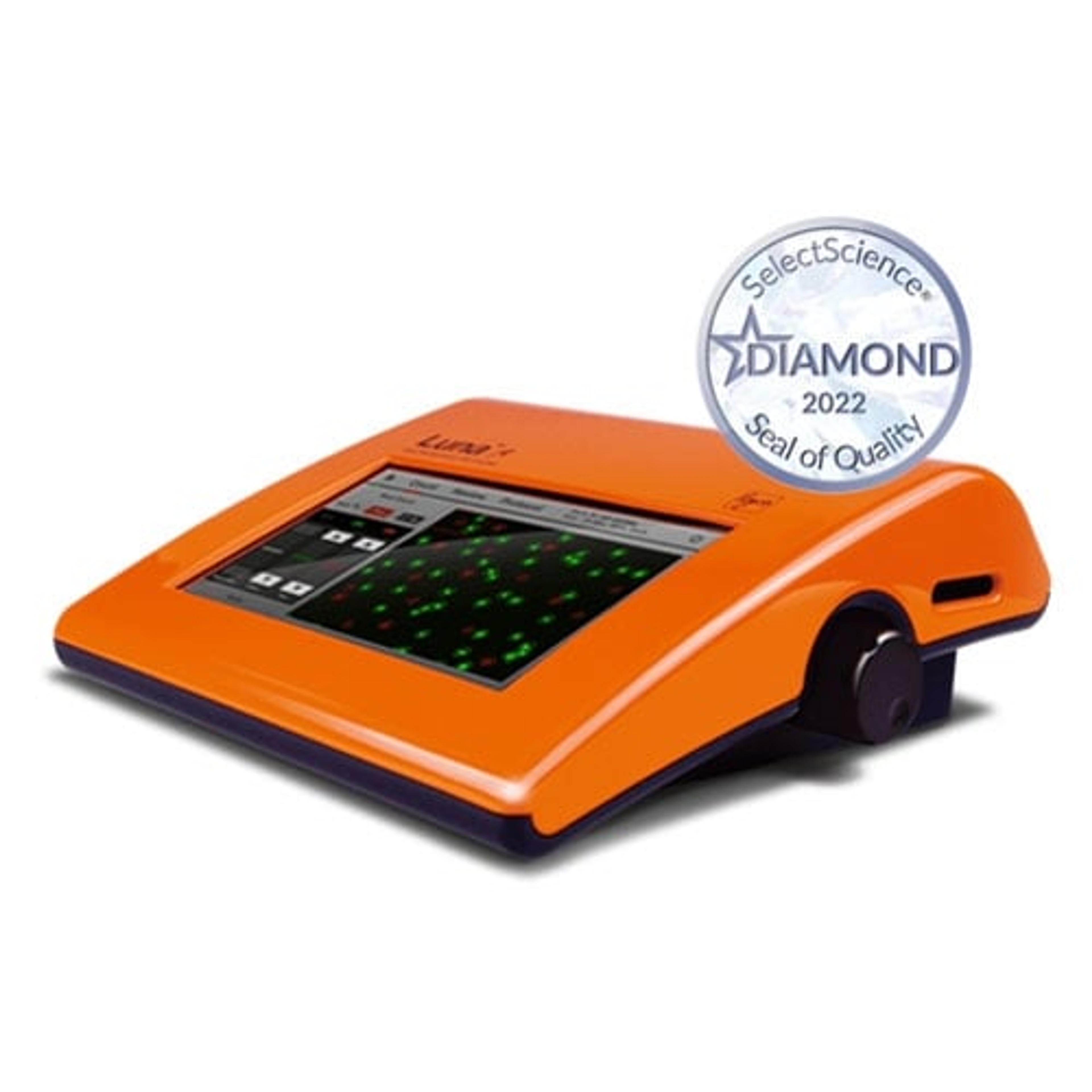
LUNA-FL™ Dual Fluorescence Cell Counter
Logos BiosystemsAccurate cell counts in as little as 7 seconds: Fast, accurate, and affordable automated cell counters from Logos Biosystems.