Electron microscopy enters a new era of high-speed discovery
Momentum-resolved STEM, phase retrieval, and neural-network models are redefining how we see and measure matter down to the subatomic level
20 Jan 2026
Prof. Knut Müller-Caspary, Ludwig-Maximilians-University (LMU) Munich
Since the development of the electron microscope in the 1930s, huge steps in science and technology have allowed researchers to see and quantify ever smaller components of organic and inorganic samples. A pioneer in 4D scanning transmission electron microscopy (STEM), electric field imaging, and phase retrieval, Prof. Knut Müller-Caspary’s work, supported by collaborations across physics, materials science and biology, is helping to advance quantitative electron microscopy.
Müller-Caspary is a professor at the faculty of chemistry and pharmacy at Ludwig-Maximilians-University (LMU) Munich in Germany, and is the 2025 recipient of the Ernst Ruska Award, granted by the German Society for Electron Microscopy for outstanding achievements in the field of electron microscopy.
The evolution of multidimensional electron microscopy
As a postgraduate at the University of Bremen, Müller-Caspary was inspired by Prof. Andreas Rosenauer’s clear explanations of the complex concepts of transmission electron microscopy (TEM) and wave optics.
“I learned the value of simulation as an important tool to quantitatively interpret experimental results in microscopy,” said Müller-Caspary. “We don't use microscopes to record images; rather we perform measurements, for example the quantity of charge required to form bonds between atoms, or the amount of strain in a crystal lattice. We then relate these numbers to physical properties.”
Since Müller-Caspary’s days in Bremen, electron microscopy, particularly STEM, has evolved to become a multidimensional approach, using developments in hardware and software to combine signals, creating images and expressing them as a function of time.
“The evolution allows us to measure electrical properties down to the atomic and subatomic scale,” said Müller-Caspary.
Momentum-resolved STEM: Bringing in a fourth dimension
Also known as 4D STEM or MRSTEM, momentum-resolved STEM generates a 4D dataset from a 2D convergent-beam electron diffraction pattern carried out at each pixel of a 2D STEM raster. This enables the measuring of magnetic and electric fields down to the subatomic level.
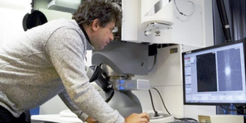
Prof. Knut Müller-Caspary at the electron microscope at LMU Munich
“4D STEM with the newest detectors provide information about where the electrons interact with the specimen, how they scatter, and in which directions the scattering occurs, so combining real and diffraction space information,” said Müller-Caspary. “This allowed us to build on research from a number of different groups to carry out center of mass imaging, ptychography and inverse multislice ptychography so as to get 3D information.”
Ptychography adds super resolution and aberration correction after acquisition by collecting diffraction information from overlapping regions to reconstruct sample phase properties. Müller-Caspary and his team are working on fast processing algorithms that select and reconstruct the regions that are of interest by incorporating physics constraints.
Using TorchSlice to match experiment and theory
The TorchSlice software was developed at LMU as part of a 2020 collaboration with Munich-based mathematicians who specialized in inverse problems. It allows researchers to get an insight into how simulation and experiment agree.
“In electron microscopy we can record the intensity but lose the phase, so the information we need to get the position of atoms or the distribution of the potential landscape in the specimen is not there. The 4D approach allows us to carry out phase retrieval in an iterative manner. Since we created TorchSlice, it has been further developed to include many of the prevalent reconstruction algorithms for the inverse problem,” explains Müller-Caspary. “We want TorchSlice to be freely available. We have published a paper on an application that was based on ferroelectric measurements at structural distortions, and this included a publication of the source code.”
The multislice algorithm simulates the elastic scattering of an electron beam. Christopher Koch of Humboldt University of Berlin and his team recognized that this model can be expressed as a neural network. Müller-Caspary’s team implemented this into a software framework, which supports TorchSlice’s speed. While TorchSlice is not yet used in live processing as it does not have an interface to the data output of the time-based detector, this may be something that the team will look at in the future.
Collaborating to build on the power of microscopy
Some labs build their own TEM setups, sourcing components from a variety of different providers to create a bespoke system. Others, including Müller-Caspary’s lab, work with companies that have established workflows and components with well-defined interfaces, as well as the experience and capabilities to carry out the engineering work required to house the components. One of the ways that the team selects companies to work with is their science focus, product quality, and staff with scientific backgrounds, for example Surface Concept, PNDetector and Quantum Detectors.
“We have worked with Quantum Detectors for nearly a decade, and the speed of their recent Timepix4 detector and its ability to capture a single event makes a real difference to our work. Previous detectors capture a few thousand frames per second with a scan region of 256 by 256 scan pixels. The Timepix4 detector easily allows for scan regions of 1000 by 1000 pixels, and with a one microsecond dwell time it can capture a million images or diffraction patterns in one second, which is a new generation of data acquisition for us,” said Müller-Caspary.
As part of their collaboration, Quantum Detectors created a prototype for Müller-Caspary’s team, and the team provided input on software handling, data formats and processing.
The application of electron microscopy to structural biology research
During his time at the Forschungszentrum Jülich, Müller-Caspary worked with Carsten Sachse and researchers from Thermo Fisher Scientific to apply differential phase contrast (DPC) imaging in STEM to biological specimens. This produced contrast at in-focus conditions.
“This was exciting because, if you can work in focus, you can make use of all the electrons, whereas if you have to defocus, for example in standard cryo-TEM imaging, you have a lot of oscillations of the contrast transfer. If you don't have these contrast reversals, nearly all the electrons you shine on the specimen will produce useful contrast,” said Müller-Caspary.
The 4D-BioSTEM imaging development collaborative project, with Prof. Sachse from Forschungszentrum Jülich, Prof. Stahlberg from Dubochet Centre at EFPL in Lausanne and Müller-Caspary’s team at LMU, aims to create a cryo-EM approach with maximized contrast and resolution. The team secured an ERC Synergy grant to explore, develop, and apply the 4D STEM technique to biological specimens.
“Biological samples, for example cell membranes, can be relatively thick, with multiple scattering events. One of the benefits of 4D STEM is that it allows the use of different scattering models within the inverse multislice technique. We are developing algorithms as part of the 4D-BioSTEM project for these experimental setups,” said Müller-Caspary.
The transition from conventional STEM to multidimensional analysis
Müller-Caspary’s advice for researchers considering using 4D STEM is firstly to consider whether it is the best approach, and whether the vast quantities of data generated will actually be useful.
“The scientists need to think about the scope of the 4D STEM with respect to their application – for example, if it's for strain mapping, the answer is yes since nano-beam diffraction is robust and usually very accurate. If it's for looking at electric fields or electrical property mapping, it needs to be on a case-by case basis. We mapped electric fields in nitride semiconductors, which went well, but when we intended the same thing for ferroelectrics, it failed. It’s not a black box where you put in data and get out resolved images,” said Müller-Caspary.
Looking to the future, Müller-Caspary predicts that event-based comparison of different signals in spectroscopy, diffraction, energy loss spectra and X-ray spectra at the microscope will be a powerful technique to get the most information from individual electrons, a methodology driven forward by Prof. Johan Verbeeck at the University of Antwerp.