Can kappa free light chains simplify multiple sclerosis diagnostics for laboratories?
New laboratory tools are prompting a rethink of how intrathecal inflammation is assessed beyond oligoclonal bands
12 Jan 2026
Nick Armfield, MSc, MIBMS, senior biomedical scientist, The Neuroscience Laboratories, The Walton Centre NHS Foundation Trust
Assessment of intrathecal inflammation remains a core function of neuroimmunology laboratories, particularly in the investigation of multiple sclerosis (MS). Oligoclonal band testing in paired cerebrospinal fluid (CSF) and serum has long been embedded in diagnostic pathways, but its technical complexity and reliance on subjective interpretation continue to pose challenges for many laboratories.
In a recent SelectScience® webinar, Nick Armfield, MSc, MIBMS, senior biomedical scientist, at The Neuroscience Laboratories of The Walton Centre NHS Foundation Trust, examined the analytical and clinical utility of CSF kappa free light chains, focusing on how this quantitative marker could be implemented within routine laboratory practice.
CSF, barriers, and immune activity
CSF plays a vital physiological role within the central nervous system (CNS), providing protection, nutrient transport, and immunological surveillance. Its composition is tightly regulated by the blood-brain barrier and the blood-CSF barrier, which restrict molecular exchange between the periphery and the central nervous system. When these barriers are disrupted by inflammation or immune activation, changes in CSF composition become detectable through laboratory testing.
“Free light chains serve as a biochemical footprint of immune activation in the central nervous system compartment, and understanding the barrier permeability and this physiology helps us interpret kappa free light chain results,” Armfield explains. For laboratory professionals, this principle underlines the importance of interpreting CSF results in the context of barrier integrity rather than relying on absolute concentrations alone.
Intrathecal inflammation is not specific to multiple sclerosis and is observed in a range of neurological and neuroinflammatory conditions. As a result, laboratory markers of immune activation must be integrated with clinical and radiological findings to support diagnosis.
Multiple sclerosis and laboratory markers
Multiple sclerosis is a chronic immune mediated demyelinating disease characterized by inflammatory lesions within the central nervous system. Patients may present with relapsing remitting disease, primary progressive disease, or secondary progressive disease, each with distinct clinical trajectories.
Laboratory evidence of intrathecal immunoglobulin synthesis has traditionally been provided by oligoclonal band testing and IgG index calculations. These tests remain clinically relevant but present operational limitations.
“Oligoclonal bands and IgG index are established but do have their limitations – that they’re subjective, labor-intensive, and sometimes can be a bit ambiguous,” says Armfield. For laboratory managers, these limitations translate into longer turnaround times, specialist staffing requirements, and restricted access outside tertiary centers.
Kappa free light chains as a laboratory marker
Free light chains are produced during immunoglobulin synthesis, with a small excess normally released into circulation. Under physiological conditions, concentrations in CSF are extremely low.
“An increased presence in the CSF actually reflects intrathecal immunoglobulin synthesis, and specifically kappa free light chains (κFLC) are actually acting as a surrogate marker for CNS immune activation, similar to oligoclonal bands without that labor-intensive process that we have beforehand,” Armfield explains.
From a laboratory perspective, this positions kappa free light chains as a quantitative alternative that can complement established tests while reducing reliance on manual, interpretive techniques.
Measurement methodology and paired sampling
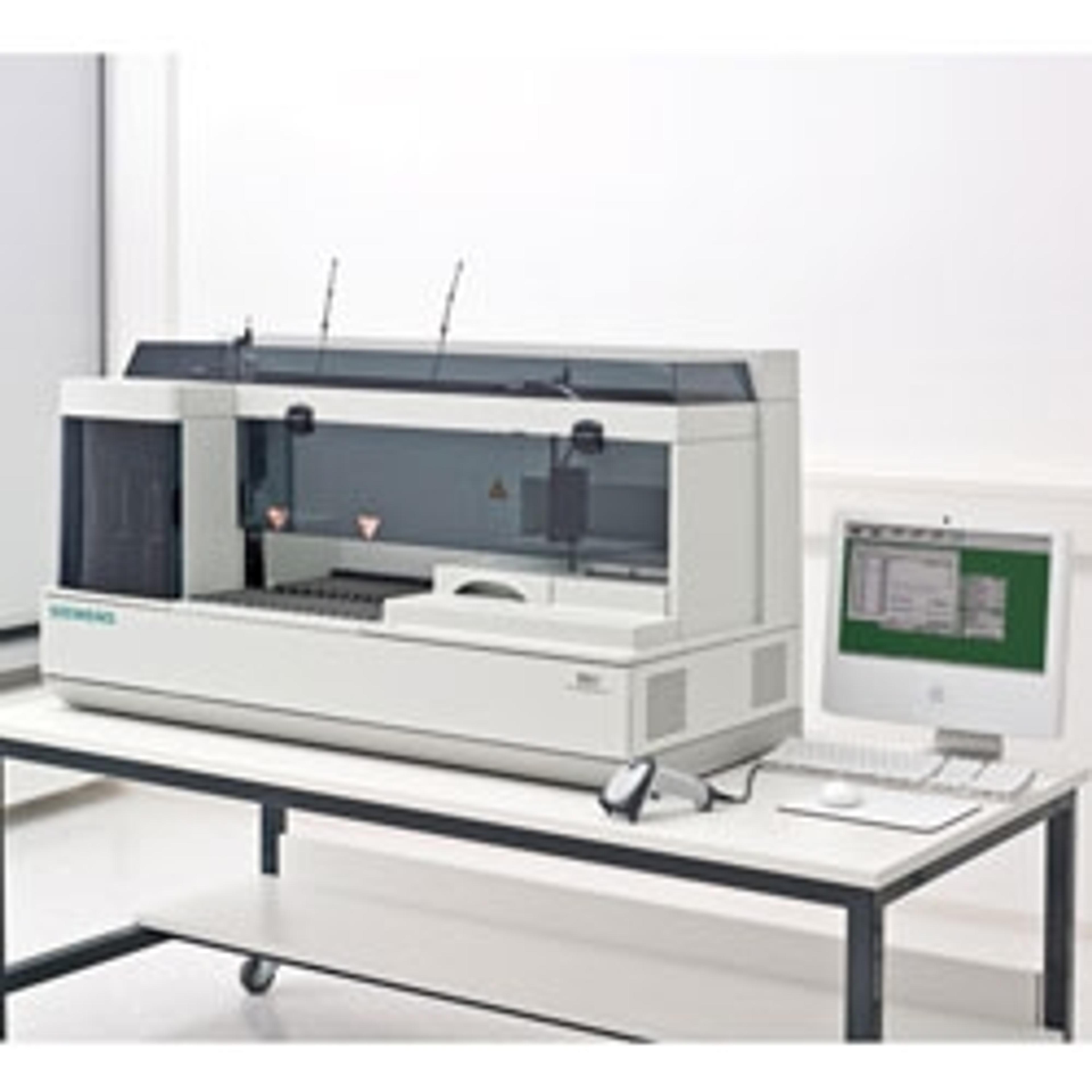
Kappa free light chains are measured using quantitative immunoassays, commonly by nephelometry. Crucially, analysis requires paired CSF and serum samples, with results expressed as a kappa free light chain index that incorporates albumin to account for barrier permeability.
“Kappa free light chains are measured from paired serum and CSF samples. We must have the serum there,” Armfield says. Without a peripheral denominator, results risk misclassification due to systemic inflammation rather than true intrathecal synthesis.
As an alternative to the κFLC index, laboratories may also use hyperbolic reference curves, known as Reibergrams, to visualize results against reference populations. These tools support interpretation but must be understood in terms of their sensitivity and specificity characteristics.
Comparative study with oligoclonal bands
Armfield presented findings from a laboratory study involving 104 paired CSF and serum samples analyzed within a tertiary neuroimmunology service. Results were compared with oligoclonal band patterns, IgG index, and clinical diagnoses.
“Type 1 oligoclonal bands are negative. They don’t display any evidence of intrathecal inflammation and, thus, have very low kappa free light chain indices,” Armfield notes. Clear separation was observed between negative patterns and type 2 and type 3 patterns associated with intrathecal synthesis.
The study highlighted the importance of paired analysis. “CSF kappa free light chain alone, just on its own, with no peripheral denominator, had significant differences between type 1 and type 4, yet didn’t have any significant differences between type 1 and type 4 for kappa free light chain index,” explains Armfield. Incorporating serum measurements shifted the clinical relevance of results.
Type 6 oligoclonal band patterns, which represent a recognized gray area, demonstrated how kappa free light chain data could add interpretive value when standard results are inconclusive.
Reibergrams, cutoffs, and interpretation
Reibergram analysis showed strong agreement with inflammatory oligoclonal band patterns, particularly for type 2 and type 3 samples. However, few false positives were observed among negative patterns.
“So, there’s a possibility that these Reibergrams might be presenting with some false positives here, but still a very, very useful tool,” Armfield notes, particularly for their negative predictive value.
κFLC index cutoffs derived from the study aligned with published literature. “We settled on this cut-off at 16 or more, which was most reflective of our oligoclonal band results and is also reflective of a meta-analysis,” Armfield says. He adds, “I still think that meta-analysis of 2.4 to 20 should be used as an equivocal range for kappa free light chain index.”
For laboratories, this reinforces the need for local validation and clear reporting guidance rather than reliance on a single universal cutoff.
Clinical performance and limitations
When correlated with clinical diagnoses, oligoclonal bands retained the highest positive predictive value, while Reibergrams performed best for excluding intrathecal inflammation. Kappa free light chain index showed strong agreement with oligoclonal band results but did not demonstrate superior specificity.
“Kappa free light chain [testing] in CSF is not more specific to MS but is a more accessible technique and less subjective to interpretation,” states Armfield. This reflects the broader reality that intrathecal inflammation is a shared feature of multiple neurological diseases.
Kappa free light chain index also did not significantly outperform IgG index in this cohort, likely due to historical reliance on IgG-based markers in defining multiple sclerosis.
Implications for routine laboratory practice
From an operational standpoint, kappa free light chain testing offers clear workflow advantages. “Kappa free light chain testing was faster, and it was less subjective. It was an empirical piece of quantitative data comparable to bands and IgG index,” concludes Armfield.
While kappa free light chains do not replace oligoclonal band testing, they provide a practical, scalable tool that can support screening, reflex testing, and longitudinal monitoring. When implemented with paired serum sampling and appropriate interpretive frameworks, they offer laboratories a more accessible approach to assessing intrathecal inflammation within evolving multiple sclerosis diagnostic pathways.
The latest revision of the McDonald diagnostic criteria for multiple sclerosis from 2024 officially adds the κFLC index to the existing biomarkers for demonstrating intrathecal immune response serving as a surrogate or equivalent to oligoclonal bands.
Watch the full ACCENT®- and PACE®-accredited webinar on demand for practical laboratory perspectives on CSF kappa free light chains.
Related products
Request Quote for All Products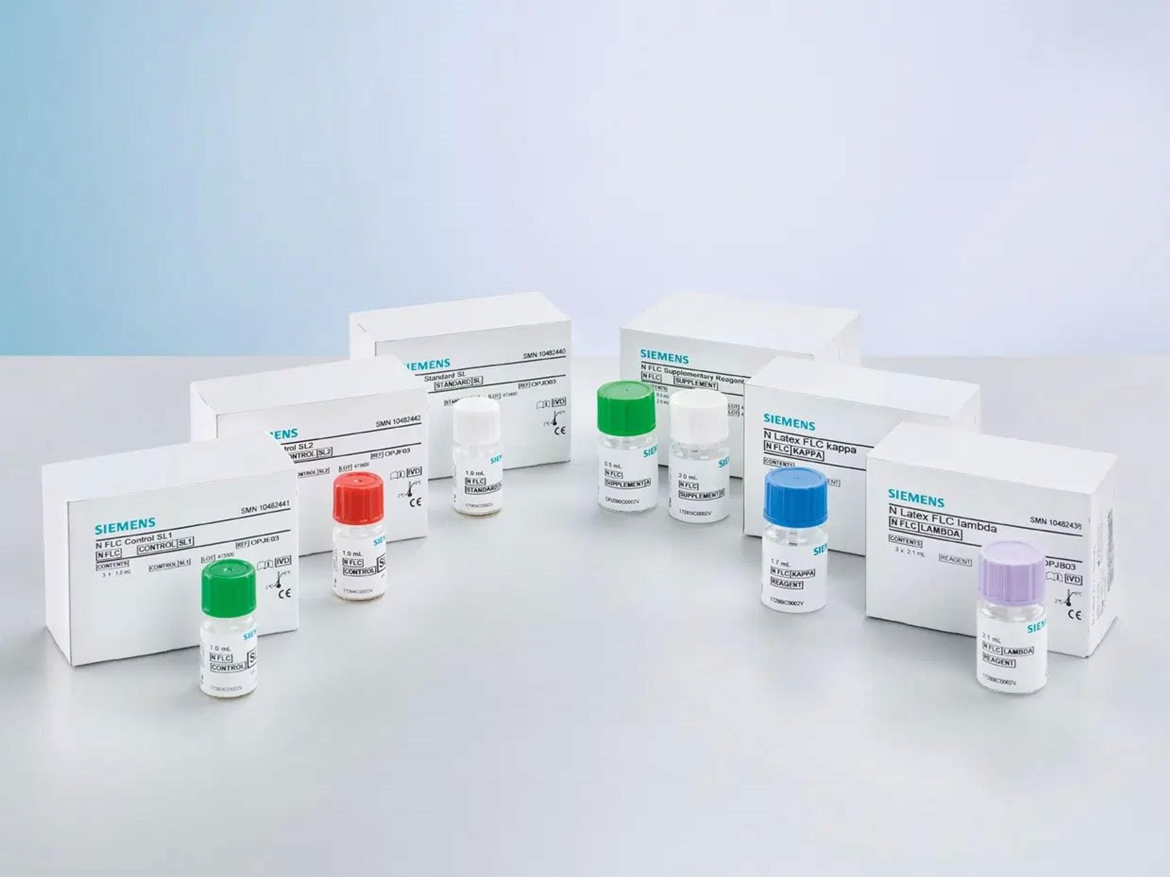
N Latex FLC kappa and N Latex FLC lambda Assays
Siemens HealthineersAdd confidence to screening and monitoring of monoclonal gammopathies.