3D Imaging of Optically Cleared Spheroids in Corning Spheroid Microplates

31 Mar 2019

The use of three-dimensional (3D) cell cultures for in vitro drug discovery assays has increased dramatically in recent years because 3D cell culture models more accurately mimic the in vivo environment compared to traditional two-dimensional (2D) monolayer cultures. However, current imaging-based analysis of these 3D cultures relies upon techniques originally developed for 2D cell culture, and as such, has significant limitations. Specifically, the light scattering inherent with thick microscopy specimens prevents imaging the entirety of 3D spheroids, which are typically >100 µm in diameter. This technical limitation introduces a sampling bias in imaging analysis in which only the exterior cells of a spheroid can be imaged. To accurately survey the cellular environment and response of the spheroid, the field needs new techniques to overcome the sampling bias.

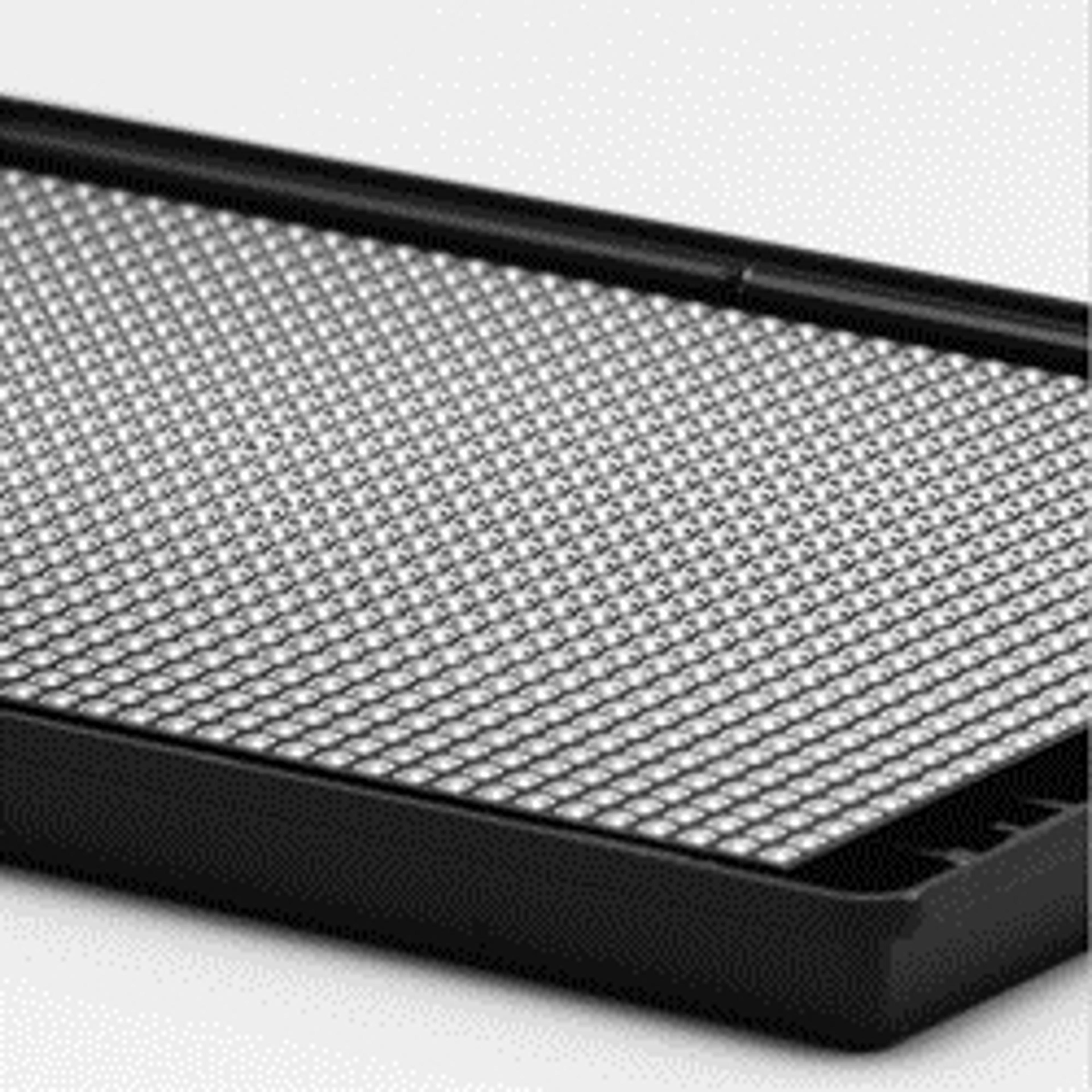
Corning 1536-well Spheroid Microplates

Corning Life Sciences

With their novel and proprietary design, these microplates are ideal for generating and analyzing 3D multicellular spheroids in the same microplate. The Ultra-Low Attachment (ULA) surface enables uniform and reproducible 3D multicellular spheroid formation. The black opaque microplate body shields each optically clear, round bottom well from well-to-well cross-talk.

(1)

Links

Tags

Cell / Tissue CultureCell culture or tissue culture is used to study the biology of cells or tissues and to isolate cellular products in an environment which can be manipulated and well defined. Accurately control your culture environment with bioreactors or culture incubators, bind your cells to a surface or together with an extracellular matrix. Distinguish cell types with differential media or proliferate cells with certain characteristics using selective media. Enrich your media with supplements such as growth factors, sera and vitamins. Find the best cell and tissue culture products, kits and equipment in our peer-reviewed product directory: compare products, check customer reviews and receive pricing direct from manufacturers.MicroplatesMicroplates are multi-well plates used to increase the throughput of biological investigations. The number of wells microplates contain can range from 6 to 3243 wells, with the 96-well format being the most commonly used. Microplates can come tailored for a range of applications including cell culture, PCR, filtration, storage, non-binding surface, protein crystallization, as well as pre-coated, deep well and normal Standard microplates. Additionally, options for microplate colors include clear , black, white or black with clear bottom and white with clear bottom for absorbance microplate reader applications. Find the best microplates for your lab in our peer-reviewed product directory: compare products, check customer reviews and receive pricing direct from manufacturers.ImagingImaging techniques are essential for obtaining visual representations of samples to understand structures, processes, and function in biological, chemical, and physical research. These tools range from traditional light microscopy to advanced imaging modalities like MRI and electron microscopy, providing researchers with valuable data for diagnostics, drug discovery, and material analysis. Explore imaging solutions in our peer-reviewed product directory to compare products, check reviews, and get pricing directly from manufacturers.High ThroughputHigh throughput experiments allow the simultaneous processing of several samples. This parallelization reduces the cost per experiment and increases reproducibility and output volume of data.SpheroidsSpheroids are clusters of cells that have been grown in 3D culture to be used as <i>in vitro</i> model systems. These 3D microtissues can be used for toxicology testing, DMPK studies and many other applications involving cell analysis.