
SelectScience Interviews
Videos
25
SelectScience Interviews
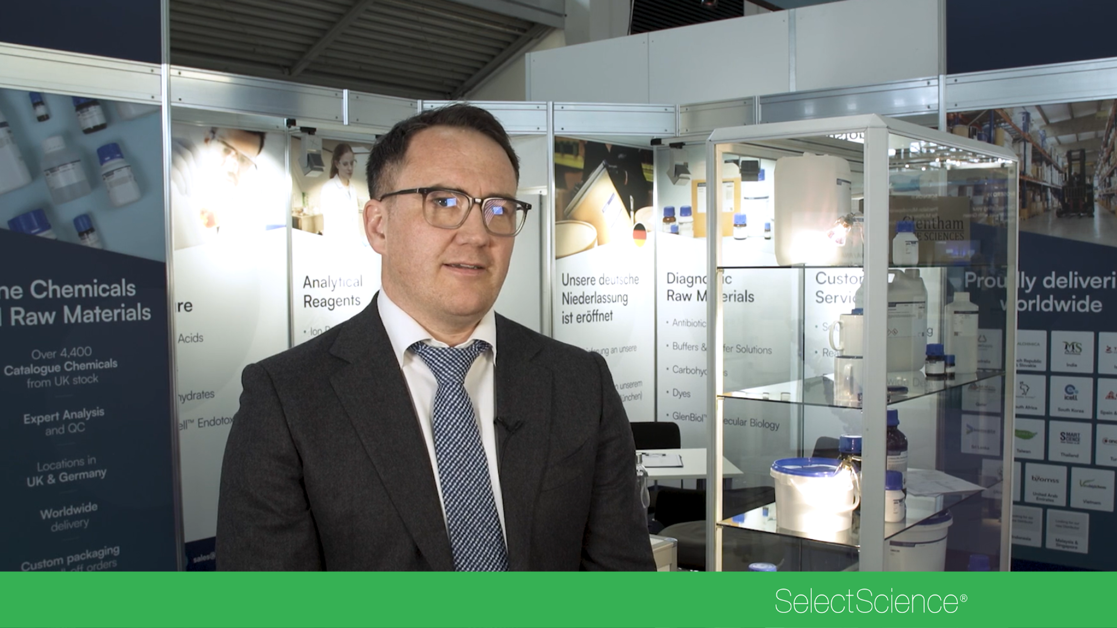
Why high-quality chemical supply and security matters for modern research

SelectScience Interviews
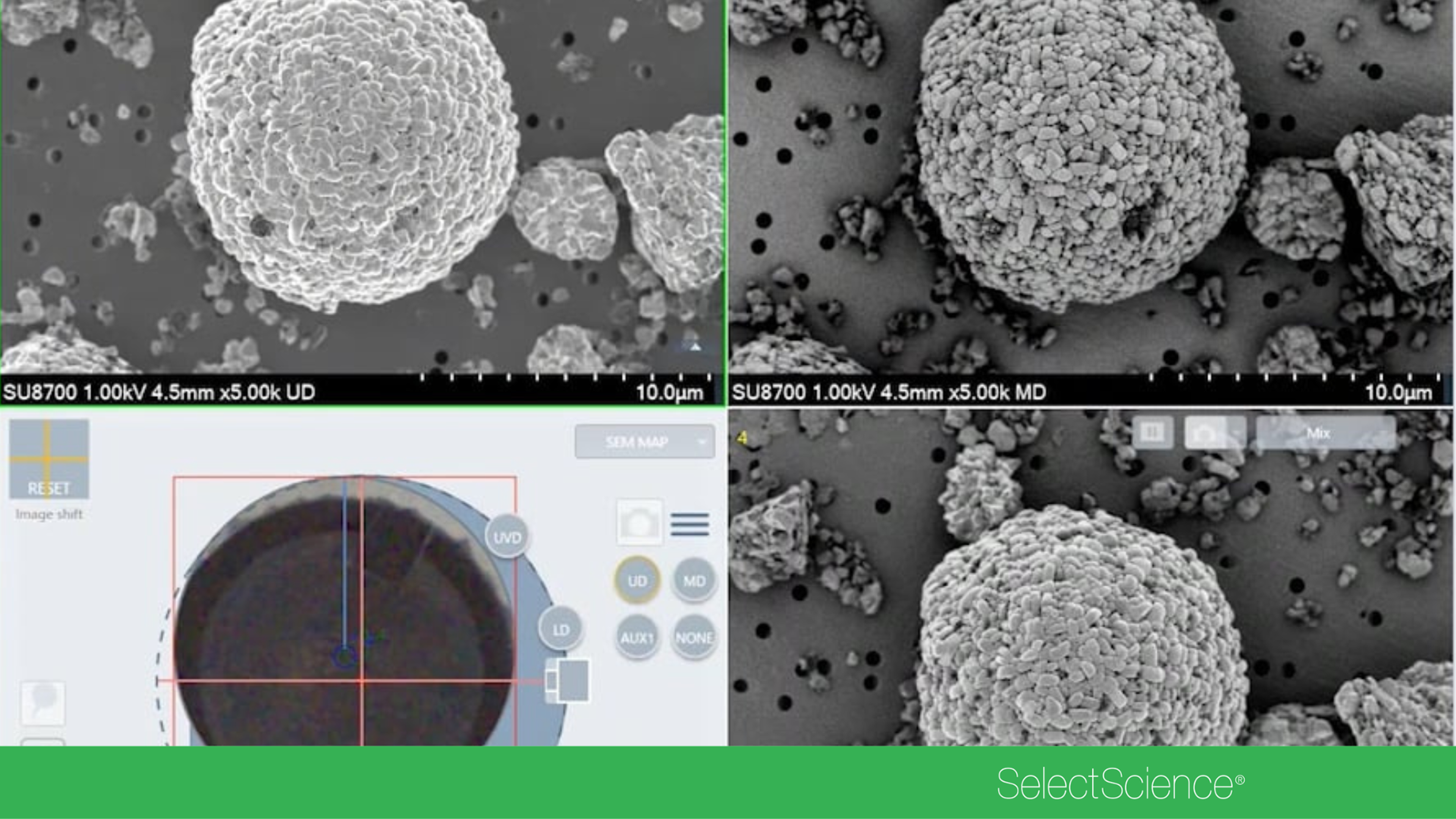
Automated SEM workflows driving faster, cleaner and more consistent analysis

Product News
Advancing AI equity and the future of laboratory governance with Bamidele Farinre
Discover how award winning scientist Bamidele Farinre is transitioning from virology to shaping UK parliamentary policy on artificial intelligence for Lab Professionals Week 2026

SelectScience Interviews
Championing lab innovation and the scientists cracking the code of biology
Dr. Asha S. Collins, Vice President and General Manager at Thermo Fisher Scientific honors laboratory professionals during Lab4Life Week and reaffirms a commitment to streamlining scientific discovery
Product News
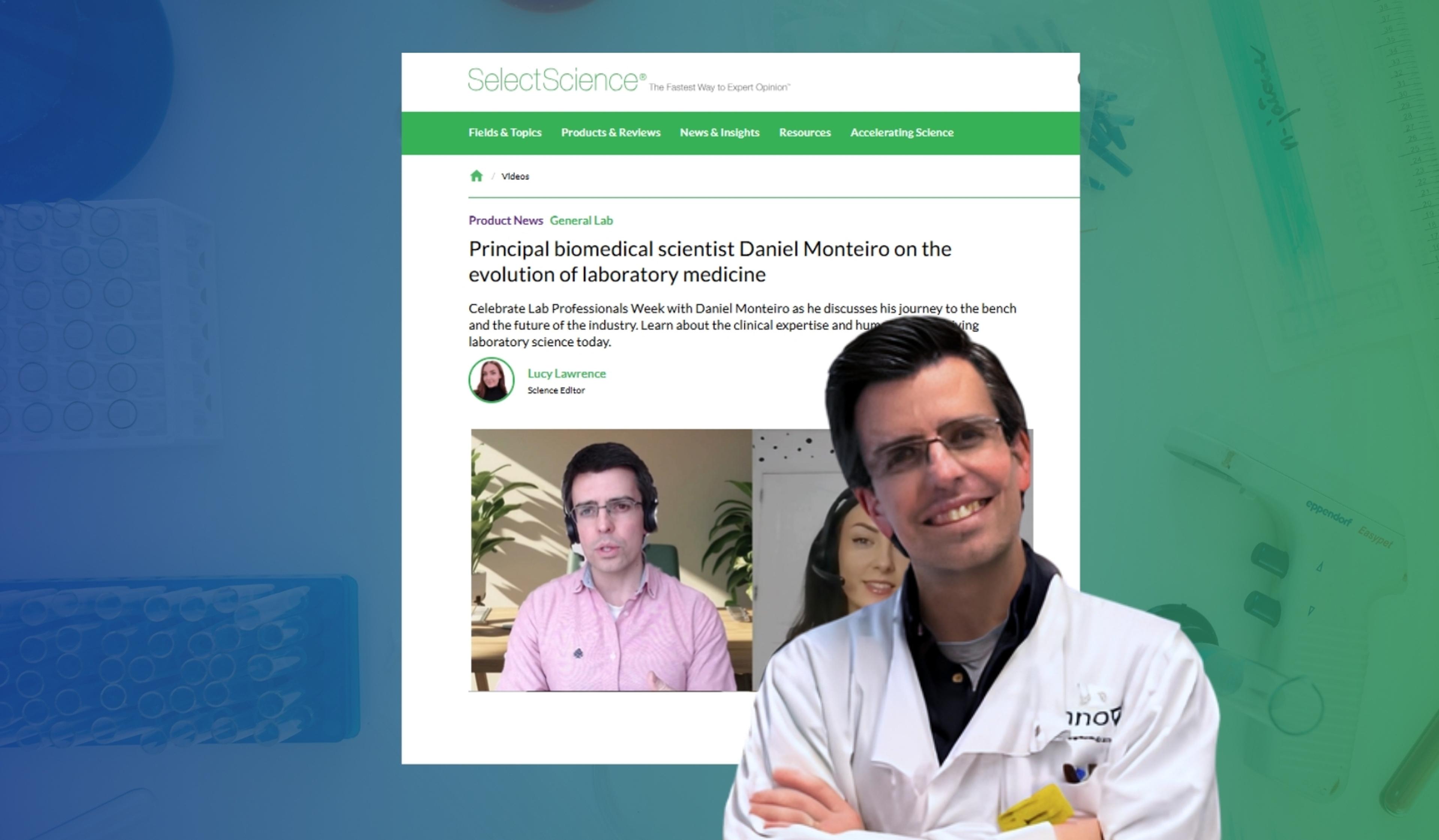
Principal biomedical scientist Daniel Monteiro on the evolution of laboratory medicine
Celebrate Lab Professionals Week with Daniel Monteiro as he discusses his journey to the bench and the future of the industry

Product News
Making lab work effortless with the SpinPro 6 R

Product Demonstrations
A guide to the AccQ•Tag Ultra Manual Derivatization workflow for amino acid analysis

Product Demonstrations
How KT Premium Slides improved histology operations at HCA Mid-America Division

SelectScience Interviews
Unlocking 4D metabolomics with timsMetabo™

Product Demonstrations
Fabric inspection methods and standards

Product Demonstrations
How to test potency of pharmaceutical products

Product Demonstrations
Instrument overview of the DS-11 Series Spectrophotometer / Fluorometer
Product Demonstrations
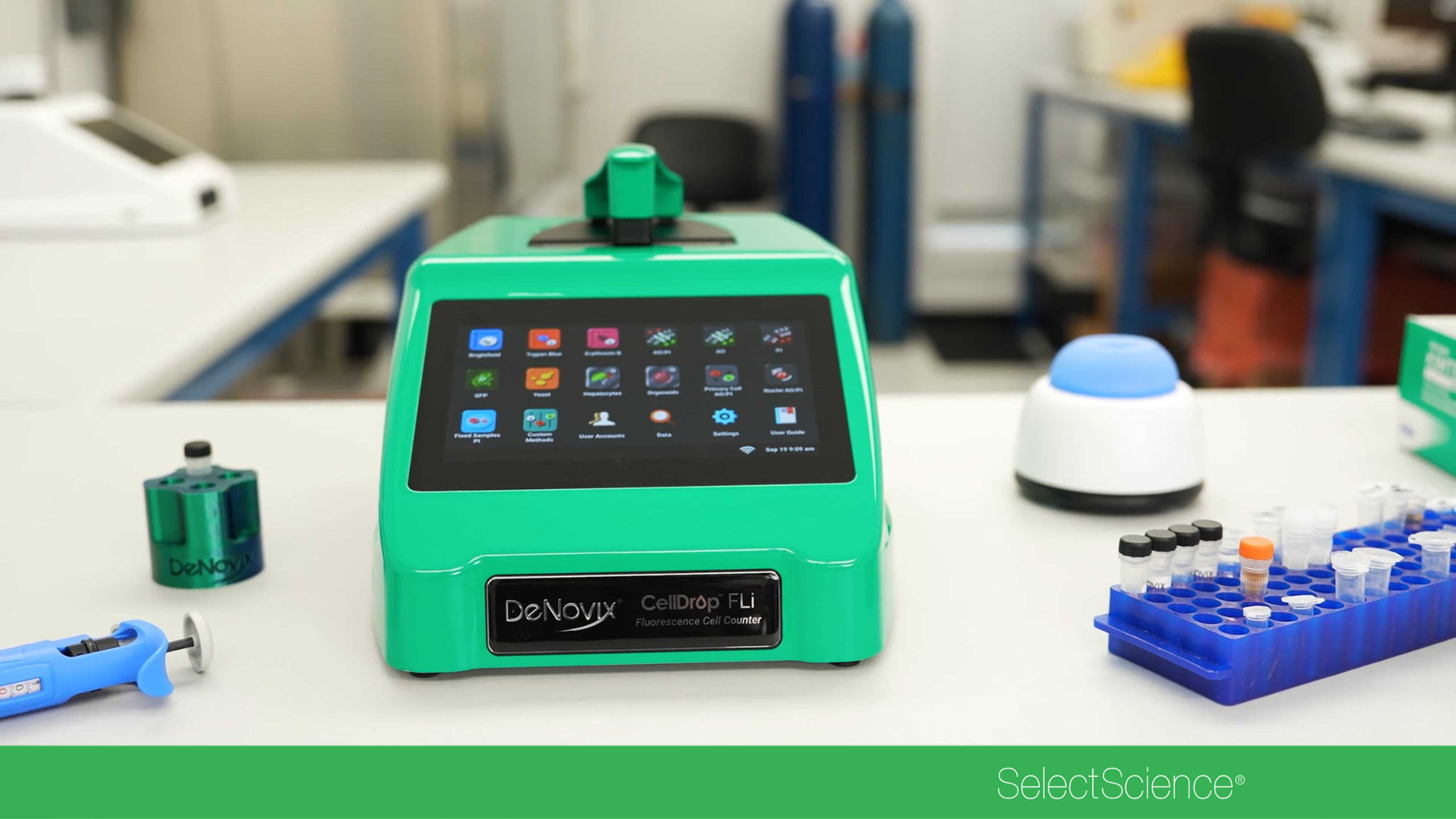
See the CellDrop Automated Cell Counter in action

Product Demonstrations
An introduction to the DS-8x Spectrophotometer

Product Demonstrations
Learn how to install the SPME MultiGuide on a SPME fiber tool


Product Demonstrations
See the PAL Method Composer in action

SelectScience Interviews
What PFAS exposure means for the fetus and why regulation must catch up


Product News
Accurate food nutritional analysis explained

Product Demonstrations
Why laser temperature stability is critical for quantum computing

Product Demonstrations