Chemistry Nobel Prize 2014: Super-Resolution Fluorescence Microscopy
31 Oct 2014
SelectScience would like to congratulate Eric Betzig, Stefan W. Hell and William E. Moerner on winning the 2014 Chemistry Nobel Prize 'for the development of super-resolved fluorescence microscopy'.1
Previously, confocal microscopy was used by many scientists to view living systems. However, by using a single beam of light it was difficult to focus on a specific area, and the light has the detrimental effect of degrading the sample over time. Recently, the Nobel Prize winners have developed a new way of viewing living systems using light microscopy, enabling scientists to view living tissues down to a molecular level (~20nm) with the resolution exceeding the diffraction limit.
PALM Microscopy
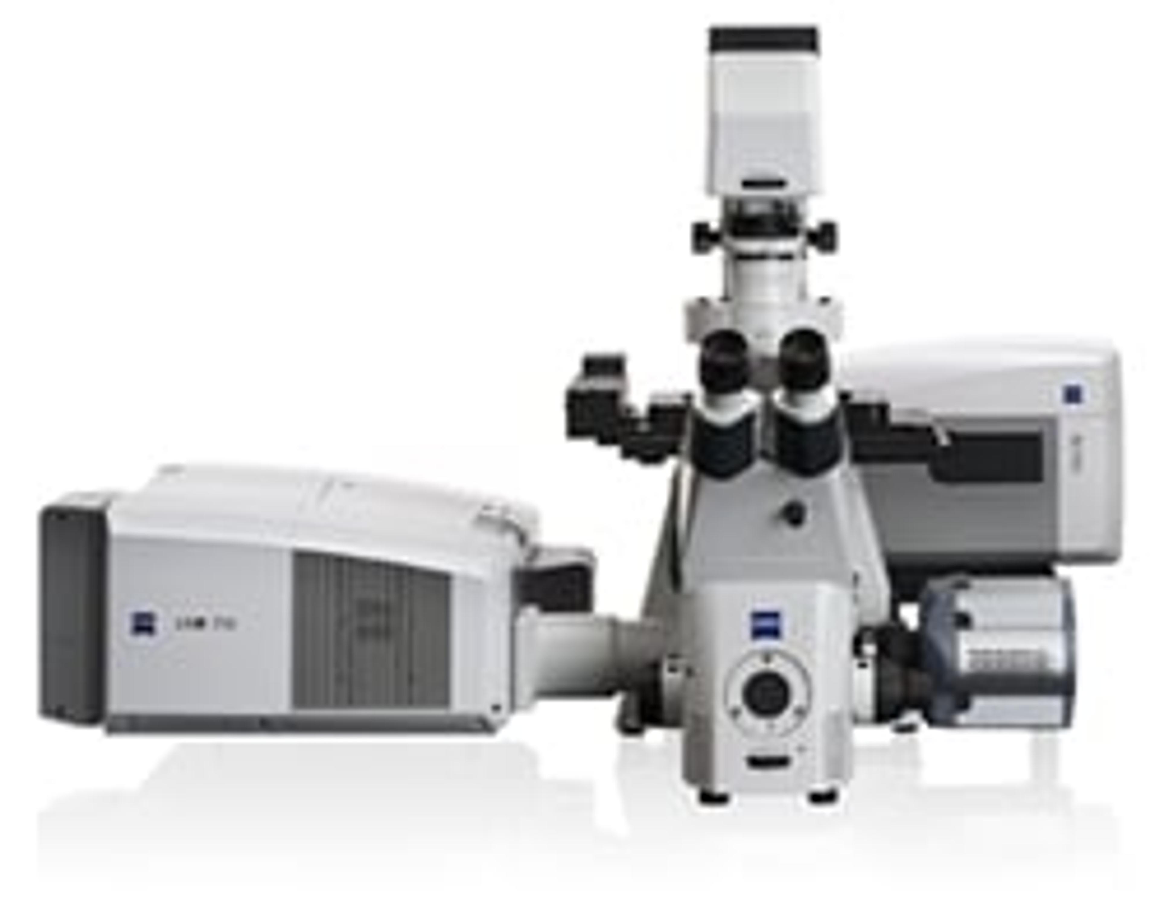
Eric Betzig of Howard Hughes Medical Institute's Janelia Research Campus, together with ZEISS, has developed the world's first commercially available 3D super-resolution fluorescence microscope. The ZEISSELYRA Super-Resolution Microscopy Systems are available in two forms;
ELYRA S.1 uses super-resolution structured illumination microscopy (SR-SIM) that enables scientists to view in fine structural details samples labeled with conventional dyes.
ELYRA P.1 uses the Nobel Prize winning photo-activated localization microscopy (PALM) that enables scientists to view endogenously-expressed photo-switchable fluorescent proteins by localizing small molecules with a resolution down to 20nm laterally and 50 nm axially.
Since winning the Nobel Prize for Chemistry, Professor Betzig has gone on to develop a new microscopy technique that enables imaging of fast-moving living systems using lattice sheets of light instead of cones.
STED Microscopy
Stefan W. Hell of Max Planck Institute, together with Leica Microsystems, has developed the world's first stimulated emission depletion (STED) microscope. The Leica TCS SP8 STED 3x enables scientists to study sub cellular architectures with nano-scale resolution.
Super-resolved fluorescence microscopy has revolutionized the way scientists can view living cells. The ability to monitor the division and growth of living cells gives in insight into the way in which cancer grows, viruses duplicate and the effect of neuro-diseases on brain tissue.
Ref: 1) http://www.nobelprize.org/nobel_prizes/chemistry/laureates/2014/ Accessed 31.10.14
Learn more about microscopy: 2014 SelectScience Microscopy Buying Guide
Related products
Request Quote for All Products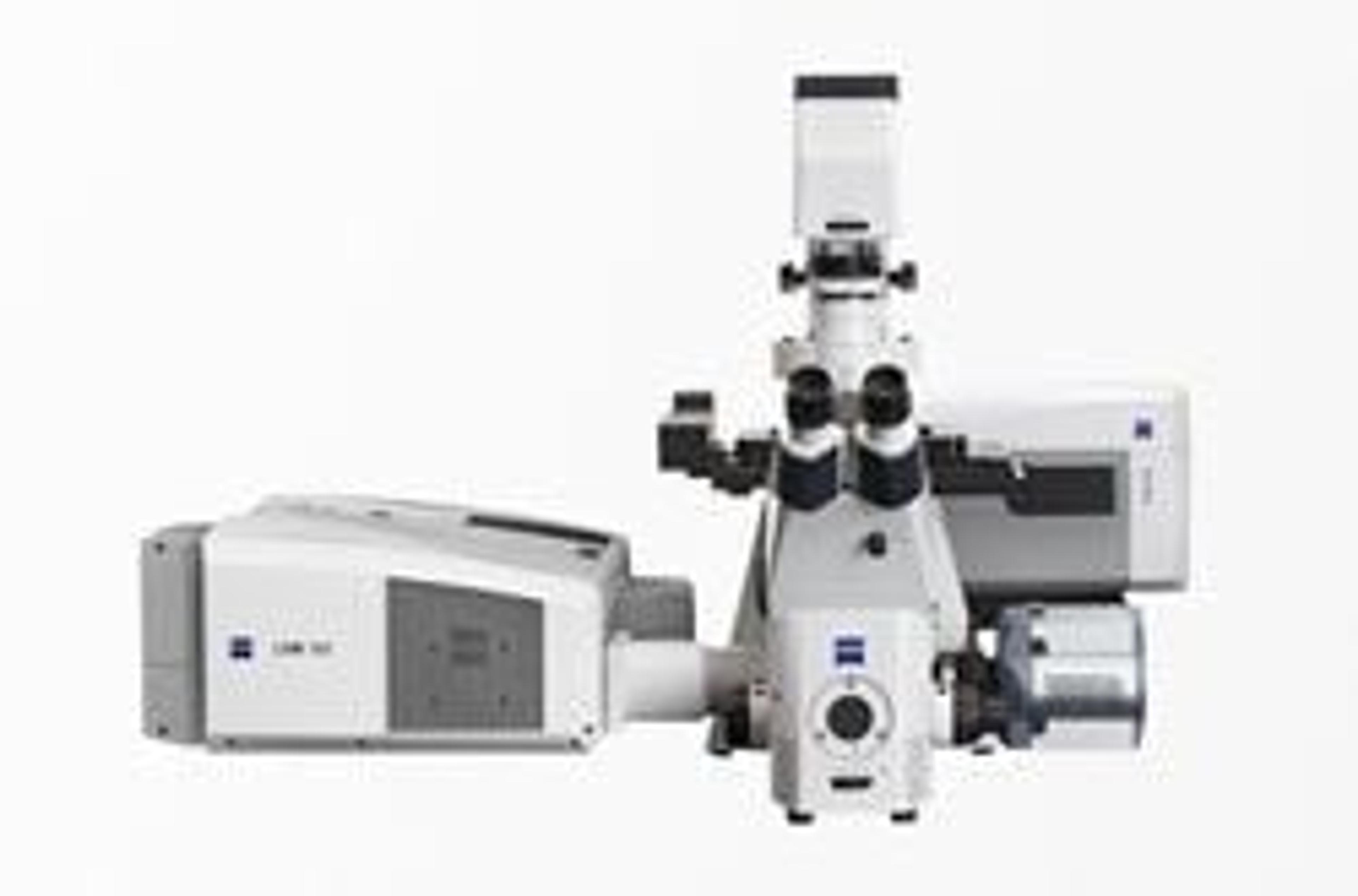
ZEISS ELYRA
ZEISS Research Microscopy SolutionsYour Flexible Imaging System for 3D Superresolution Microscopy.
ZEISS ELYRA S.1
ZEISS Research Microscopy SolutionsPut Flexibility First with Structured Illumination. ELYRA S.1 images any fluorophore – with up to twice the resolution of a conventional light microscope. Using structured illumination (SR-SIM) you reveal fine structural details while remaining free to label your samples with conventional dyes.You have invested a lot of time and energy in producing fusion proteins and multicolor staining protocols that are adapted to your experimental system. Now, with ELYRA S.1, you capture superresolution microscopy data with ease, using samples that may already be available in your lab's freezer. Specially-designed gratings give you the best resolution for each wavelength. Do you need Z-sectioning for 3D data acquisition? A fast, light-efficient detection? Then ELYRA S.1 is your ideal choice.Applications: Resolve structural detail in 3D with high penetration depth. Probe the structural organization of a whole cell. Investigate arrangement of cellular components and proteins. Explore interaction of molecules. Reveal the ultrastructure of organelles. Probe the ultrastructure of molecular assemblies. Map protein localization onto a structural context. Track many molecules and retrieve diffusion behavior. Study structural changes of slower dynamics.
ZEISS ELYRA P.1
ZEISS Research Microscopy SolutionsLocalize Single Molecules with Unrivalled Precision. ELYRA P.1 takes light microscopy to the limit. By localizing small structures and even single molecules, you are able to achieve resolutions of down to 20 nm laterally and 50 nm axially. Use ELYRA P.1 and photoactivated localization microscopy (PALM) for endogenously-expressed photo-switchable fluorescent proteins.You are interested in processes that take place near the coverslip. You want to see and measure single molecules in or near the plasma membrane like lipid rafts, receptor clustering or cell-substrate adhesion sites. With 3D-PALM you use photo-switchable proteins and profit from an excellent z capture range. The patented exclusive PALM technology of ELYRA P.1 takes you into a new world of data quality. Detection with an effective resolution down to 20 nm will show you substructure and patterns where conventional light microscopy will simply show co-localization.As a single molecule method, PALM is inherently quantitative – every image is a molecular statistics experiment.Applications: Probe the structural organization of a whole cell. Investigate arrangement of cellular components and proteins. Explore interaction of molecules. Reveal the ultrastructure of organelles. Probe the ultrastructure of molecular assemblies. Map protein localization onto a structural context. Track many molecules and retrieve diffusion behavior. Study structural changes of slower dynamics.