
How to Achieve Publication-Quality Images
7 Nov 2014
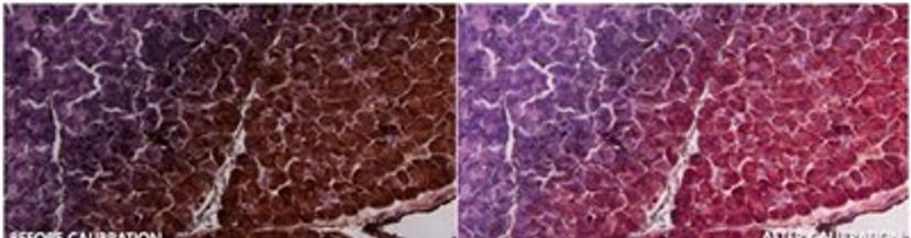
For many microscopists, images can be the only source of data for publication. Therefore, it is imperative that scientific integrity is maintained when capturing and processing images. Discover how Datacolor’s ChromaCal Color Calibration System can help you to generate publication-quality images in this guide.
1. Easily Compare Color and Brightness
The ChromaCal system automatically white balances and adjusts brightness levels to enable comparable color and brightness in calibrated images. This is essential if you wish to publish your images in reports and journal articles.
2. Avoid Data Corruption
Image editing software relies on a subjective approach to image calibration that can lead to data corruption. However, ChromaCal software uses an objective system to generate trusted calibrated images.
3. Calibrate Your Images Using Your Existing Workstation
The software is designed to work seamlessly with your existing imaging process and workflow. One image of a special calibration slide is captured any time during the imaging session to represent the color fingerprint of that session.
4. Distinguish Subtle Differences in Specimens
ChromaCal calibrated images are comparable and the software permits subtle differences among specimens, treatments, and doses of test substances to be distinguished.
5. Save Time by Processing Images in Batch
ChromaCal offers batch processing to color calibrate an entire folder of images quickly and accurately. Calibration of each individual image takes only seconds, saving you time and effort in downstream processing and editing.
Learn more in this white paper.