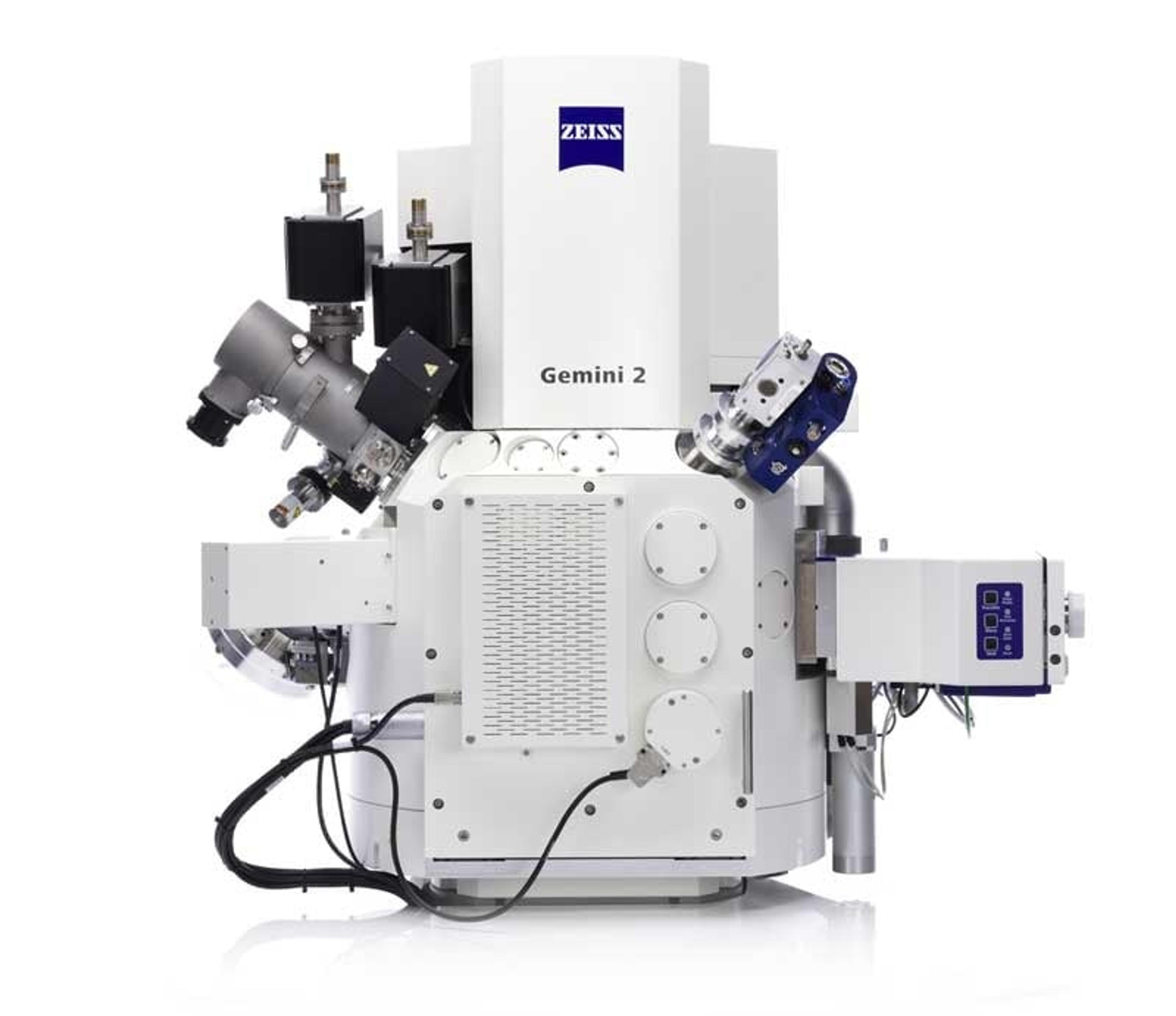
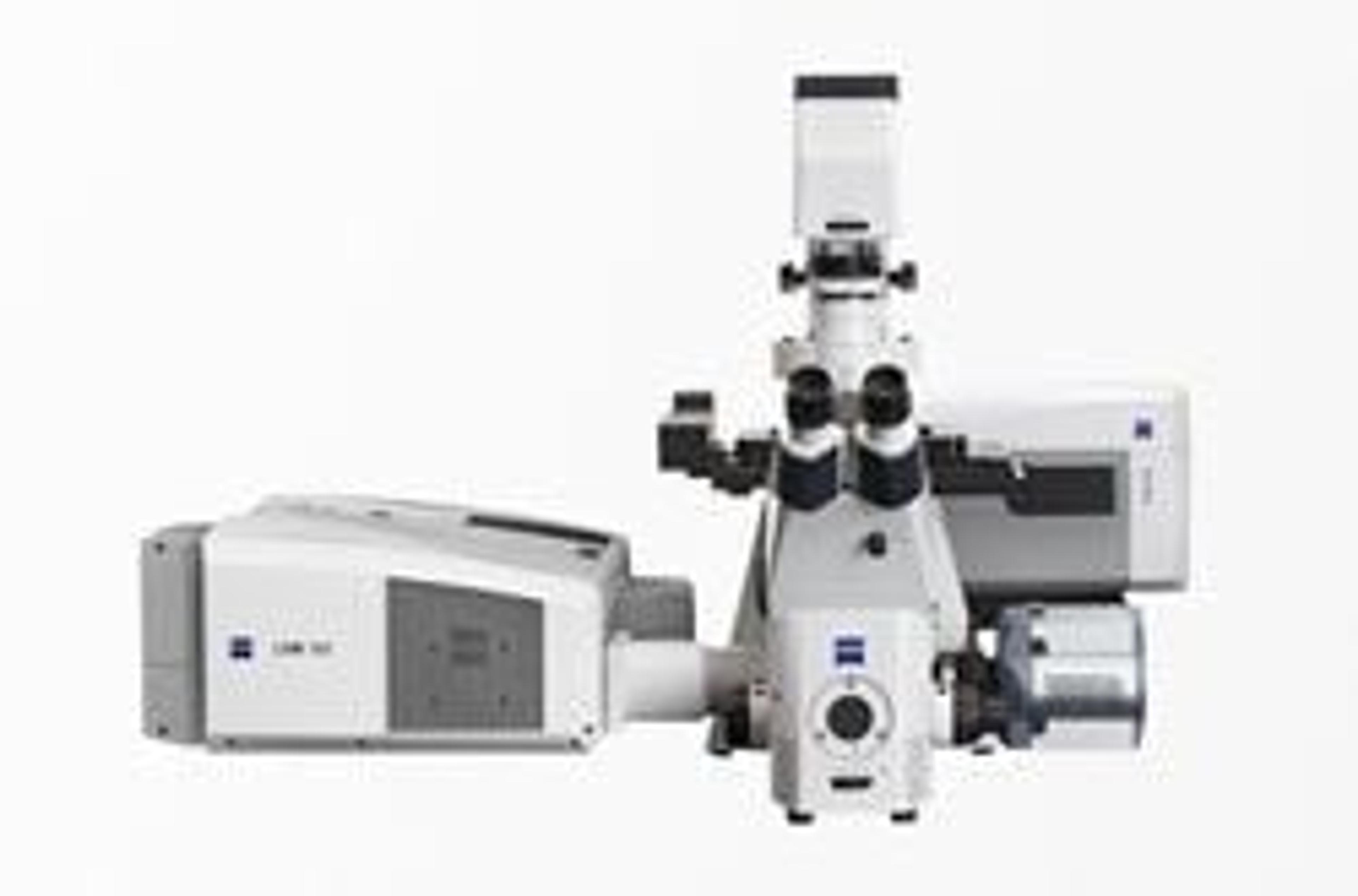
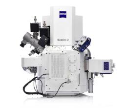
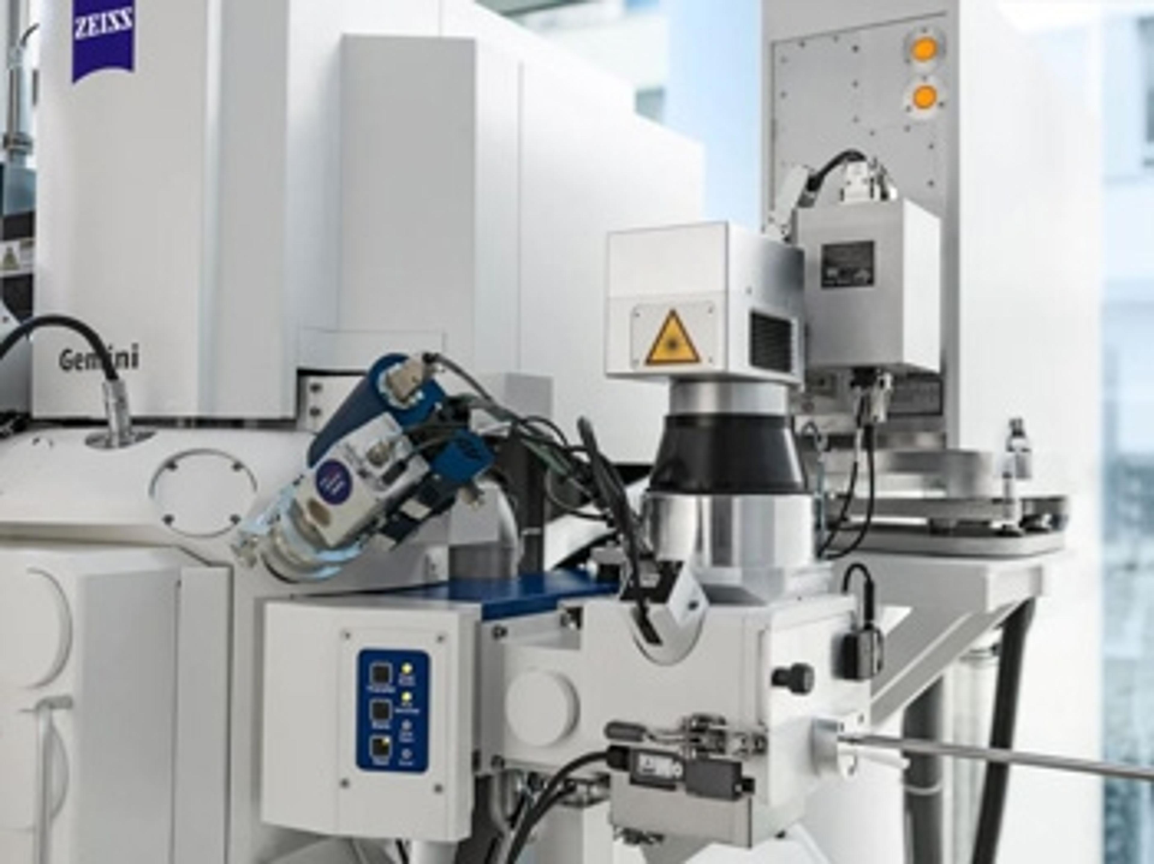
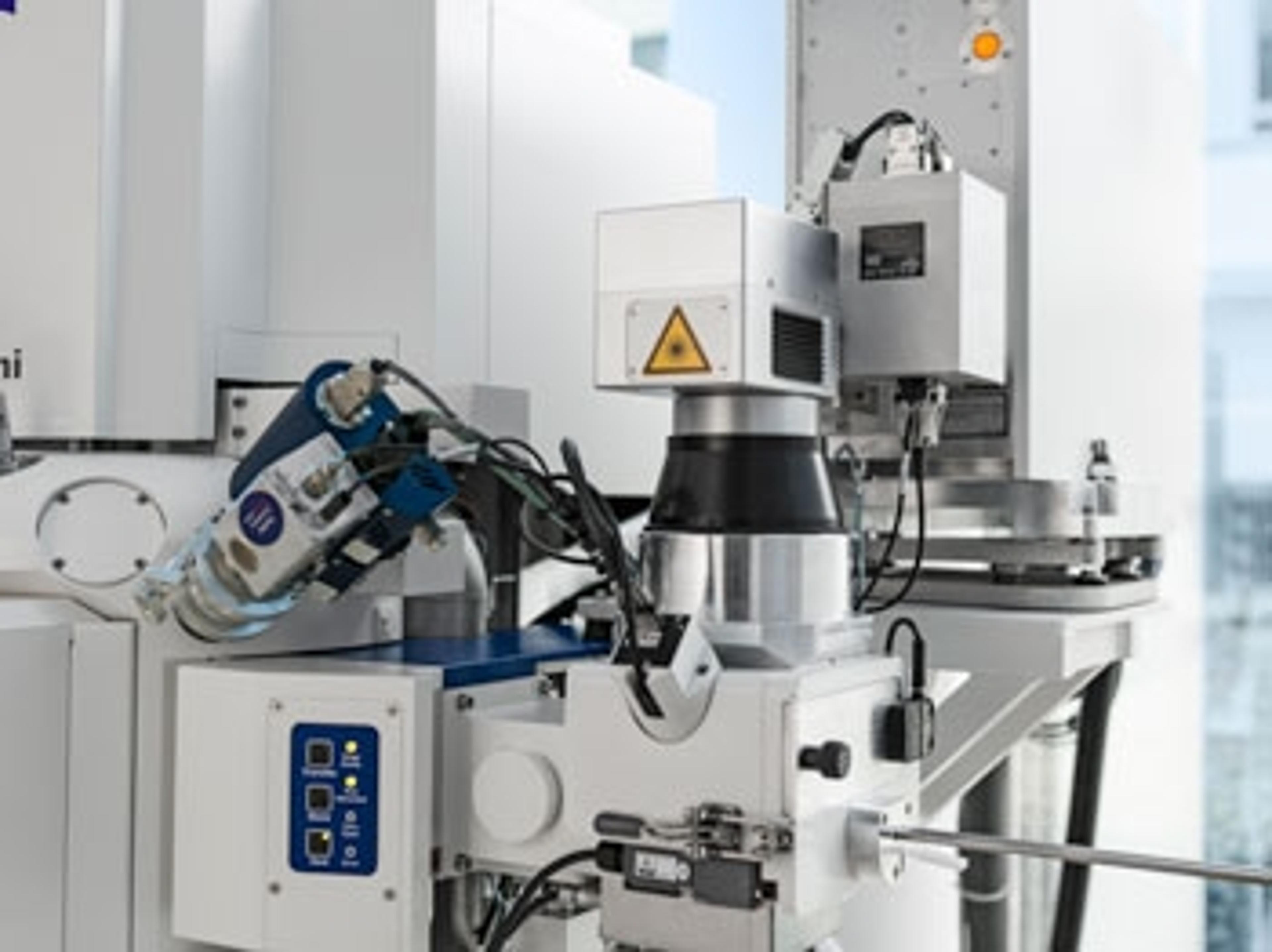
ZEISS Crossbeam Family
Within ZEISS Crossbeam Family you have the choice between Crossbeam 340 or Crossbeam 550. Exploit the variable pressure capabilities of Crossbeam 340. Or use Crossbeam 550 for your most demanding characterizations and choose the chamber size, standard or large, that best suits your samples.
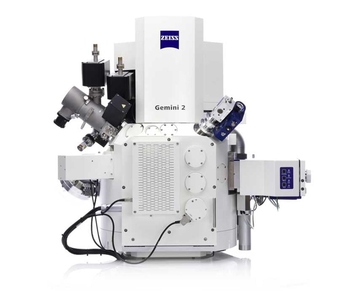
ZEISS Crossbeam 550: large chamber


Receive your quote directly from the manufacturer.
Good instrument!
Consumer goods
Very high-resolution SEM, FIB is OK.
Review Date: 23 Jul 2021 | ZEISS Research Microscopy Solutions
Good instrument, easy to use.
Tooth analysis
Good instrument.
Review Date: 26 Mar 2021 | ZEISS Research Microscopy Solutions
Great product, essential to all our products; it never stands still in our lab!
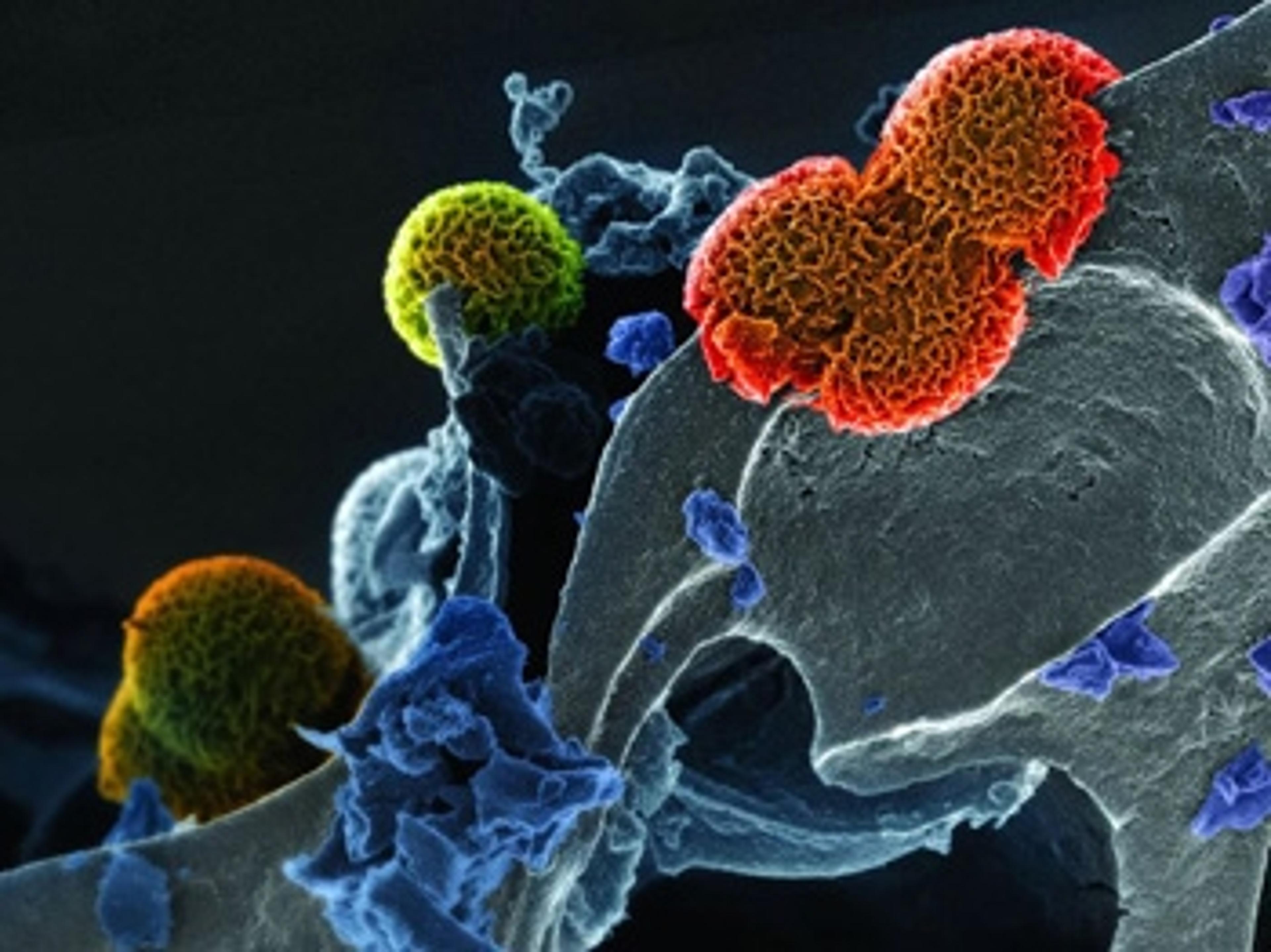
Imaging of sample surfaces and assessment of analytical information
Without high resolution imaging assessment of materials and bio-medical tissue is complicated. Images and additional analytical information permit materials and device optimization and the understanding of tissue.
Review Date: 2 Feb 2021 | ZEISS Research Microscopy Solutions
Excellent cutomer service.
Solids analysis
Excellent cutomer service, the service engineer who installed instrument was excellent.
Review Date: 12 Mar 2020 | ZEISS Research Microscopy Solutions
Maximize Your SEM Insights
Take advantage of achieving up to 30% better SEM resolution at low voltage.
Increase Your FIB Sample Throughput
Profit from up to 40% faster material removal by the introduction of intelligent FIB milling strategies.
Experience Best 3D Resolution in Your FIB-SEM Analysis
Enjoy the benefits of integrated 3D EDS analysis.
Trends in biomaterials imaging
Explore new insights from an industry-wide survey and discover answers to questions such as:
- What are the key research sectors, applications, and materials being investigated?
- What experimental outcomes are scientists looking for?
- How is constantly developing technology landscape impacting scientists?
Plus, delve into the key challenges in biomaterial analysis and the solutions that can drive your workflows forward, including automation, AI, and beyond.
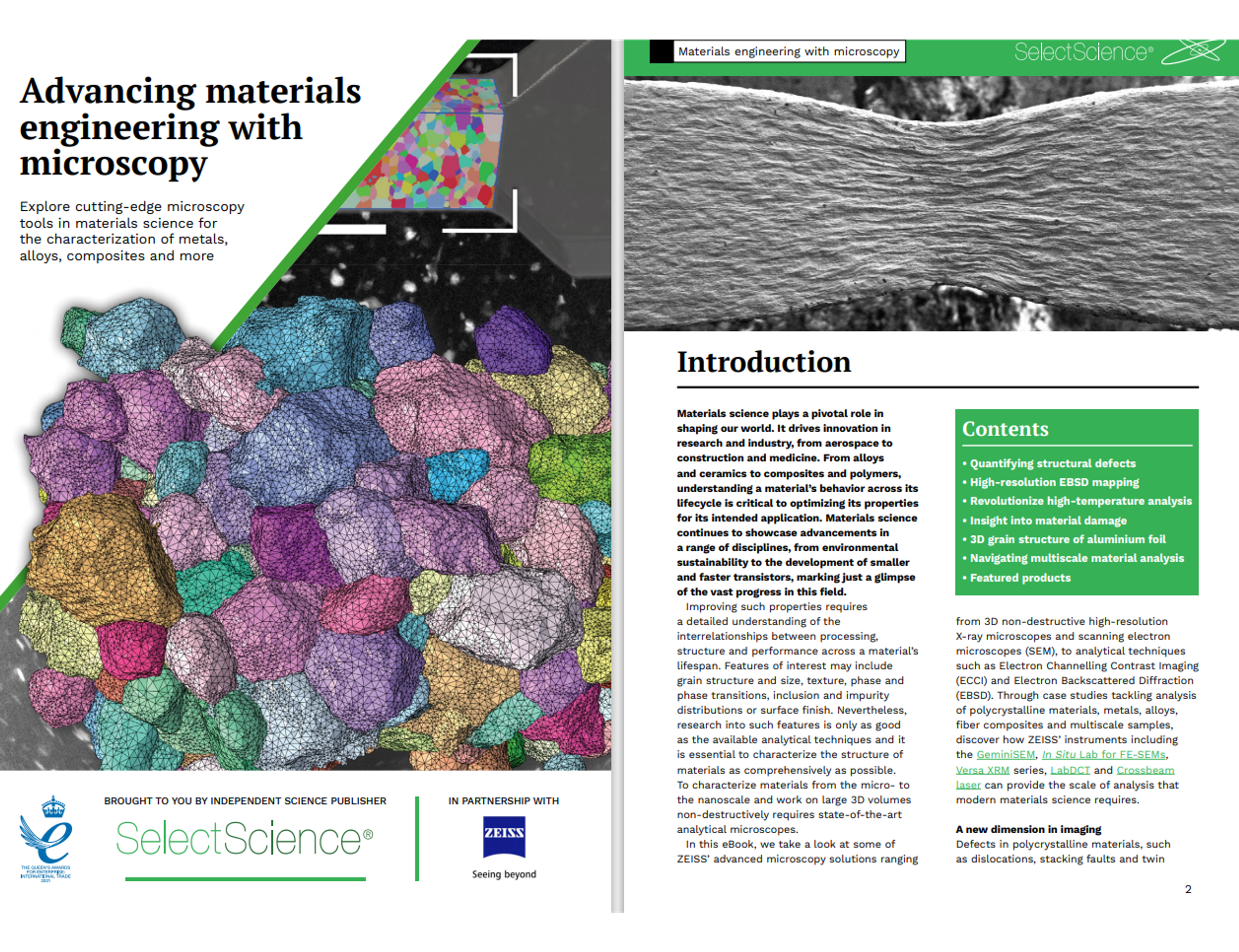
Advancing materials engineering with microscopy
Materials science is crucial in shaping our world and driving innovation across various fields like aerospace, construction, and medicine. It involves understanding the behavior of materials such as alloys, ceramics, composites, and polymers throughout their lifecycle. This knowledge is essential for optimizing their properties for specific applications.
Download this expert guide for free today and explore advanced microscopy solutions from ZEISS, including 3D non-destructive high-resolution X-ray microscopes and scanning electron microscopes (SEM). The eBook also covers analytical techniques such as Electron Channelling Contrast Imaging (ECCI) and Electron Backscattered Diffraction (EBSD).
Plus, find case studies on polycrystalline materials, metals, alloys, fiber composites, and multiscale samples, and learn about the tools essential for modern materials science analysis.

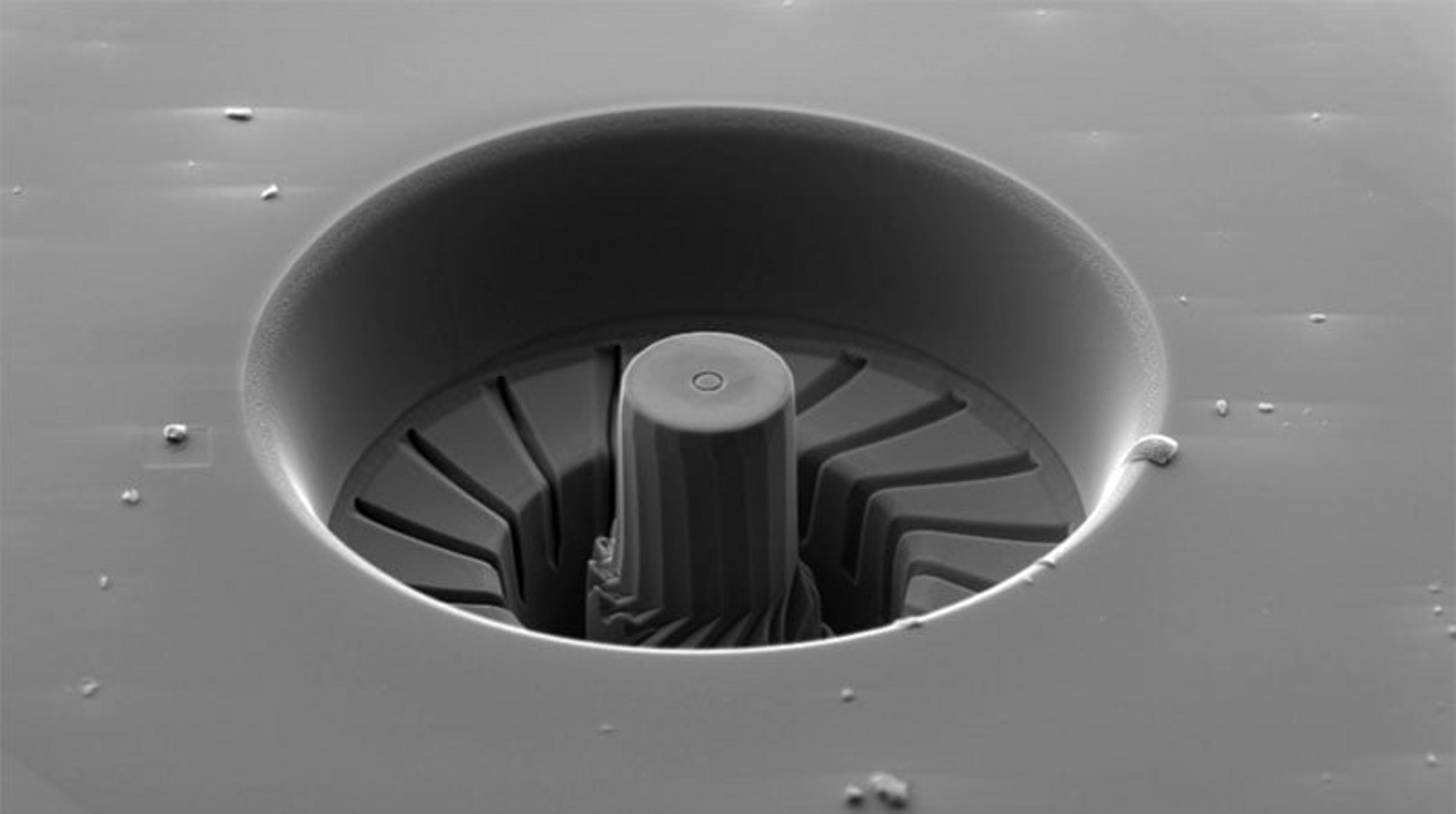
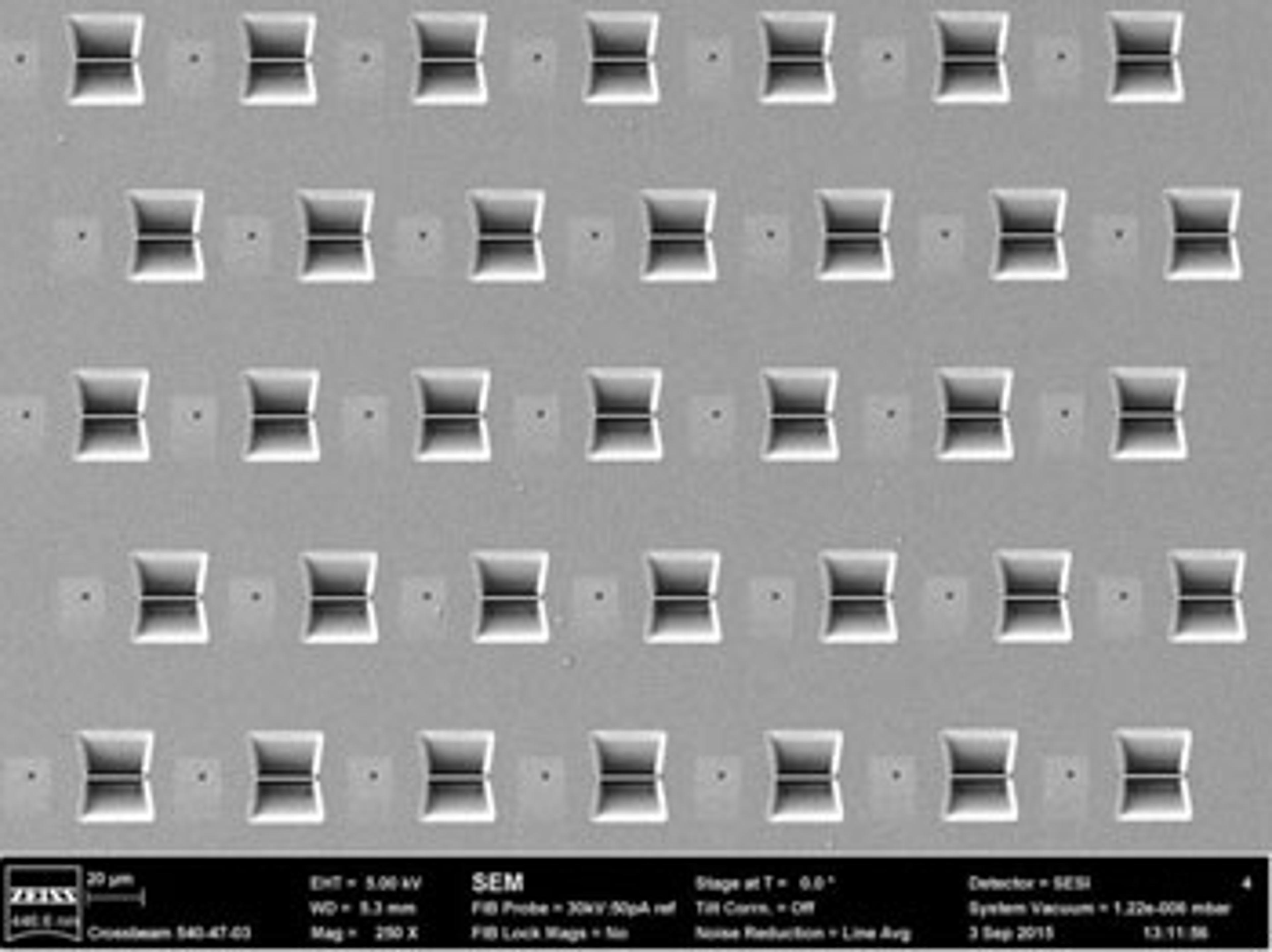
Fabrication and characterization of nanofluidic devices for DNA optical mapping
Nanofluidic lab-on-a-chip devices for the analysis of single DNA molecules were fabricated and characterized using FIB-SEM. Direct FIB nanopatterning of silicon master stamps allows fast prototyping of nanochannels of different shapes, cross sections and depths.
In this application note, ZEISS demonstrates how the Crossbeam can be an extremely useful tool for research in the field of nanofluidics and single molecule detection.
Rapid sample preparation for EBSD-analysis: Enabled by the LaserFIB
State-of-the-art preparation methods are mechanical polishing with a vibration-polish finish for large areas or focused ion beam (FIB) polishing for smaller areas and sensitive materials.
In this application note, ZEISS demonstrate how to overcome the limitations of these methods by utilizing the new femtosecond laser for ZEISS Crossbeam, to rapidly prepare cross-sections in sheets of different metals and EBSD is performed on the laser-polished surfaces.
Multi-modal chemical characterization of lithium ion battery particles
In this application note by ZEISS, a range of chemical analysis methods was applied to identical individual Li (Ni0,33Mn0,33Co0,33)02 cathode particles. The chemical studies revealed different insights and were compared side by side.
Microscopy characterization of coatings
Coatings have a broad range of applications in industries like pharmaceuticals, automotive, medical devices, textiles, oil and gas, aerospace and defense, food and packaging, and consumer optics, thanks to their role as functional, high- performance or protective materials. This paper illustrates a case study on how optical and confocal microscopy, focused ion beam (FIB), and correlative microscopy are used by ZEISS R&D to make sure new customer demands are met while keeping the quality and great performance of coatings.
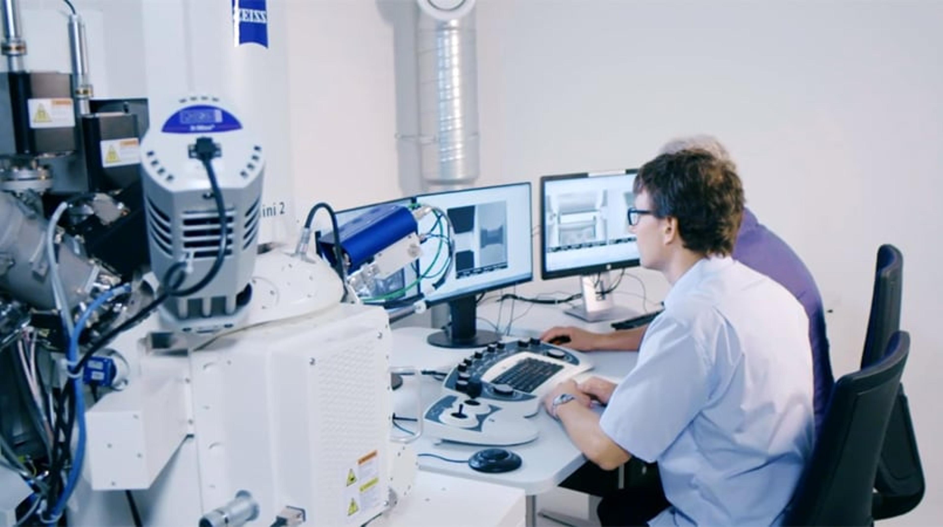
High Throughput Imaging with ZEISS Crossbeam 550
For more than 13 years ZEISS has been known as a manufacturer of two-beam FIB-SEM systems – Crossbeam instruments. During these years, Crossbeam performance in terms of image resolution and contrast, ease of use, workflow automation and throughput has improved continuously. This development culminates in today’s ZEISS Crossbeam 550.
ZEISS Microscopy Solutions for Steel and Other Metals
ZEISS Microscopy offers multi-modal characterizations and advanced analysis options for industry and research. This application note provides a variety of applications of ZEISS products to analyze steel and other metals.
FIB-SEM Investigations of the Microstructure of CIGS Solar Cells
Efficiency of thin-film copper indium gallium selenide (CIGS) solar cells is above 22 percent reaching the performance levels of modern polycrystalline silicon solar cells. Strong development efforts are ongoing to further increase cell efficiency. In this context, electron microscopy plays a crucial role because it allows the analysis of the microstructure of the solar cell through all relevant length scales and with very high resolution.
In this application note, high-efficiency thin-film solar cells based on Cu(In,Ga)Se2 were studied extensively by focused ion beam – scanning electron microscopy (FIB-SEM) on a ZEISS Crossbeam. Cross sections of cells on different substrates were prepared by FIB to reveal the internal structure of the cell, allowing detailed characterization of the electrodes and microstructure throughout the cell by STEM, EDS and SIMS.
High Resolution Imaging of Non-Conductive Specimen Benefits From Local Charge Compensation
In this application note, high-resolution SEM imaging is executed on non-conductive samples by the integration of a charge compensation system.
Battery centric workflows for next-generation battery developments
Tuesday, July 28 at 16:00 BST | 17:00 CEST | 11:00 EDT | 08:00 PDT
Want to know how to design battery-centric workflows, handle air-sensitive samples, and connect glovebox, FE-SEM, and XRM?
The development of high-energy-density chemistries, recycling methodologies, and the optimization of lithium-ion systems rely on characterization methods to provide a feedback loop between microstructure, processing, and electrochemical behavior.
In this SelectScience® webinar, we will examine how today’s microscopy workflows address battery research, including Li- and Na-based solid-state batteries. You will learn how to design 'battery-centric' workflows that connect destructive and non-destructive methods to specific research questions, including:
- What is the microstructure of an electrode, including its 3D nanoscale architecture, and how is that linked to electrochemical performance?
- How are components and microstructures arranged and aligned inside an assembled battery, and how do they evolve during cycling?
- How can battery and electrode microstructures be assessed coherently across different length scales?
- How can battery recycling be optimized from the efficient characterization of black mass powder in multiple dimensions?
The first part of this webinar will focus on handling air-sensitive materials using air-free transfer workflows that bridge glovebox environments with multiple electron microscopy modalities, enabling investigation of microstructure, while preserving the native state as much as possible.
The second part will highlight X-ray microscopes (XRM) as a non-destructive, minimal-preparation technique for 3D multiscale characterization of intact cells and complex samples such as black mass from recycling processes.
The overall focus will be on research-driven case studies and practical workflow strategies, illustrating how combined destructive and non-destructive microscopy approaches can provide deeper insight into battery behavior, degradation, and lifetime for the battery circular economy.
Certificate of attendance
If you attend the live webinar, you will automatically receive a certificate of attendance, including a learning outcomes summary, for continuing education purposes. If you view the on-demand webinar, you can request a certificate of attendance by emailing editor@selectscience.net.
Webinar details
- Cost: Free to attend
- Location: Online
- Duration: 60 minutes
Registration is required to secure your place. If you register but can’t attend live, you will receive a link to the on‑demand recording once it becomes available.

Microscopy with ZEN core: A unified approach to imaging and analysis
April 29, 2025 - 09:00 BST / 10:00 CEST / 16:00 CST / 17:00 JST
Join us for an informative webinar sponsored by ZEISS, where we will introduce ZEN core, a sophisticated software solution designed to enhance your microscopy workflows. This session will focus on how ZEN core provides a streamlined user interface that accommodates both light and electron microscopy, allowing users to transition seamlessly between modalities.
During the session, you will discover how ZEN core's intuitive user interface adapts to the unique needs of your laboratory allowing users of all experience levels to efficiently image, analyze, and manage data. We will explore the advanced imaging capabilities, automated workflows, and powerful AI features that make ZEN core the command center for both light and electron microscopes.
Join us for a live demonstration of ZEN core and take part in our Q&A session to address your specific inquiries. Register today to learn how ZEN core can support your microscopy research and analysis needs.
Key learning objectives:
- Understand how ZEN core fosters seamless collaboration in connected laboratory environments
- Explore the integration of multiple microscopy techniques within a unified platform
- Learn how ZEN core ensures data consistency and standardization across instruments and locations
Who should attend?
This webinar is ideal for researchers in both academia and industry looking to improve their microscopy practices and enhance productivity.
Certificate of attendance
All webinar participants can request a certificate of attendance, including a learning outcomes summary, for continuing education purposes.
If you view the on-demand webinar, you can request a certificate of attendance by emailing editor@selectscience.net.
TEM lamella preparation with ZEISS Crossbeam
In this video, learn how to prepare a TEM sample using the ZEISS Crossbeam.
How ZEISS Microscopes are Shaping the Future
In this video, learn how ZEISS is developing technologies for use at the cutting edge of science, consumer electronics, energy, construction, and more.
ZEISS Legacy Continues: Advancing Microscopy at M&M 2017
Peter Lander discusses the legacy and development of ZEISS microscopy and the importance of conferences such as M&M. Its collaboration with scientists has enabled ZEISS to provide instrumentation that satisfies their needs, with the aim to develop simplified software that enables complementary and overlaying analyses over a range of its instrumentation.
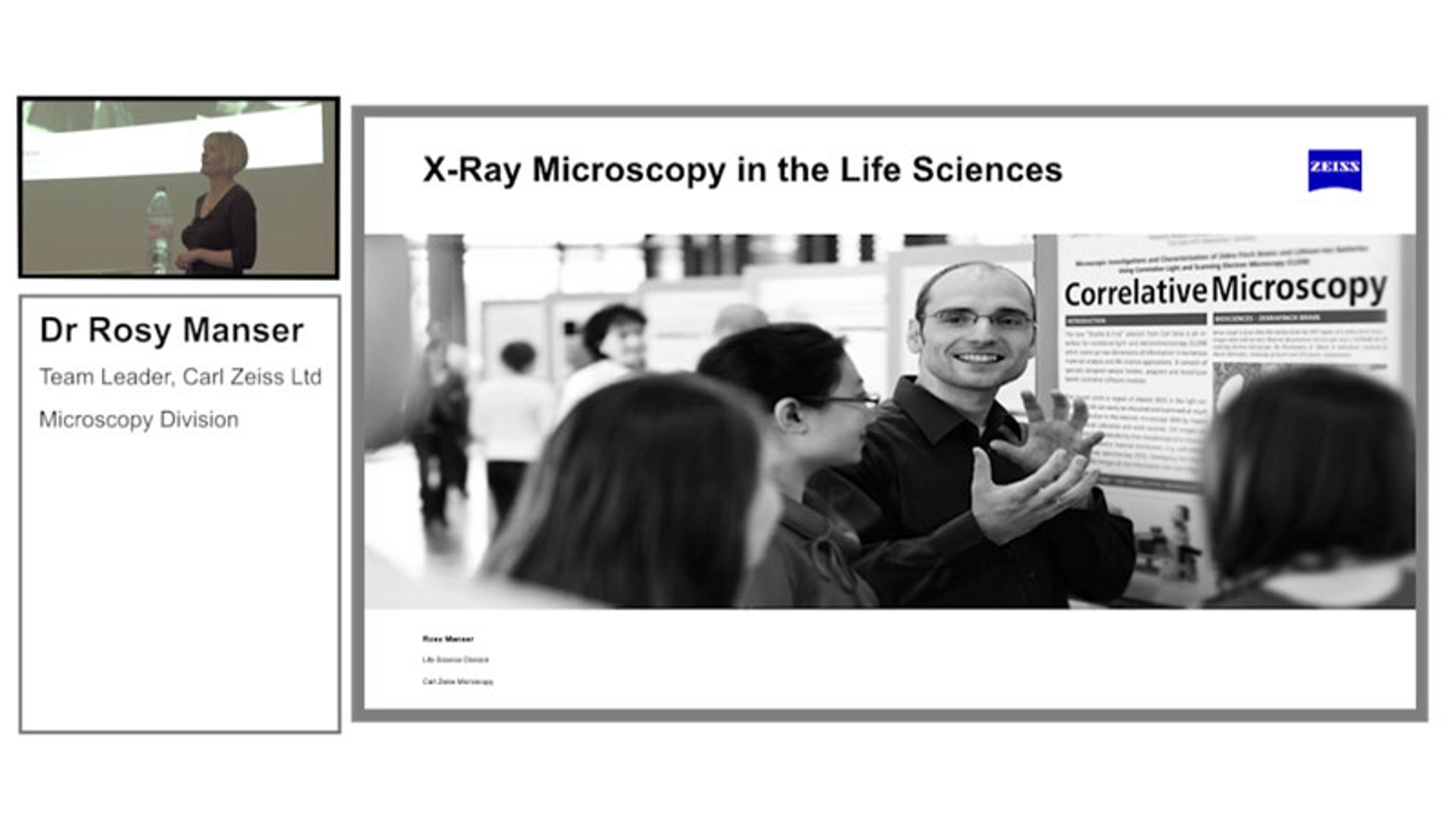
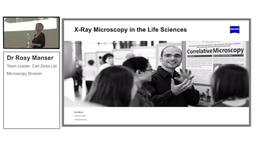
Presentation: X-Ray Microscopy in the Life Science
Dr Rosy Manser, Carl Zeiss Microscopy GmbH, Germany, discusses new X-Ray Microscopy applications enabled by the latest ZEISS technology, including the imaging of unstained tissue using the ZEISS VERSA XRM. Also, learn how the technology can be used as part of a correlative microscopy workflow.

7 top new resources for materials characterization research
Exclusive interviews, new methods, free downloads and much more to help advance materials characterization research and development

Analytical chemistry round-up for 2019
Take a look back through 2019 at the best content from our analytical community
ZEISS enhances efficiency in multi-scale and multi-modal workflows
Researchers benefit from faster FIB-SEM sample preparation, more accurate 3D tomography, and greater integration in data reporting
ZEISS enhances efficiency in multi-scale and multi-modal workflows
Researchers benefit from faster FIB-SEM sample preparation, more accurate 3D tomography and greater integration in data reporting

ZEISS Introduces Enhanced Capabilities for Ion Beam Microscopes
Upgrade opens new opportunities in materials science and covers advances in analytics, tomography, sample preparation, and data integrity
Major improvements in workflow offerings for material sciences core facilities
Promise of enhanced analytics, tomography, sample preparation and data integrity

Nanomaterials and Particle Size Analysis: Special Feature
Discover a range of cutting-edge applications and new technologies, plus, don't miss our exclusive video interview with a Nobel laureate

Analytical Science Highlights of 2018: From Hurricane Heroics to Cars of the Future
We look back at the biggest stories and the best content from our analytical chemistry communities
Automate FIB Processing for High-Throughput TEM Lamella Preparation
Creating TEM lamellae with the new Ion-sculptor FIB column of ZEISS Crossbeam