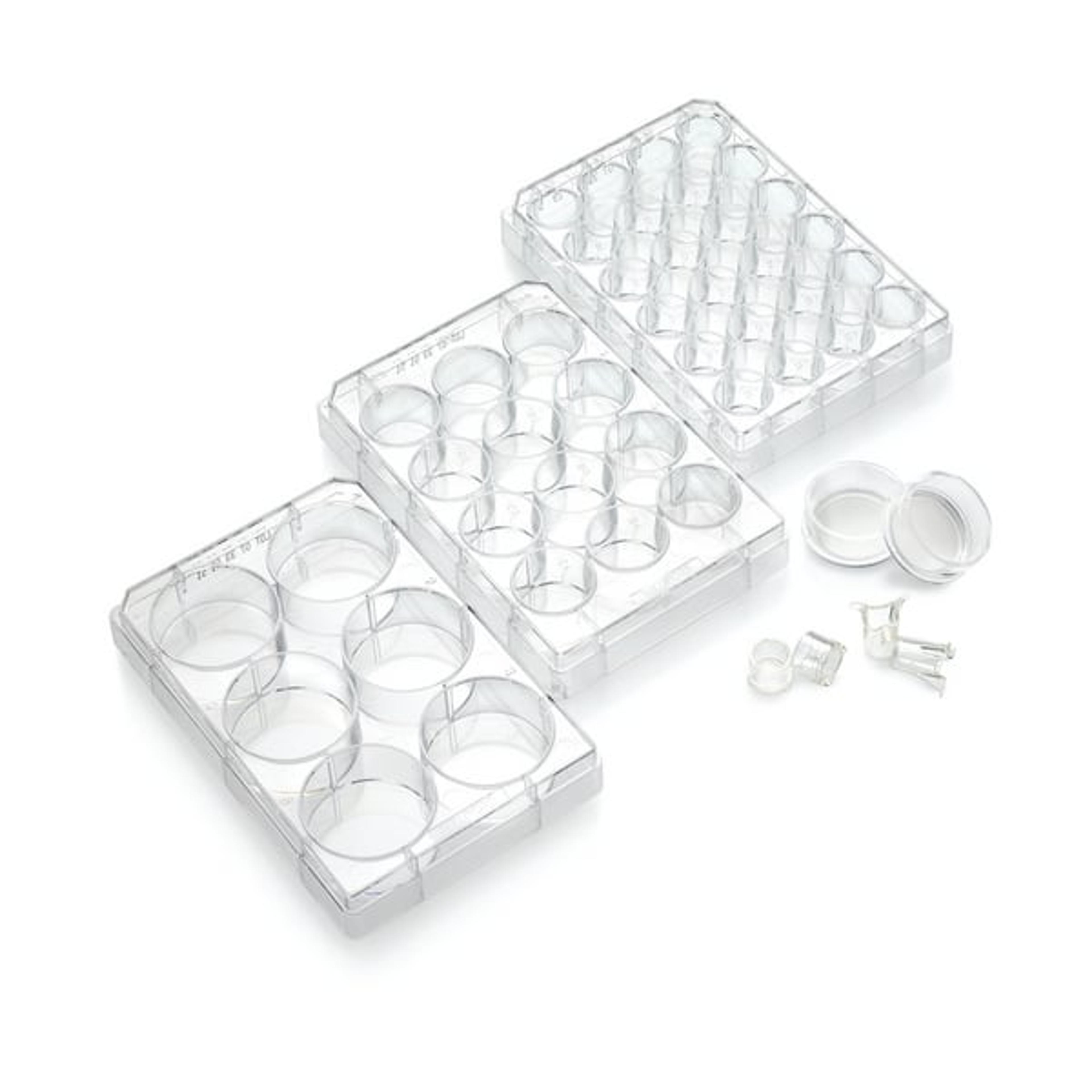
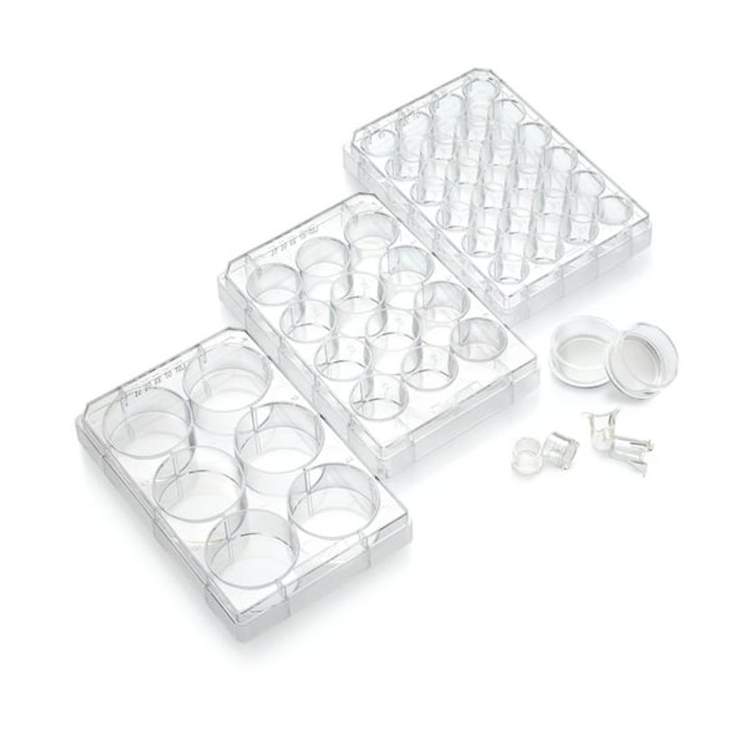
Millicell® Cell Culture Inserts
With Millicell inserts, attachment or suspension cells can access media from both their apical and basolateral sides. Cell growth, structure, and function more closely mimic what occurs in vivo. In addition, Millicell inserts make it possible to study both sides of the cell monolayer. Millicell inserts are available for 24-, 12-, or 6-well plates.

Receive your quote directly from the manufacturer.
Great brain cultures!
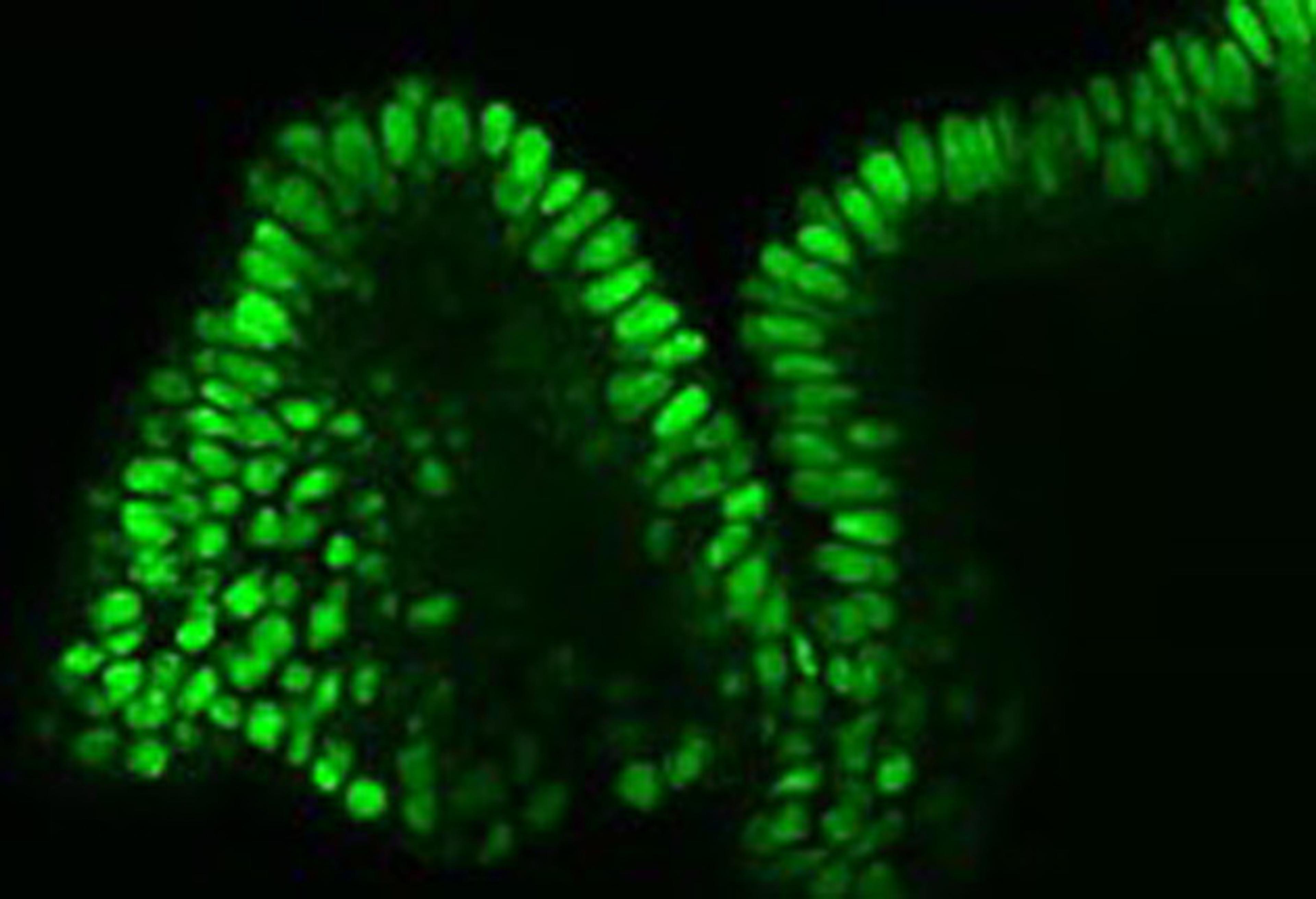
Organotypic brain cultures
I've used the inserts for organotypic brain cultures from different rat brain regions in 6-well plates. The quality of the insert is very high, like the quality and viability of the cultures. The only disadvantage is the price, in fact for numerous experiments thy could be expensive.
Review Date: 14 Apr 2017 | Merck
With Millicell inserts, attachment or suspension cells can access media from both their apical and basolateral sides. Cell growth, structure, and function more closely mimic what occurs in vivo. In addition, Millicell inserts make it possible to study both sides of the cell monolayer.
Millicell inserts are available for 24-, 12-, or 6-well plates. The inserts are easily prepared for SEM and TEM visualizing techniques, and they are compatible with cellular and/or fluorescent stains.

2.5D cell culture models for more predictive in vitro research
2.5D cell culture is an emerging approach for creating more physiologically relevant in vitro models, overcoming key limitations of conventional 2D systems. Using microporous membrane inserts restores apical-basolateral polarity, supports barrier formation, and enables air-liquid interface (ALI) culture—critical for differentiated epithelial models with improved predictive value for ADME/Tox, drug transport, and disease research.
2.5D culture bridges simple monolayers and complex 3D systems. It allows organoid-derived cells to be grown on permeable membranes, combining biological relevance with accessibility for TEER measurement, permeability, migration, and wound healing assays. Tools such as Millicell® inserts and plates support applications from GI barrier and drug absorption studies to skin and respiratory models, enabling more predictive epithelial and tissue-representative systems.
Download this SelectScience guide to explore:
- How to culture colon organoid monolayers for GI barrier research
- Small intestine organoid-derived monolayers and physiologically relevant alternatives to Caco-2 models
- Optimizing MDCK cell culture for high-throughput TEER and permeability assays
- The principles behind barrier formation and permeability assays using cell culture inserts
- Generating organotypic air-liquid interface 3D skin cultures
- Modelling respiratory lung diseases using bronchial epithelial cells at ALI
Resource details:
- Document type: SelectScience guide
- Page count: 47
- Read time: 85 mins
- Edition: 1st

More like life: Microporous membrane-based culture systems
The value of cell culture experiments lies in how well they represent the native microenvironments of in vivo systems. In this application note, Millipore Sigma explores the advantages of using microporous membranes, compared to 2D plastic surfaces, to provide more in vivo-like cell growth environments for cell culture experiments.
More like life: Microporous membrane-based culture systems
Membrane-based culture systems foster a more in vivo-like cell growth environment and support a range of applications including primary and secondary screening, transport assays, toxicity screening, cell signaling, cell proliferation and ADME/toxicity drug studies. In this application note, Millipore details the features of the Millicell® cell culture product family and reveals how membrane-based cultureware can closely recapitulate in vivo conditions.

Latest Advances in Cell-Based Assays: Special Feature
From flow cytometry and kinetic analysis to porous membranes and liquid handling, find out the latest techniques and technologies advancing cell-based assays