Agilent BioTek Gen5 Software for Detection
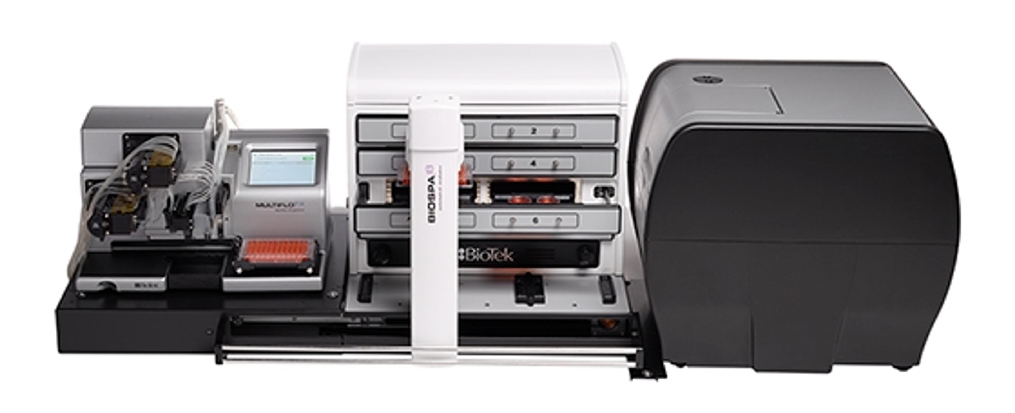
Agilent BioTek Gen5 software for Agilent BioTek multimode and single-mode microplate readers is an integrated tool for endpoint, kinetic, spectral scanning, and well area scanning. It controls all the functions of the plate reader and has powerful data analysis capabilities for a broad range of applications. Gen5 microplate reader software is also used to integrate BioTek plate readers to BioTek BioStack and other automated…


Receive your quote directly from the manufacturer.
Great software for quick and easy, multiplexed cell analysis!
Cell biology
The Gen5 software facilitates an easy and fast multiplexed analysis for experiments in a multi-well format. With this application, it is possible to analyze fluorescence, luminescence, and absorbance signals simultaneously. Unfortunately, the software is still quite expensive. We use the software to analyze single-cell 2D as well as 3D co-cultures measuring the viability, motility, cell death, and other biological measures. The automated output helps to obtain the data fast without post-experiment data processing and image analysis.
Review Date: 7 Mar 2021 | Agilent Technologies
Received help from manufacturer.
Gen5
Helped me a lot with kinetic read challenges. They solved my problem.
Review Date: 28 Jan 2020 | Agilent Technologies
Detection of bacterial antigens and antibodies
Produces many results for analysis
Review Date: 8 Jun 2017 | Agilent Technologies
Protein Biochemistry
Gen5 software is really easy to use and only takes me a few minutes to train up new users. Our after sales experience has been really good and they always respond immediately to any requests. The information I received before I purchased the system was very comprehensive and allowed me to compare all the specs with other models on the market at the time. Having a demo disk was really valuable because it allowed me to trial the software to make sure it met our needs. The online support and updates, and the TekTalk emails I have received have also been very useful and informative, and have helped my understanding of the full capabilities of the instrument and software. I have already recommended this product to colleagues on more than one occasion.
Review Date: 12 Oct 2015 | Agilent Technologies
Biochemistry/Cell Biology/Molecular Biology
This is definitely a 5 star software! It is very easy to use for running our BioTek plate reader and for programming results analysis. Instant raw results transformation is very convenient and in a minute you get your results in a mode ready to use. The tech service is excellent and if they do not have the answer right away, they will find a way and get back to you with an idea for a new program/analysis. I would definitely recommend this software in an academic environment where many junior trainees are evolving.
Review Date: 7 Nov 2014 | Agilent Technologies
amyloid quantity
The repeatability of fluorescent value in parallel experiments are poor.
Review Date: 31 Jul 2014 | Agilent Technologies
Cellular And Molecular Biology, Biochemistry
It is the best investment I made in the lab. The versatility and the number of results we can obtain with the microplate reader is so good there isn’t another piece of equipment that can compete. Right now other labs come to use it too. We have other microplate reader in core equipment, but by far this is the equipment of choice for my colleagues.
Review Date: 23 Jul 2014 | Agilent Technologies
CRO
It is a great work horse for the applications we use it for. It’s resilient and has excellent quality data along with standards.
Review Date: 17 Jul 2014 | Agilent Technologies
Clinical Research
This software is extremely powerful, and is capable of a wide range of assays and data analysis. A fairly significant time investment is required in order to effectively program the machine beyond the basic templates. After sales support is good for most things, but can be lacking when it comes to rarer situations and questions.
Review Date: 16 Jul 2014 | Agilent Technologies
Pharmaceutical
Their instrument is easy to use and the after sales services are great.
Review Date: 4 Jul 2014 | Agilent Technologies
Agilent BioTek Gen5 software for Agilent BioTek multimode and single-mode microplate readers is an integrated tool for endpoint, kinetic, spectral scanning, and well area scanning. It controls all the functions of the plate reader and has powerful data analysis capabilities for a broad range of applications.
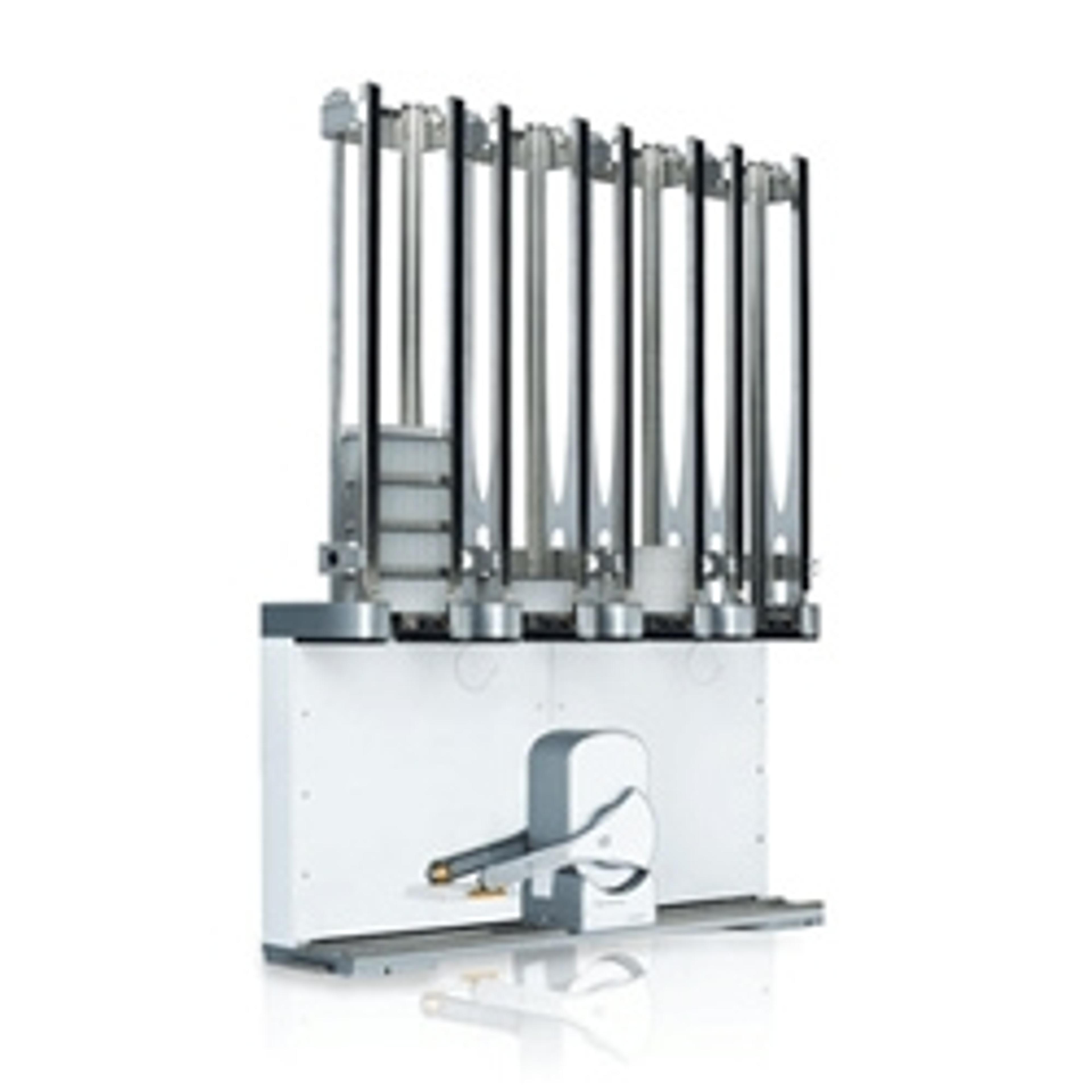
Gen5 microplate reader software is also used to integrate BioTek plate readers to BioTek BioStack and other automated systems.

High-throughput platform for fluorescence image-based neurite outgrowth analysis
The evaluation of neurite outgrowth using high-throughput image-based analysis provides an essential tool for neurobiologists to advance basic, translational, and clinical research in neuroscience. Explore the BioTek Gen5 neurite outgrowth module from Agilent Technologies that offers flexible, automated, multichannel analysis of neurite outgrowth across multiple fluorescence channels and modalities to support a broad range of neurite outgrowth approaches. The module is compatible across Agilent BioTek imaging instruments to provide a single platform solution for neurite outgrowth assays.
Automated viral plaque assay workflow using the Cytation Cell Imaging Multimode Reader
Plaque assays remain the standard method for determining viral titers for lysogenic viruses. The assay relies on determining the number of plaque-forming units (PFU) created in a monolayer of virus-infected cells. In this application note, Agilent presents an assay that provides an accurate and efficient method for determining plaque counts and calculating viral titers in a range of microplate densities, including 6-, 24-, 48-, and 96-well formats.
Detection and automated imaging of Regions of Interest when performing Whole Slide Imaging
In this application note, BioTek highlights two examples that use a combination upright and inverted automated image acquisition system to demonstrate the ability to rapidly image entire vessel surfaces at low resolution, select only those regions of interest, and capture high resolution images of the ROIs using automated imaging methods.
Calculate cell count/volume using automated label-free image analysis
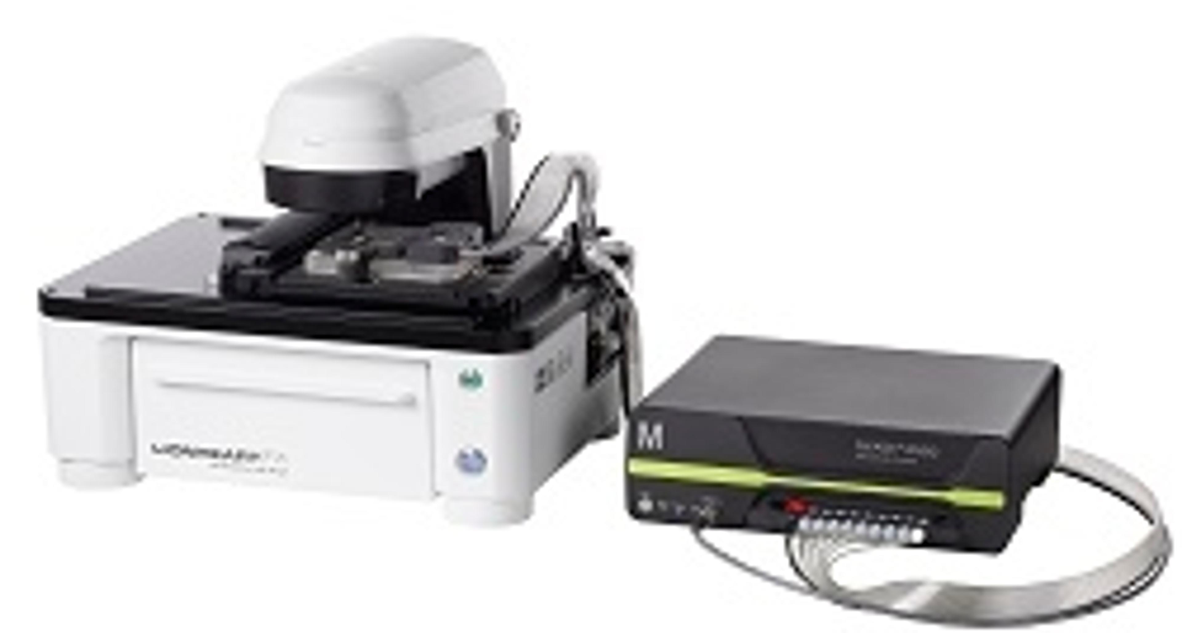
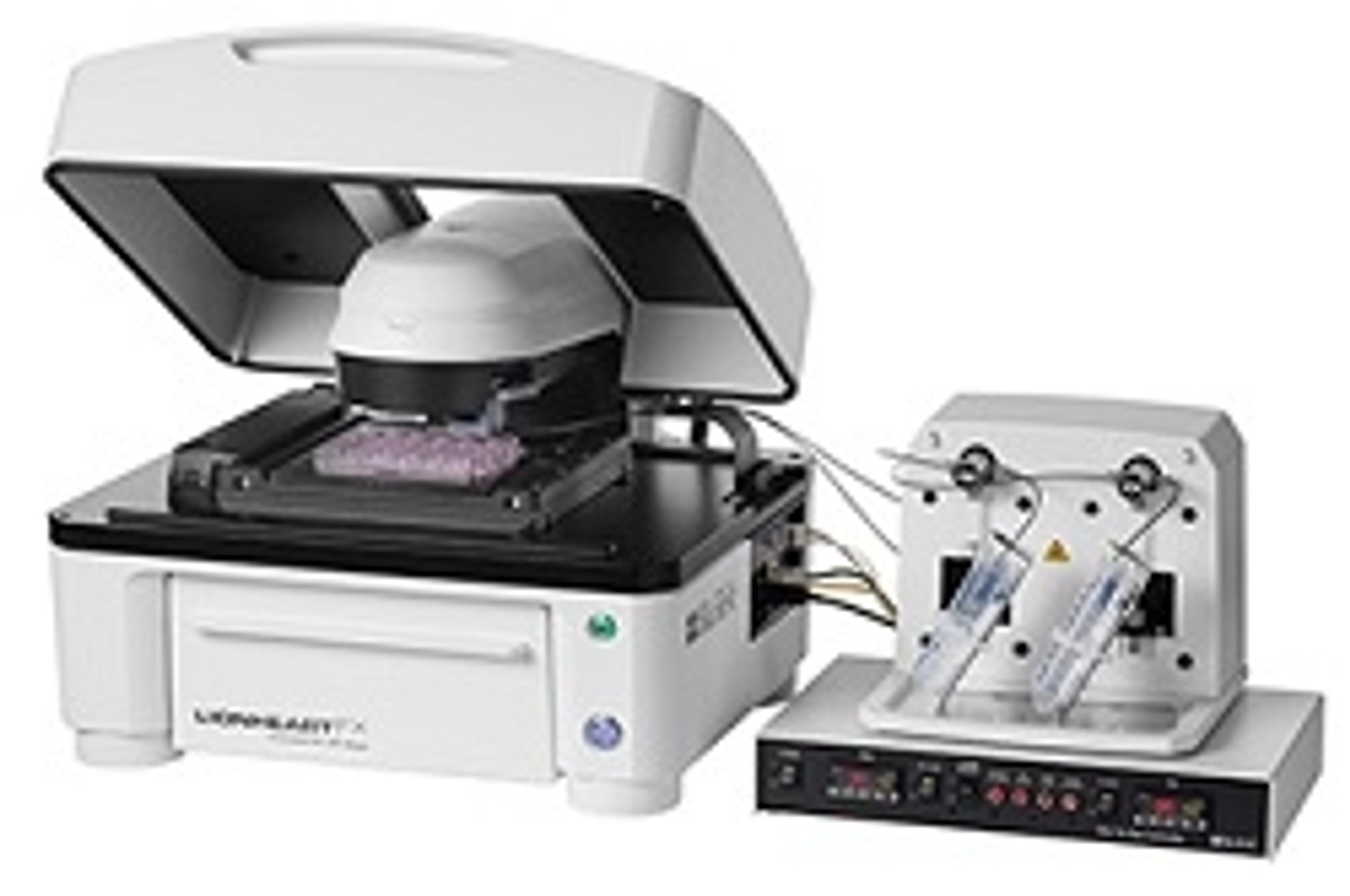
With BioTek’s Lionheart FX Automated Microscope and Gen5 Image Analysis Software, total cells/volume can be calculated for almost any type of cell, using digitally captured and analyzed images, independent of probes or stains. This process is demonstrated in this free infographic that shows how to quantify pre- and post-fermented yeast cells in corn mash.
Nuclear translocation of RelA in stimulated macrophages
As critical components of the innate immune system, macrophages respond to microbes by recognizing molecules, such as the gram-negative bacteria product lipopolysaccharide (LPS), via Toll-like receptors. Receptor activation stimulates a complex signaling network that involves, among others, the NF-κB pathway. Here we quantitate the nuclear translocation of the protein RelA, a component of the NF-κB transcription factor complex, in macrophages using image-based analysis.
Brightfield and fluorescence imaging using 3D PrimeSurface ultra-low attachment microplates
3D cell culture has become a well-established in vitro experimental approach as it realistically mimics an in vivo environment. The use of clear U-bottom ultra-low attachment microplates, that minimize cell adherence, has become a standard for applications such as spheroid proliferation. In this application note, BioTek presents data generated with the Cytation™ 5 Cell Imaging Multi-Mode Reader, using PrimeSurface ULA plates to develop simple and robust spheroid assays for brightfield and fluorescence imaging.
Automated image screening of zebrafish embryos exposed to developmental toxins
In this application note, BioTek demonstrates high-throughput screening and analysis of zebrafish embryos treated with several developmental toxins. Here, BioTek exposes embryos to ethanol, retinoid acid, and cyclopamine, then capture brightfield images of the embryos in multi-well plates with Lionheart™ Automated Microscope. The images are analyzed for size and shape using Gen5™ software. The combination of high-throughput imaging and analysis using a vertebrate model makes this an attractive method for toxicology screening.
Using Acridine Orange to Measure Cell Death in Ethanol Treated Zebrafish Embryos
Ethanol exposure during development can have devastating effects on the fetus. In this application note, zebrafish are used, an excellent model to study both normal development and toxicity, in order to determine the effects of ethanol exposure during development. Zebrafish embryos were treated with ethanol during the first 24 hours of development and the effects of ethanol treatment on cell death was assessed using acridine orange staining. It was found that exposure of embryos to ethanol results in a dose-dependent increase in cell death overall in the embryo.
Autophagy Analysis Using Object Spot Counting
Autophagy is critical for the maintenance of cellular homeostasis. However, dysregulated autophagy can lead to death of healthy cells and survival of cancerous cells. Here we describe the use of CYTO-ID® Autophagy Detection Kit in combination with automatedobject-based spot counting to quantitatively assess the effects of starvation and rapamycin on cellular autophagy by determining the size and number of autophagosomes per cell.
Automated Hepatotoxicity Testing Using in vitro 3D Bioprinting
To meet the demand for increased hepatotoxicity testing, automation has been incorporated to streamline the procedure and reduce the need for large-scale manual manipulations. This application note describes how to combine liquid handling with a novel cell imaging multi-mode reader to perform automated 3D hepatotoxicity studies.
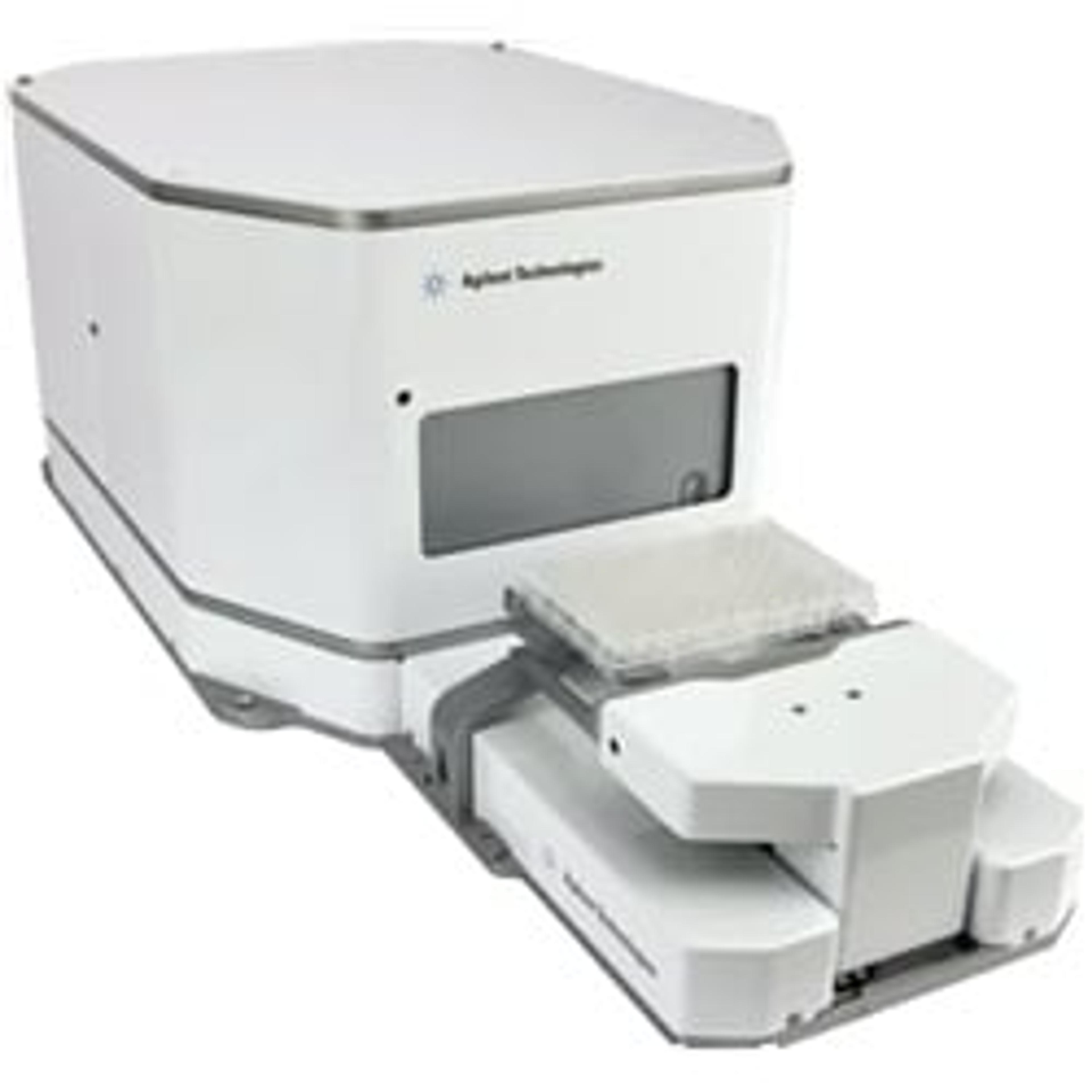
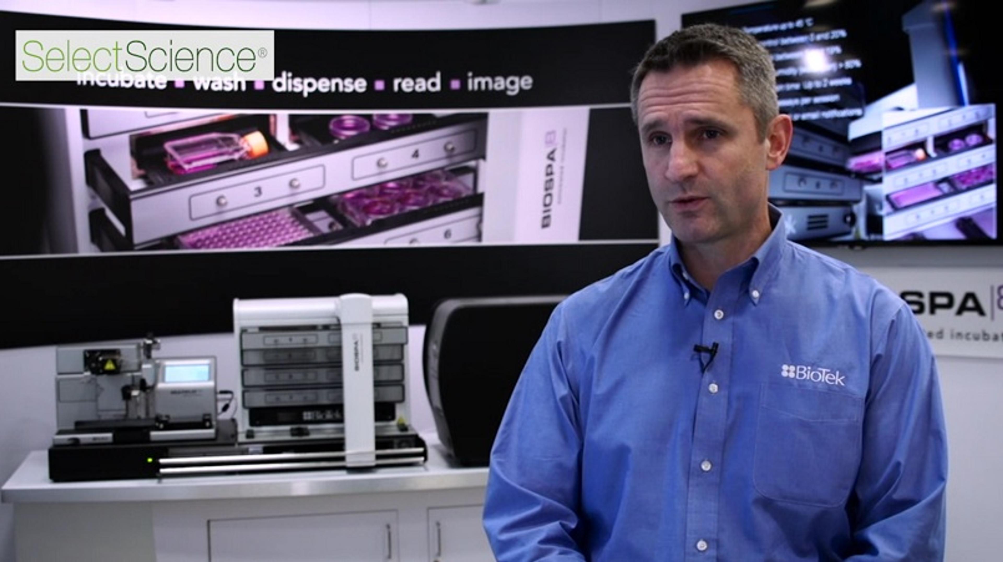
Integrated Live Cell Imaging Solutions from BioTek Instruments, Inc.
Watch this video to discover how BioTek Instruments, Inc. could enhance your cell imaging workflow. Learn how the Cytation™ 5 Cell Imaging Multi-Mode Reader, combined with Gen™ 5 Data Collection & Analysis Software and the BioSpa™ 8 Automated Incubator facilitate live cell imaging automation, with versatile assay detection, optional imaging modes and environmental controls for long-term imaging.
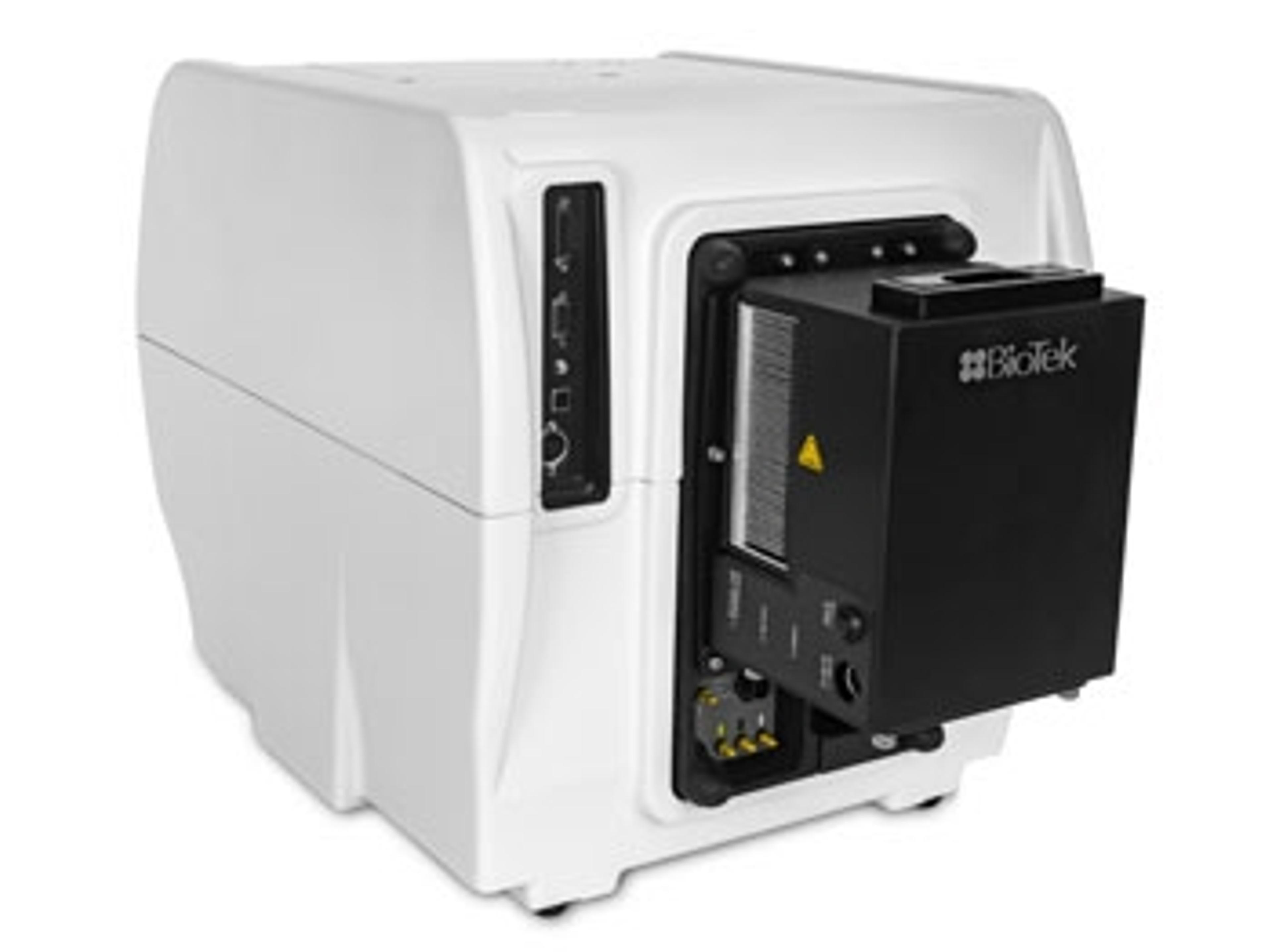
Optimizing Conditions for Temperature Sensitive Assays
New Peltier cooling module by BioTek prevents temperature fluctuations in multimode cell imagers
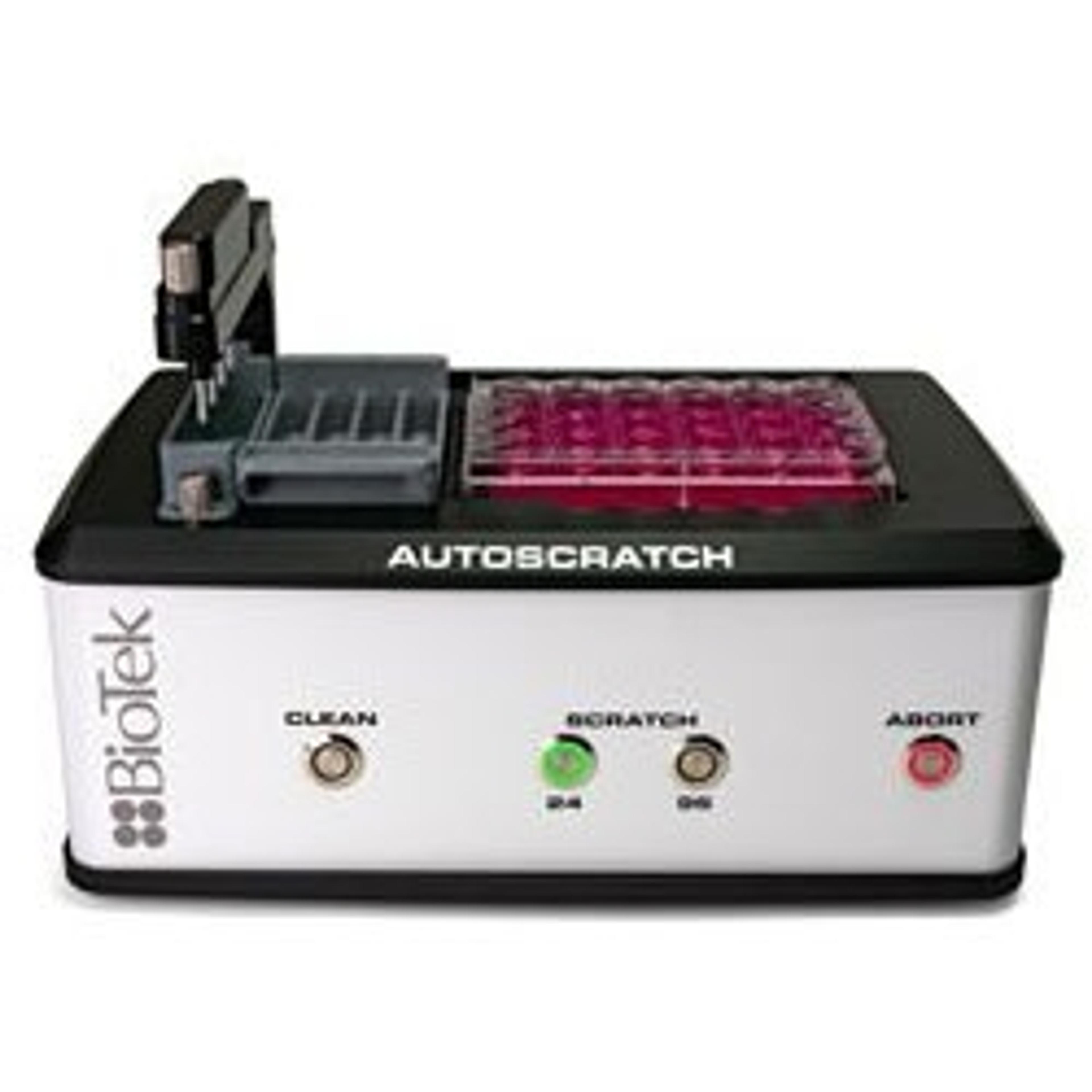
New Wound Making Tool Enhances Reproducibility and Efficiency
AutoScratch tool to facilitate 2D collective cell migration and invasion applications


Advancing Cancer Research Using High Throughput Screening in Cancer Cell Lines
Learn about cutting-edge drug discovery techniques at Southern Methodist University, Dallas
BioTek Introduces New Lionheart™ FX Automated Live Cell Imager
Capturing live cell images and transforming them into intelligent information
Discover the Latest 3D Cell Culture News and Methods
Don't miss these new products and methods