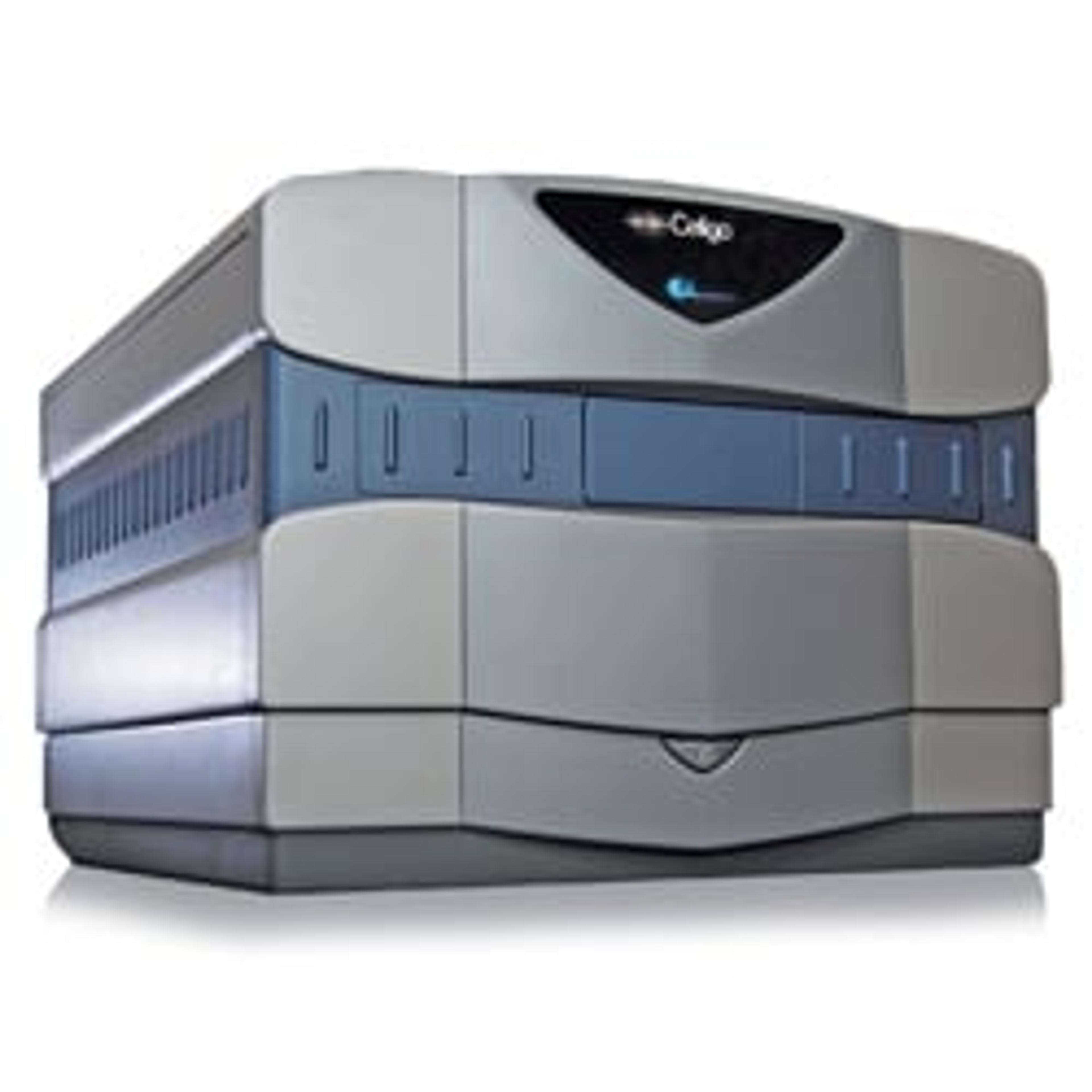
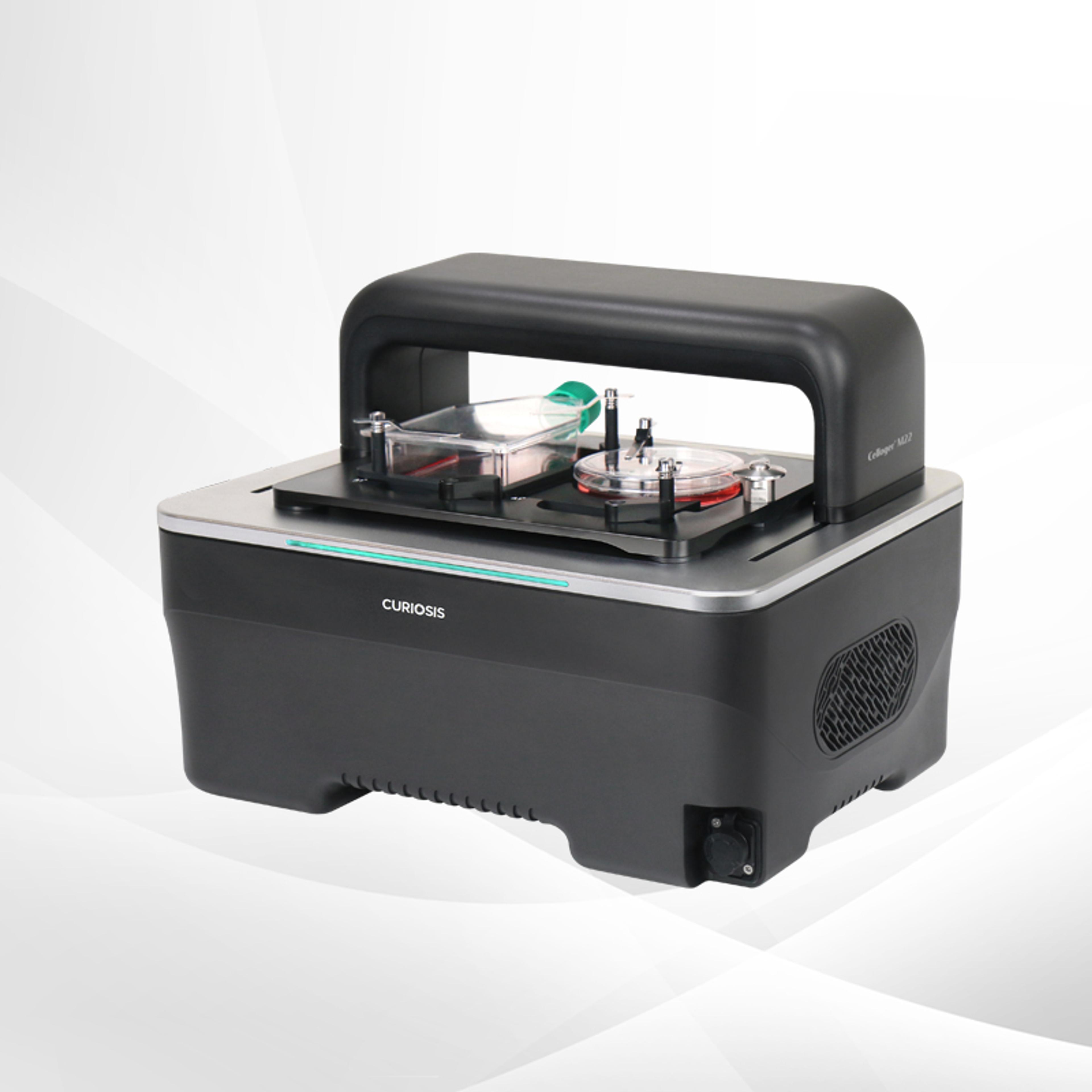
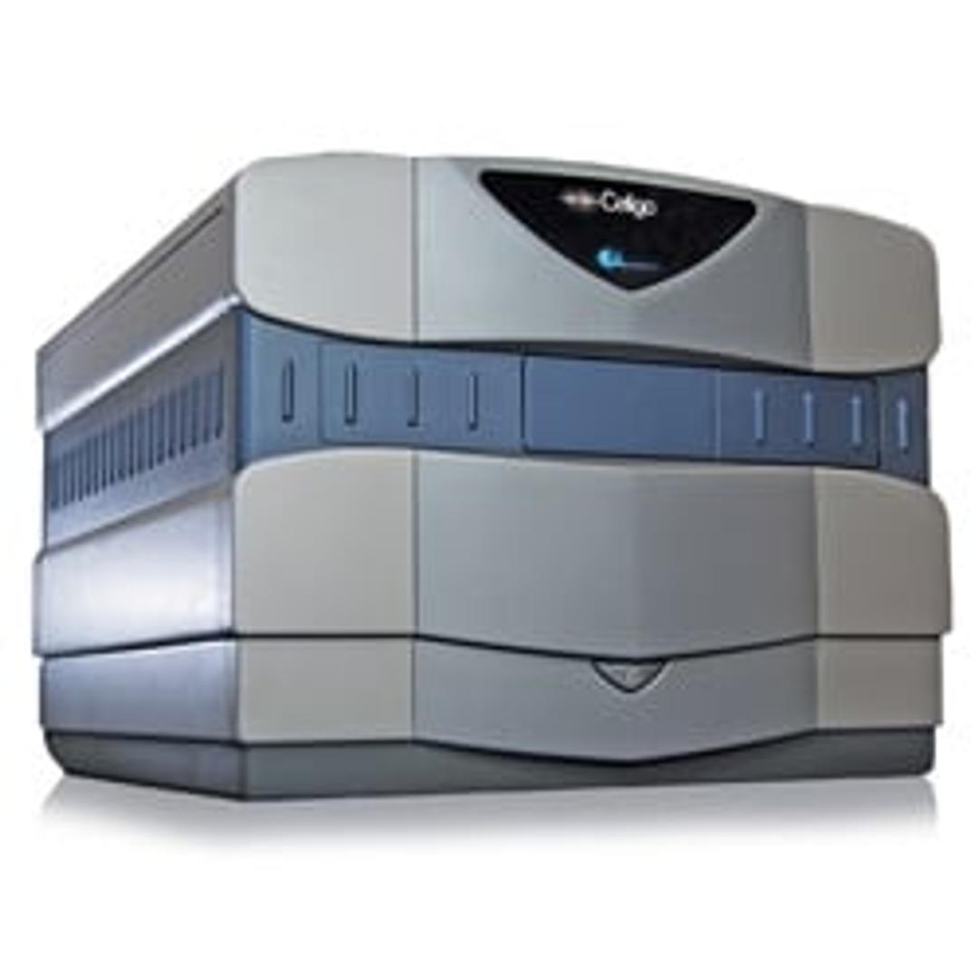
Celigo Image Cytometer - 5 Channel
A micro-well, plate-based multi-channel bright field and 4 fluorescent channels imaging cytometer for 2D and 3D culture using both adherent and suspension cells.

Not just an instrument, but a collaborator.
Immunooncology
My lab utilizes the Celigo Image Cytometer for a variety of in-vitro assays, including cell count and viability, cell migration and cytotoxicity. The after-sales care is excellent, and the reason we have an affinity for the Celigo Image Cytometer. Nexcelom has a great technical team, their assigned technical staff is very knowledgeable and often spends time not only trouble-shooting the occasional technical kinks, but also brainstorming ideas and new applications with us as we come up with new scientific hypotheses and questions.
Review Date: 8 Feb 2021
Great results. It saves us having to do fixed time points.
Cell biology
A very friendly, personal approach and custom-built sales model for us. There was good training provided and the imager performed as expected, allowing brightfield imaging at specific timepoints, without fixation of cells. This did require removal from the incubator at 24hours, 48hours, and 72hours, and scanning for 15mins (384-well plates).
Review Date: 26 Feb 2020
Expensive but it has saved me so much time! Brilliant support.
Immuno-oncology
Love this instrument and has saved me so much time with my assays. I can't thank the Nexcelom support team enough as they have helped me to optimize and set up my experiments which has saved me a lot of time. Thank you!
Review Date: 17 Apr 2019
The Celigo S Image Cytometer offers best-in-class bright field imaging capabilities with 4 channels of fluorescent imaging: Blue (Ex 377nm / Em 470nm), Green (Ex 483nm / Em 536nm), Red (Ex 531nm / Em 629nm) and Far-Red (Ex 628nm / Em 688nm). It is ideal for multiplexed assays and provides high-speed, fully automated imaging and quantification of suspension, adherent, tumor spheroids, iPSC and cancer stem cell colonies. The system images every cell in every well, without any well edge effect. A 96-well plate will take less than 5 minutes to image. System is compatible with automation platforms for full integration.

Rapid, Label-Free Counting and Characterization of Live Embryoid Bodies
This application note explores the benefits of a fast, automated method for embryoid body (EB) analysis. The Celigo™ Colony Counting: Embryoid Body Application by Nexcelom Bioscience quickly and accurately analyzes EB populations to assess the number, size, and shape of live EBs within multi-well plates.
Automated Cell Growth Tracking for Cytotoxicity and Proliferation
The Growth Tracking application automatically integrates label-free cell counts of the same well/flask from different time points to provide direct measurement of growth rates and doubling times, which is also a good overall assessment of cell health.
Monitoring iPSC Reprogramming, Stem Cell Pluripotency and Differentiation
The Celigo adherent cell cytometer, by Nexcelom Biosciences, combines microscopic imaging with flow cytometry to enable both qualitative and quantitative cell analysis. This application note demonstrates the ability of Celigo to monitor iPSCs over time, tracking the reprogramming of multiple colonies in a much faster time that of similar assays.
Automating Cell Imaging and Counting with Celigo
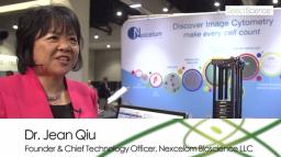
Dr. Jean Qiu, founder and Chief Technology Officer at Nexcelom Bioscience LLC, discusses the new automated imaging and cell counting platform: Celigo. Celigo is an innovative, high-throughput system which enables integrated image and data acquisition from a variety of microplates. High-speed whole well image acquisition coupled with a bright field and 4 florescent channels enables Celigo to perform a multitude of 2D and 3D assays.