
Olympus CKX53 Inverted Microscope for Cell Culture

1 Feb 2016

Lynsey Forsyth
Post Doc / Research Fellow

Product news

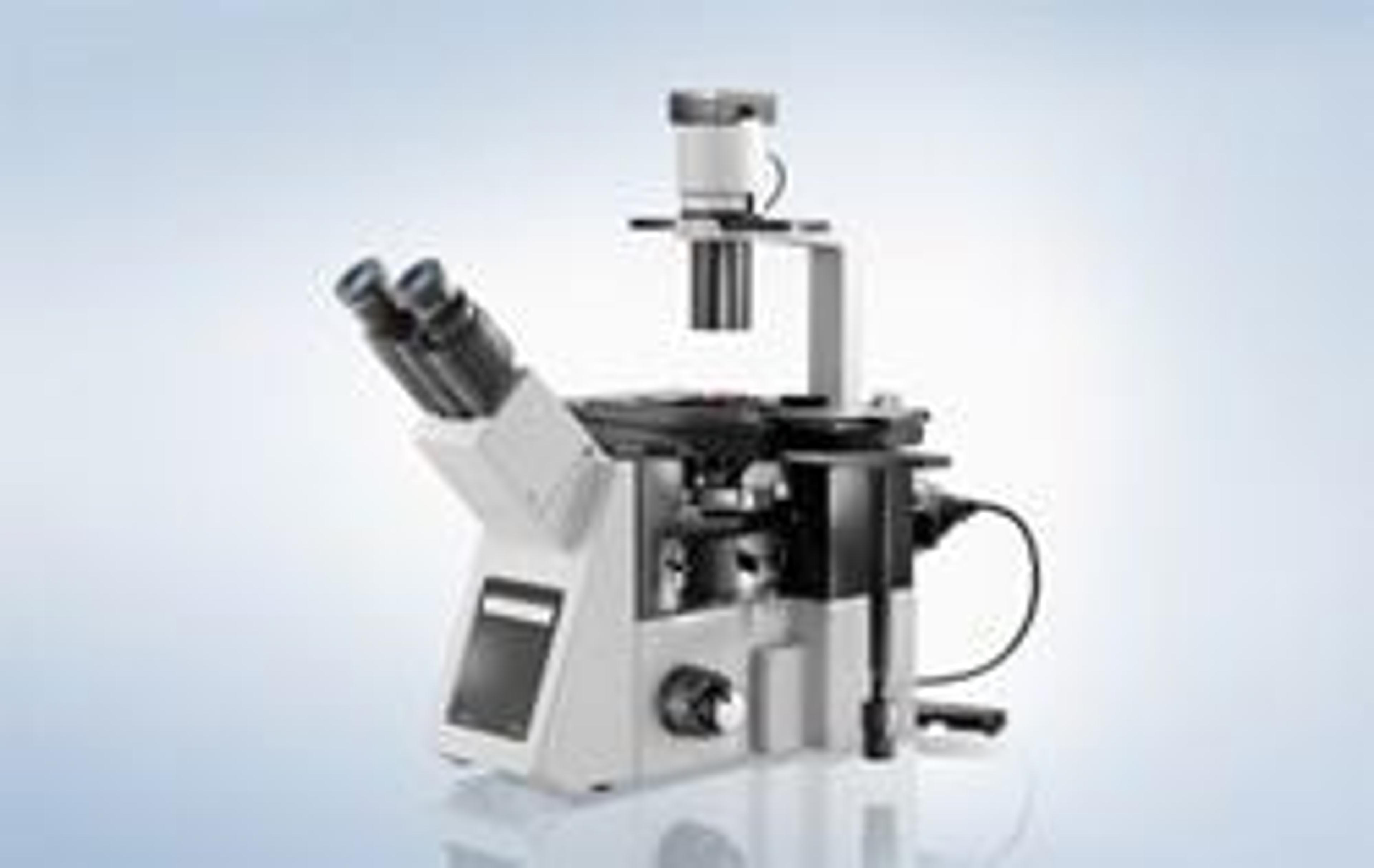
Merging novel phase contrast technologies, high quality imaging and user-friendly handling, the CKX53 is optimised to enhance a variety of cell culture applications – from live cell observation and documentation to cell sampling and handling.

Olympus’ renowned optical expertise has given rise to the new CKX53 microscope, tailored to the cell culture process. At the core of the CKX53 are the superior Olympus UIS2 objectives with a large field number of 22, providing the basis for excellent image quality. Using the 2X objective, observation is possible over areas of up to 11 mm in diameter. This wide field of view saves processing time through allowing quick screening of cell culture samples without the need to move the stage - and is ideal for observing large embryonic and induced pluripotent stem cell colonies in a variety of different microtiter plate formats. Fast processing is vital to minimise the time precious live cells are removed from optimal incubation conditions, and this is further facilitated by the new Olympus unique integrated Phase Contrast (iPC) system. Removing the need to prepare or centre the objectives when moving between 4X and 40X magnification, Phase Contrast observations are fast and efficient with the CKX53. The quality of Phase Contrast observation is also enhanced with the novel Olympus IVC technique, enabling 3D views devoid of halos or directional shadows. In addition to Phase Contrast and brightfield, high contrast fluorescence imaging is possible for multiple signals, utilising the advanced technology of the successful IX3 inverted microscope series.

Further streamlining the cell culture workflow, operational and ergonomic design has been optimised, with different configurations tailored to suit every need. The systems are compact and UV light resistant, and can remain inside the clean bench environment with sliding pads to allow easy one-handed repositioning. Multiple holder types accommodate a range of sample formats and even multi-layer tissue flasks, while significantly higher illumination intensity and uniformity is also achieved with minimal maintenance, thanks to long-life LED illumination.

The CKX53 is the latest addition to the growing Olympus portfolio of cell culture solutions, and complements the new cell counter model R1, which simplifies and shortens the cell counting process to just 15 seconds. These advanced solutions combined with Olympus’ expertise strengthens support to drive cell culture applications, ranging from regenerative medicine to drug development.

Key Features:

Quality UIS2 optics with FN22 provides a comprehensive view
Brightfield, contrast and fluorescence imaging capabilities
Fast and efficient Phase Contrast with iPC
Novel IVC optical technique
Small, ergonomic workflow-optimised design
Bright LED illumination requires minimal upkeep

Relevant Applications:

Cell cultivation
Stem cell research
Life science research
Regenerative medicine
Drug development
Links

Tags

Cell / Tissue CultureCell culture or tissue culture is used to study the biology of cells or tissues and to isolate cellular products in an environment which can be manipulated and well defined. Accurately control your culture environment with bioreactors or culture incubators, bind your cells to a surface or together with an extracellular matrix. Distinguish cell types with differential media or proliferate cells with certain characteristics using selective media. Enrich your media with supplements such as growth factors, sera and vitamins. Find the best cell and tissue culture products, kits and equipment in our peer-reviewed product directory: compare products, check customer reviews and receive pricing direct from manufacturers.Cell-Based AssaysCell-based assays are used to monitor the presence, quantity and activities of a desired cellular analyte including drug molecules or biomarkers. This can reveal information on cell health (apoptosis, cytotoxicity, viability and proliferation assays), cell metabolism, cell migration and cell signaling mechanisms. Find the best cell-based assay products, kits and equipment with our peer reviewed product directory: compare products, check customer reviews and receiving pricing direct from manufacturers.Light MicroscopyLight microscopes or optical microscopes are used to visualize microscale objects under magnification, including cells, clinical specimens and materials. Lab equipment for light microscopy includes confocal microscopes, fluorescence microscopes, zoom and stereo microscopes. Microscope slides and imaging reagents are available for visualizing samples, as well as various microscope stages and incubators for large or temperature-sensitive samples. Find the best light microscopes in our peer-reviewed product directory: compare products, check customer reviews and receive pricing direct from manufacturers.In VitroIn vitro refers to experiments conducted outside living organisms, often in controlled lab environments such as petri dishes or test tubes. In vitro models are widely used in drug testing, cell biology, and disease research. Explore in vitro research tools in our peer-reviewed product directory; compare products, check reviews, and get pricing directly from manufacturers.Cell ImagingCell imaging can be achieved using a number of techniques including confocal microscopy, transmission electron microscopy, atomic force microscopy, and light sheet microscopy.Fluorescence MicroscopyFluorescence microscopy has become an essential tool in biology, as well as in materials science. The application of many fluorochromes has made it possible to identify cells and sub-microscopic cellular components with a high degree of specificity. Using multiple fluorescence labels, different probes can simultaneously identify several target molecules.Live Cell ImagingLive cell imaging is the study of living cells using microscopes and high-content imaging systems. This technique provides in-depth insight into fast and complex biological processes, by allowing dynamic imaging of living cells instead of acquiring an individual image at a single point in time.MicroscopyMicroscopy is a technique used to observe small objects in detail, from cells to materials, using light or electron microscopes. It enables researchers to examine structures with high resolution, aiding in fields such as biology, medicine, and materials science. With advanced microscopy techniques, scientists can gain insights into cellular processes, tissue structures, and material properties. Explore the best microscopy solutions in our peer-reviewed product directory, compare products, read customer reviews, and get pricing directly from manufacturers.Cell BiologyCell biology studies the structure, function, and behavior of cells. Understanding cellular processes is essential for research in areas such as cancer biology, stem cell research, and neurobiology. Techniques like flow cytometry, microscopy, and cell culture enable researchers to explore cellular mechanisms in detail. Browse our peer-reviewed product directory to find the best cell biology tools and equipment, compare products, check customer reviews, and get pricing directly from manufacturers.