Identification and Characterization of Cancer Risk Factors Using High Throughput Cell Screening

Dr Kirk McManus describes how 3D cell culture is advancing his cancer research

16 Jun 2016
Lynsey Forsyth
Post Doc / Research Fellow

Editorial article

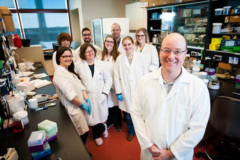
The McManus Lab Image: Courtesy of Dr Kirk McManus

Dr Kirk McManus, Associate Professor in the Department of Biochemistry & Medical Genetics at the University of Manitoba and a Senior Scientist within the Research Institute in Oncology at CancerCare Manitoba, spoke to SelectScience® about his cutting-edge cancer research, which aims to identify cancer causing genetic abnormalities and screen potential therapies.

Dr McManus’ research is focused on two main areas:


Integration of the BioSpa™ 8 enabled us to do more long-term, live cell imaging, … which provides a lot more information than standard fixed end-point analysis.

Dr. Kirk McManus  University of Manitoba


The identification of aberrant genes that can predispose to cancer development or contribute to its progression

Screening putative cancer therapies that can exploit these genetic defects

The team use a variety of cell based assays and imaging methodologies to identify and investigate genetic risk factors that cause genome instability and predispose carriers to the development of cancer, as well as testing potential modifiers for therapeutic potential.

Chromosome instability and cancer therapy

Mutations that cause genome instability, are known risk factors for the development of several cancers, including colorectal, ovarian and breast. Dr McManus’ research group is working to identify and characterize genetic mutations that predispose to chromosome instability. Such genes are often associated with DNA repair mechanisms and, if silenced or mutated, can result in abnormal chromosome number, a feature associated with tumor growth and development and poor patient prognosis. Synthetic lethal interactors are modulators able to exploit the defects in these risk genes that regulate chromosome instability and are putative therapeutic targets for killing cancer cells.

Screening cells for chromosome instability

Dr McManus and his team have identified genes associated with chromosome instability using a cell based screen. Multiplex, high content assay screening of cells is performed using the Cytation 3 Cell Imaging Multi-Mode Reader from BioTek Instruments Inc. Cell phenotypes of abnormal chromosome number, such as enlargement of the nuclear area or formation of extranuclear micronuclei, can be examined using this imaging system. The team is now using fluorescence microscopy to validate a subset of the putative risk genes and to investigate cellular mechanisms of chromosome instability. The system is also being used to screen candidate synthetic lethal interactors by imaging changes in cell numbers in response to the putative gene modulators.

3D tumor models

Dr McManus’ lab has integrated BioTek Instruments Inc., EL406 Combination Washer Dispenser, BioSpa™ 8 Automated Incubator and Cytation 3 Cell Imaging Multi-Mode Reader, for complete cell culture workflow that allows seeding, maintenance and imaging of cells. In previous studies, cells were fixed for end-point analyses but with the addition of the BioSpa™ 8 Automated Incubator, long-term, live cell imaging analyses of 3D spheroid cell cultures can be performed.

The team is now using the integrated system to screen modulators of genes associated with chromosome instability in 3D tumor spheroids. These 3D spheroid cultures are more biologically relevant than traditional 2D cell cultures and also far more cost effective than animal models, so are ideal for identifying therapeutic candidates for further investigation. Using BioTek’s integrated systems, the team are able to observe formation and growth of tumor spheroids, as well as monitor real-time dynamics of the spheroids, in response to different effector molecules. These data are more physiologically relevant and informative than fixed cell end-point analyses.

Future work

Dr McManus now aims to examine the effects of silencing the previously identified risk genes, to investigate underlying mechanisms that give rise to chromosome instability. In another clinical collaboration, tumor cells isolated from ovarian cancer patients will be used to generate 3D spheroid cultures, which will enable chromosome instability and drug interventions to be examined in real cancer cells. The findings from these studies are not only relevant to ovarian cancer but will be applicable to other cancer types, including colorectal and breast.

Visit the website to find out more about the McManus Lab.

Do you use any BioTek Instruments Inc. products? Tell us what you think, leave a review today.

Links

Tags

Cell / Tissue CultureCell culture or tissue culture is used to study the biology of cells or tissues and to isolate cellular products in an environment which can be manipulated and well defined. Accurately control your culture environment with bioreactors or culture incubators, bind your cells to a surface or together with an extracellular matrix. Distinguish cell types with differential media or proliferate cells with certain characteristics using selective media. Enrich your media with supplements such as growth factors, sera and vitamins. Find the best cell and tissue culture products, kits and equipment in our peer-reviewed product directory: compare products, check customer reviews and receive pricing direct from manufacturers.MicroplatesMicroplates are multi-well plates used to increase the throughput of biological investigations. The number of wells microplates contain can range from 6 to 3243 wells, with the 96-well format being the most commonly used. Microplates can come tailored for a range of applications including cell culture, PCR, filtration, storage, non-binding surface, protein crystallization, as well as pre-coated, deep well and normal Standard microplates. Additionally, options for microplate colors include clear , black, white or black with clear bottom and white with clear bottom for absorbance microplate reader applications. Find the best microplates for your lab in our peer-reviewed product directory: compare products, check customer reviews and receive pricing direct from manufacturers.High-Throughput ScreeningHigh-throughput screening (HTS) is an automated drug discovery technique for identification of active compounds against a compound library. Use HTS readers and integrated assay preparation / analysis workstations to screen your compounds. Identify active compounds against various HTS libraries, including membranes, proteins and peptides and HTS cell lines. Find the best high-throughput screening products in our peer-reviewed product directory: compare products, check customer reviews and receive pricing direct from manufacturers.Cell Lines Stem Cells and Primary CellsPrimary cell cultures, established cell lines and stem cells are vital for <i>in vitro</i> and <i>ex vivo</i> experimentation. High-quality cells, optimized for your applications, alongside optimized cell substrates, growth medium and supplements, are critical for experimental success. Explore a range of cells suitable for your applications, including isogenic cell lines, competent cells, induced pluripotent stem cell (iPSC)-derived cell lines, fungal/bacterial/mammalian cell lines, stem cells and cancer cell lines. Find the best cells for your research in our peer-reviewed product directory: compare products, check customer reviews and receive pricing direct from manufacturers.Microplate Readers / DetectorsMicroplate readers are used to automate the detection and analysis of labeled or label-free components in microplates during assays or live-cell monitoring. Microplate readers are generally distinguished by their mode of detection. Types include absorbance, luminescence, fluorescence intensity, fluorescence polarization, TRF / FRET and multimode microplate readers. Microplate readers deliver a high throughput of samples by reading multiple wells simultaneously, with the 96-well format the most commonly used. As a result, microplate readers are often used in the drug discovery, bioassays, research and pharmaceutical industries for screening applications. Microplate loading can also be automated, with robotic microplate stackers to increase throughput. Find the best microplate readers in our peer-reviewed product directory: compare products, check customer reviews and receive pricing direct from manufacturers.Cell-Based AssaysCell-based assays are used to monitor the presence, quantity and activities of a desired cellular analyte including drug molecules or biomarkers. This can reveal information on cell health (apoptosis, cytotoxicity, viability and proliferation assays), cell metabolism, cell migration and cell signaling mechanisms. Find the best cell-based assay products, kits and equipment with our peer reviewed product directory: compare products, check customer reviews and receiving pricing direct from manufacturers.High-Content ScreeningHigh-content screening (HCS), also known as high-content analysis (HCA), is a high-throughput technique used in drug discovery to identify substances that alter the phenotype of cells. HCS uses fluorescent microscopic imaging and automated image analysis to investigate cellular events such as apoptosis, cell viability, GPCR activation, oxide production, neurite outgrowth, and cell signaling. Find the best fluorescent labeling reagents, cellular assays, and high-content imaging systems in our peer-reviewed product directory: compare products, check customer reviews and receive pricing direct from manufacturers.BiomarkersBiomarkers are biological markers which can be measured and evaluated to indicate a biological state. The use of biomarkers in research and diagnosis can indicate a normal or disease state or drug response of cells / tissues. Biomarkers include genetic markers, cell surface markers such as antigens, antibodies or receptors and secreted molecules such as cytokines. An assay system is required for identification of biomarkers. :Plate ProcessingAutomated plate processing is used in microplate technologies for high-throughput, speed and efficiency. Plate processing systems include microplate handlers for moving plates, heat sealers, stackers and carousels for storage, retrieval and delivery, microplate washers and piercers. Useful features of plate processors include walk-away automation, compatibility and easy integration with microplate systems.Light MicroscopyLight microscopes or optical microscopes are used to visualize microscale objects under magnification, including cells, clinical specimens and materials. Lab equipment for light microscopy includes confocal microscopes, fluorescence microscopes, zoom and stereo microscopes. Microscope slides and imaging reagents are available for visualizing samples, as well as various microscope stages and incubators for large or temperature-sensitive samples. Find the best light microscopes in our peer-reviewed product directory: compare products, check customer reviews and receive pricing direct from manufacturers.Live Cell ImagingLive cell imaging is the study of living cells using microscopes and high-content imaging systems. This technique provides in-depth insight into fast and complex biological processes, by allowing dynamic imaging of living cells instead of acquiring an individual image at a single point in time.Drug Discovery & Development ScreeningCell Based AutomationCancer ResearchAlthough cancer is often referred to as a single condition, it actually consists of more than 100 different diseases. Microscopy, mass spectrometry, high throughput sequencing and flow cytometry are some of the most common techniques employed in cancer research labs.