Mass Spec Imaging Enables Greater Understanding of Tumors Than Ever Before
20 Feb 2018
Hear how scientists at the National Centre of Excellence in Mass Spectrometry Imaging (NiCE-MSI), National Physical Laboratory, UK, are using a combination of different mass spectrometry techniques to address the Cancer Research UK Grand Challenge to map tumors at a molecular and cellular level. This research will enable the mapping of tumor metabolism in far more detail than ever before and will help to advance both the diagnosis of cancer and the development of new therapies.
About the company

SelectScience
Established in July 1998, SelectScience is the fastest way to impartial, expert opinion about the best laboratory equipment and latest techniques.
Related products
Request Quote for All Products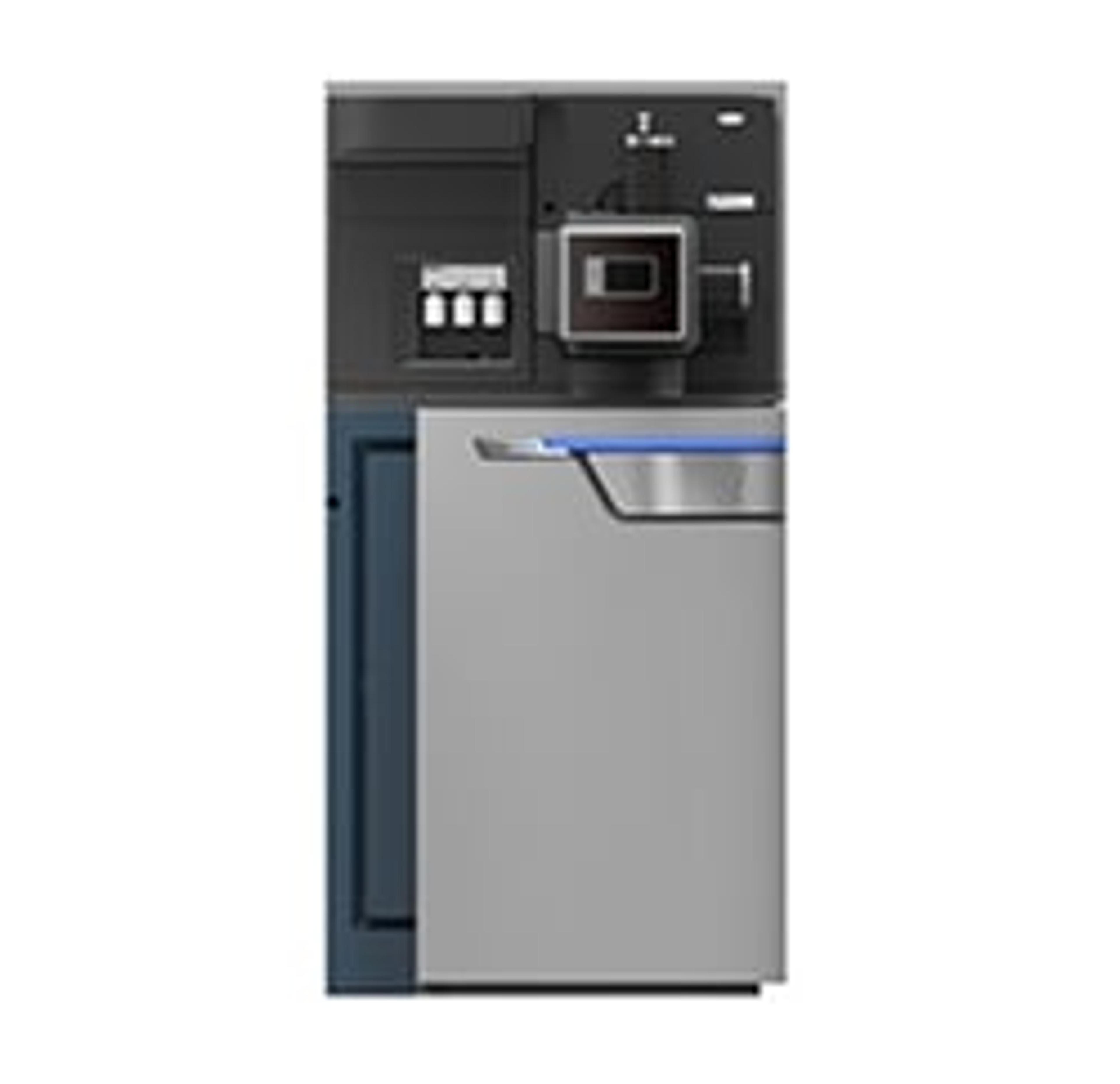
SYNAPT G2-Si HDMS
WatersThe Waters SYNAPT G2-Si High Definition Mass Spectrometry provides complete characterization of complex mixtures and molecules with unique levels of MS performance, industry leading informatics and unparalleled platform versatility.Through high-efficiency T-Wave™ ion mobility you can get superior separation, enhance the peak capacity, specificity and sensitivity of your analysis and transform your targeted and untargeted workflows. Ultimate UPLC/MS/MS performance, data independent and data dependant T-Wave IMS solution, CID and ETD fragmentation capabilities and a wide range of experimental options. This system gives you additional dimension of separation, based on molecular size and shape. It delivers a third dimension to your analysis, proven to transform your analytical perspective whatever the application.