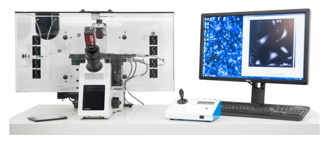
VL21 Live Cell Imaging
The Phasefocus Virtual Lens® provides non-toxic phenotypic screening for live cells, enabling long-term time-lapse assays without the need for fluorescent labels. This is an important consideration for the screening of sensitive primary cells, and stem cells.


The supplier does not provide quotations for this product through SelectScience. You can search for similar products in our Product Directory.
Exploring the use of this equipment to make novel discoveries in our research
Label-free Live Cell Imaging of Primary Prostate Epithelial Cells
We have been using the Phasefocus VL21 to carry out label-free live cell imaging of primary prostate epithelial cells. We are interested in characterising cellular heterogeneity within our cultures as well as analysing response to drug treatment. There is a dedicated team at Phasefocus that are very approachable and helpful and committed to providing customers with what they need to answer their biological questions. There is a learning curve when starting to use the equipment, but this may be because I am a novice in the live cell imaging field. There are many options to choose in terms of the type of data that can be generated by the bespoke software. With every cell being a single data point, and with several days of imaging to analyse, the amount of data that can be generated is astounding. The key is the ability to generate both morphological data about each cell (volume, size, sphericity etc) as well as kinetic data (e.g. cell speed, meandering index). Significantly, all this data is generated without any fluorescent labelling required. This equipment could be used in two ways; (1) if in a core facility, individuals could get a lot of unique information from a few strategic experiments. (2) whole projects could be dedicated to the use of the equipment to explore a model or a system in more depth. We have only begun to explore the potential of this equipment in our own research and we look forward to continuing to do so.
Review Date: 3 Feb 2017 | Phasefocus
There is great scope for devices such as the VL-21 from phasefocus in high content.
Stem cells
Quantitative Phase Imaging is an exciting technology that carries several benefits compared to traditional live cell imaging. First, the cells are exposed to very little light and thus phototoxicity; Second, a virtual zero background facilitates segmentation; Third, increased content in the form of information on dry mass and its distribution within the cell can be exploited to improve tracking and potentially used to answer defined biological questions. Phasefocus has given us the chance to use their device and we are in the process of completing its evaluation and very impressed by its potential. The company has been extremely supportive to us and we are currently hoping to be able to raise resources to acquire this instrument or the more recently launched Livecyte.
Review Date: 9 Dec 2016 | Phasefocus
there is great scope for devices such as the VL-21 from phasefocus in high content
stem cells
Quantitative Phase Imaging is an exciting technology that carries several benefits compared to traditional live cell imaging. First, the cells are exposed to very little light and thus phototoxicity.;Second, a virtual zero background facilitates segmentation; Third, increased content in the form of information on dry mass and its distribution within the cell can be exploited to improve tracking and potentially used to answer defined biological questions. Phasefocus has given us the chance to use their device and we are in the process of completing its evaluation and very impressed by its potential. The company has been extremely supportive to us and we are currently hoping to be able to raise resources to acquire this instrument or the more recently launched Livecyte.
Review Date: 6 Dec 2016 | Phasefocus
Already known for its extreme ease of use, the Phasefocus Virtual Lens enables robust long-term time-lapse assays of unlabelled cells (including sensitive primary and stem cells) with minimal interference to the cells or growth conditions.
Individual cells can be tracked over periods of many days, producing multi-parametric quantitative data for every cell at each time-point. The Virtual Lens greatly simplifies cell-based assays by removing the need to label, fix or harvest cells - they are monitored fully automatically in their growth environment.
Virtual Lens Benefits:
- Interphase Prophase Prometaphase Metaphase Anaphase Telophase Cytokinesis Chromosomes doubled Chromosomes align to middle of cell Microtubules attach to centromeres Separated chromosomes pulled apart Microtubules disappear and cell division begins Two daughter cells formed Analyse cell growth and behaviour without labels
- Perform long-term time-lapse assays on sensitive primary and stem cells that previously were only practicable with immortalised cell lines due to the need for labelling.
- Perform fully-automated high resolution measurements on statistically significant numbers of cells, and over multiple fields of view, to produce flow cytometry-type measurements without removing cells from their culture medium.
- No photo-bleaching effects during measurements or inconsistencies due to label variability result in better reproducibility of measurements.
- Save time and reduce variability in results caused by complex experimental protocols associated with labelling, fixing and harvesting of cells.
- Directly measure morphological changes in cells such as volume, thickness or dry mass - enables previously impossible assays such as contraction of primary smooth muscle cells.
- Automatically follow multiple cells individually, through multiple cell divisions, and compare multiple generations of cells to their ancestors.
- Multiplexed data from analysis provide outputs equivalent to multiple assays without the need to repeat experiments, saving time and money.
- Post-acquisition auto-focussing means that you will never suffer from focal drift issues during a time-lapse experiment again.
Quantitative images enable label-free assays, often from a single experiment, including:
- Cell count, confluence and proliferation, at multiple time-points - Allows simple yet robust cell population growth monitoring.
- Toxicitity, cell viability and apoptosis rate - Provides the kinetics of the cytotoxic response rather than just a snapshot like many colourimetric endpoint assays.
- Mitotic index vs time - Measurement at multiple time-points allows identification of changes in proliferation as a population evolves.
- Morphology such as sphericity, nucleus-cytoplasm ratio - Highlights changes in cell behaviour such as formation of neuronal processes.
- Cell motility, including scratch-wound assays - Robustly measures cell migration parameters.

Dynamic angiogenesis assays
In this application note, Phasefocus demonstrates the ability of the Phasefocus VL21 to perform dynamic angiogenesis assays.
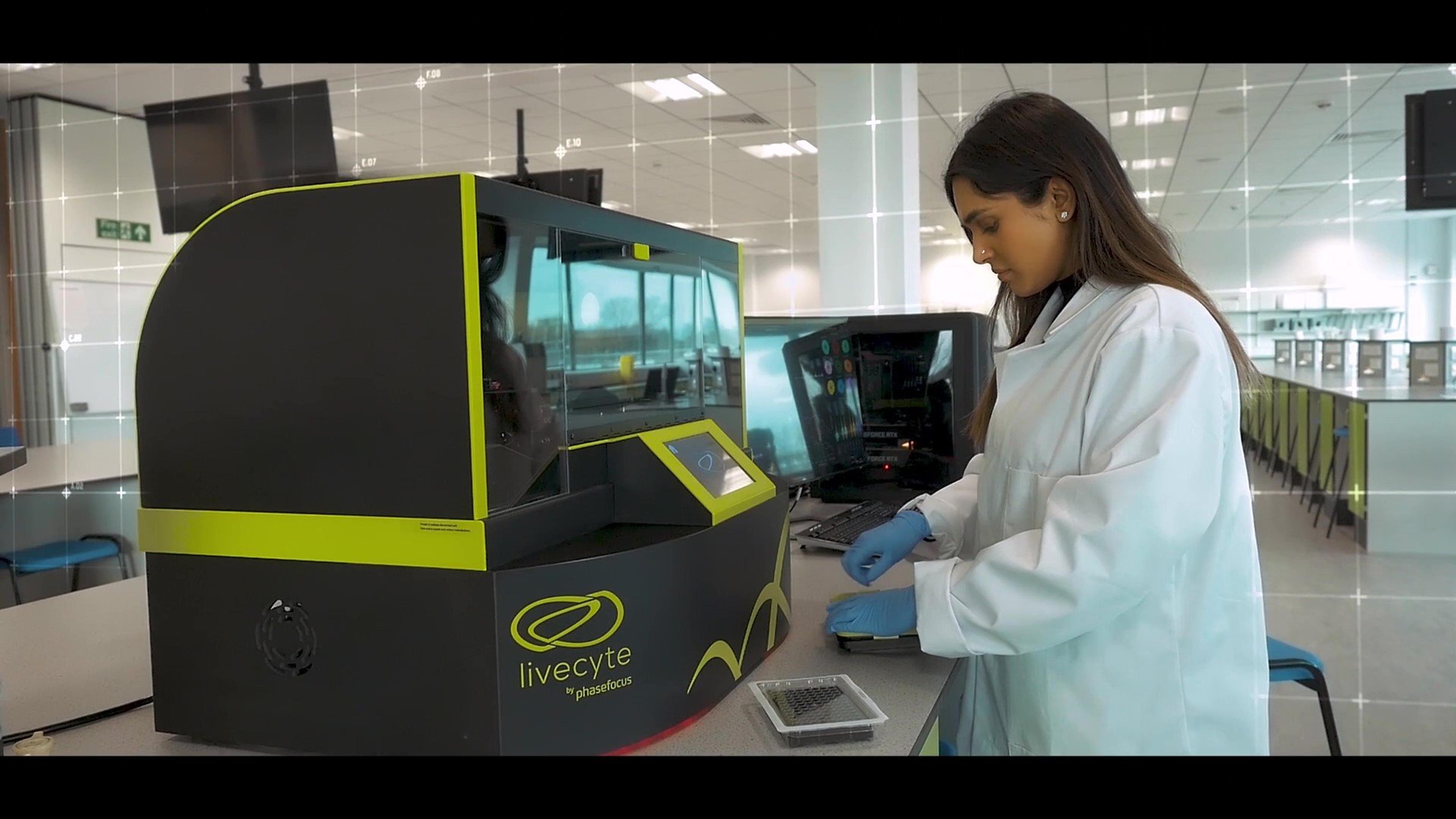
Introducing Livecyte: An automated kinetic cytometer
Livecyte is a kinetic cytometer that undertakes automated cell imaging and analysis. It uses ptychography QPI (Quantative Phasing Imagery) for high resolution and combines images to produce time-lapse movies enabling the study of cell movement. It provides a high level of detail of morphological and dynamic phenotypes while allowing the monitoring of temporal changes. Livecyte has a simple workflow with publication ready outputs that allow movement from plate, to population, to individual cell.