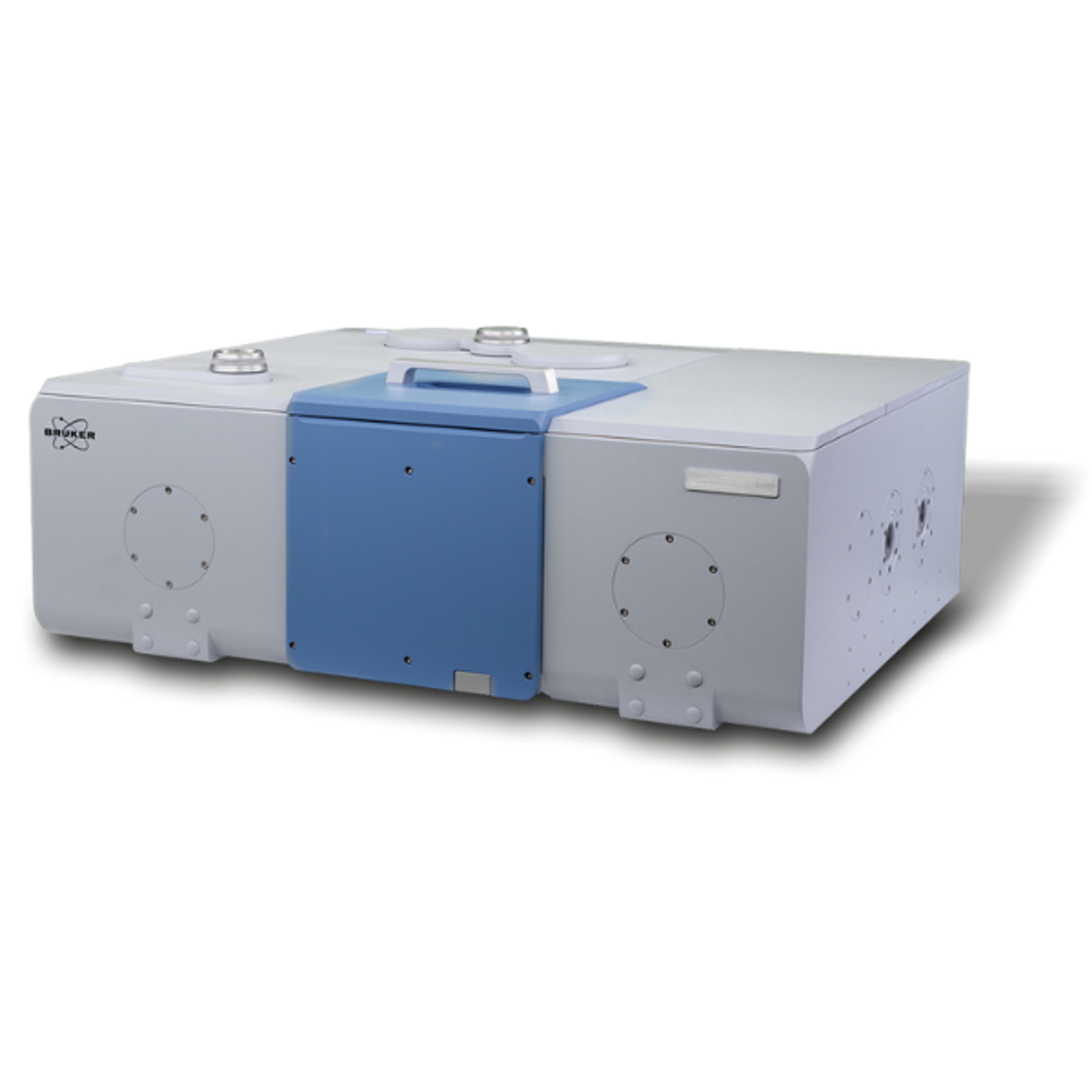
Scanning Probe Microscope Series WITec alpha300
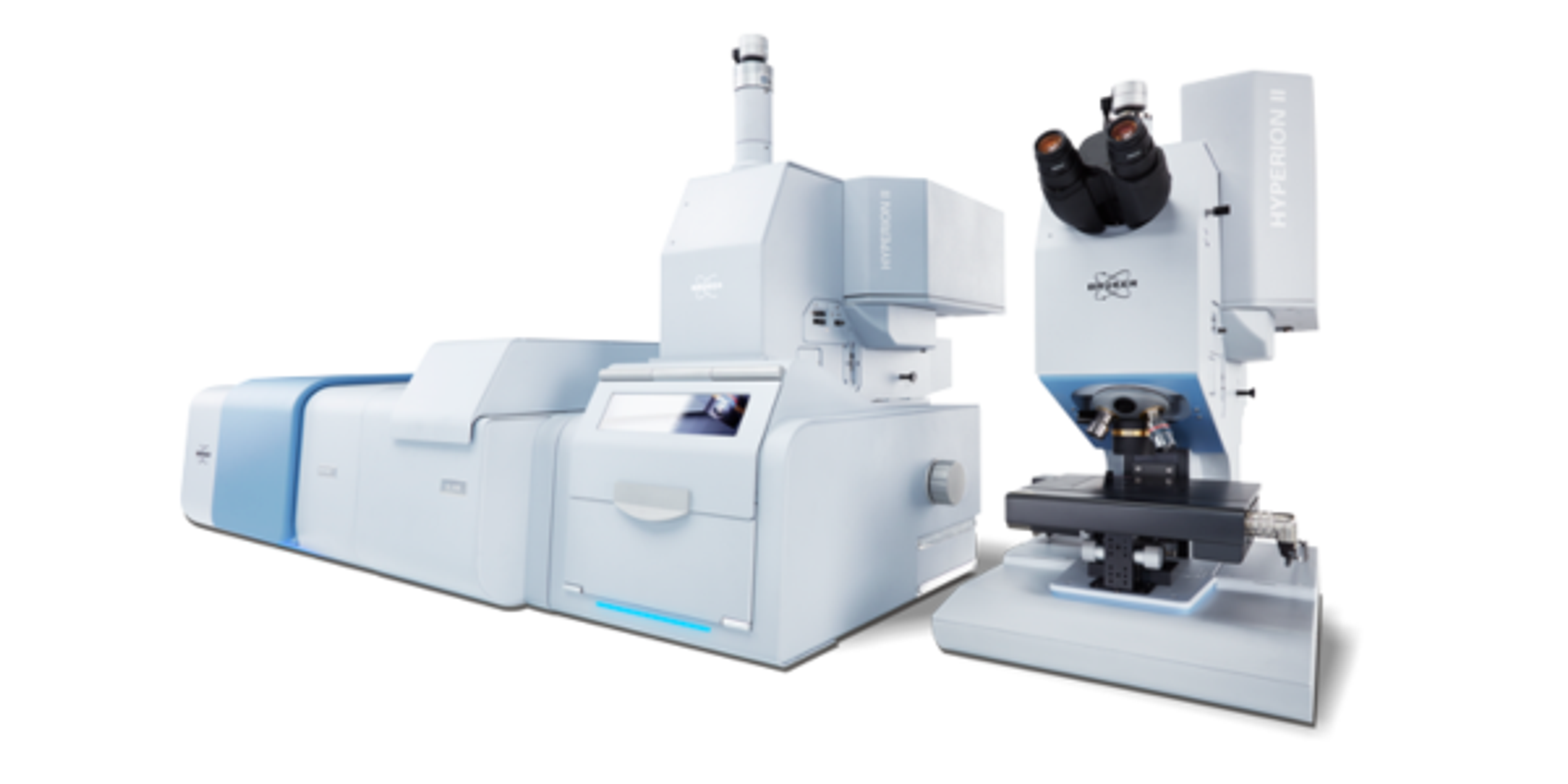
Scanning Probe Microscope Series WITec alpha300 The WITec microscopy series features scanning probe as well as high resolution optical and Raman microscopy techniques in either a single instrument or combined system configurations for the highest flexibility throughout a wide range of microscopy applications. For example, it is possible to start with Confocal Raman Microscopy and upgrade later to Atomic Force Microscopy or vic…


The supplier does not provide quotations for this product through SelectScience. You can search for similar products in our Product Directory.
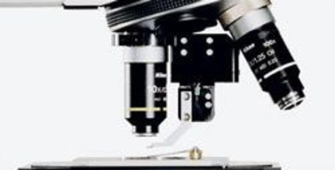
Real confocal system which allows for high-resolution Raman imaging.
Basic science
The alpha300 RS allowed us to obtained high-quality results which were published in such Journals like Analyst, J. Biophotonics or even Chemical Review. The system is for the user with challenging experimental requirements and is perfect for animal cells and tissue studies . It is a real confocal system which allows for high-resolution Raman imaging (in x,y and z).
Review Date: 17 Dec 2018 | Oxford Instruments Raman
Scanning Probe Microscope Series WITec alpha300
The WITec microscopy series features scanning probe as well as high resolution optical and Raman microscopy techniques in either a single instrument or combined system configurations for the highest flexibility throughout a wide range of microscopy applications.
For example, it is possible to start with Confocal Raman Microscopy and upgrade later to Atomic Force Microscopy or vice versa. With such a combined instrument, chemical information can be directly linked to structural AFM information from the same sample area using only one instrument. For high-resolution optical information, the system can even be equipped with SNOM capabilities.
All of these methods enable nondestructive sample analysis on the nanometer scale while requiring only minimal sample preparation, if any. This extensive modularity and ease of use facilitates a more comprehensive understanding of the sample. Typical applications are found in all fields of surface and materials science, geosciences, life science, pharmaceutical research, thin films and coating analysis as well as nanophotonics and nanotechnology.
WITec’s technological leadership and the high-quality design of the alpha300 series pushes the frontiers of high-resolution Raman- and SPM-imaging abilities even further. This will aid our customers as they endeavor to become leaders in their varied fields.

Confocal Raman Imaging, Correlative Raman-SEM, and Atomic Force Microscopy: Geo-Science Applications
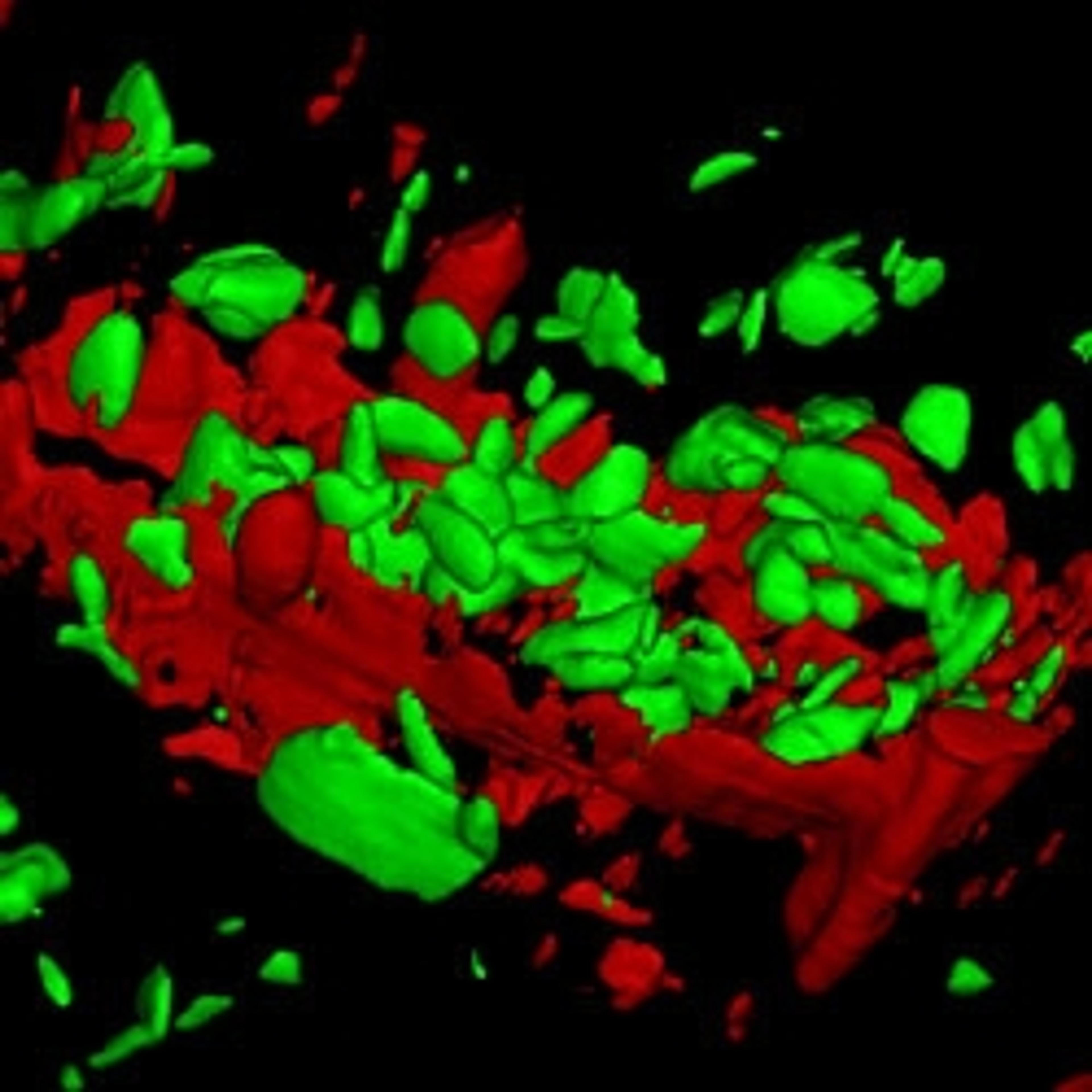
Raman spectroscopy has long been applied in geoscience, for example for the identification and characterization of minerals, or in the observation of mineral phase transitions in high- and ultra-high pressure/ temperature experiments. By means of confocal Raman imaging (CRI), such characteristics can be evaluated from large-area scans on the centimeter scale to the detailed investigations with sub-micron resolution. In this study, WITec confocal Raman microscopes of the alpha300 and alpha500 series were used in order to carry out high-sensitivity measurements on a sample of diorite.
Correlative Confocal Raman Microscopy for 2D Materials Investigation
This application note presents a variety of application of the WITec alpha300 Confocal Raman Microscope Series in material testing and investigation. It covers examples such as analysis of graphene, analysis and imaging of transition metal dichalcogenides and photoluminescence imaging of layers and defects of WS2 crystals. The alpha300 series can carry out advanced confocal Raman imaging with multiple correlative microscopy technique options, including AFM, SNOM, SEM (RISE), fluorescence and photoluminescence.
Imaging of Nano-Carbon Samples with Combined Raman-AFM, SNOM, Nearfield-Raman, and Raman-SEM (RISE)
Graphene shows immense promise in many applications: transistors, sensors, and optoelectronics, to name just a few of them. Flexible and adaptive analyzing methods can support the effective investigation of graphene and accelerate the progress in graphene research and product development. This application note provides an overview of various imaging techniques that can be applied to graphene research.
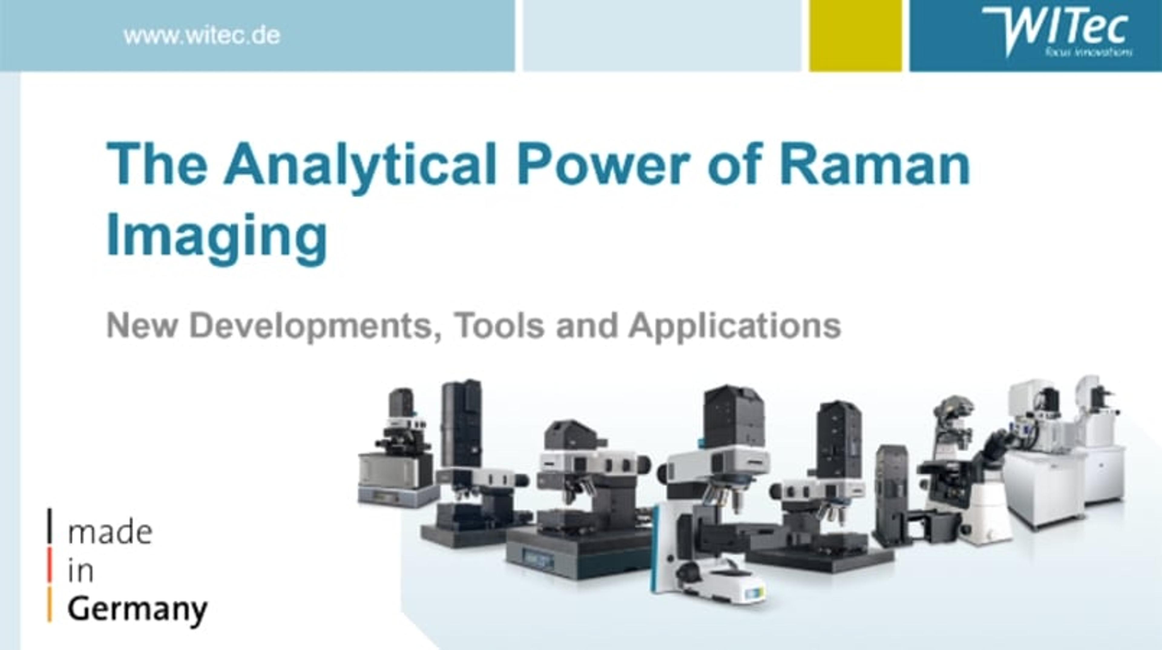
The analytical power of Raman imaging
Watch this presentation by Dr. Miriam Boehmler, Head of the Application and Support Department at WITec GmbH, titled: The analytical power of Raman imaging - new developments, tools, and applications. This talk was presented at the SelectScience® Virtual Analytical Summit 2021.

New Inverted Confocal Raman Microscope
WITec’s proven 3D Raman imaging functionality is now available in an inverted microscope