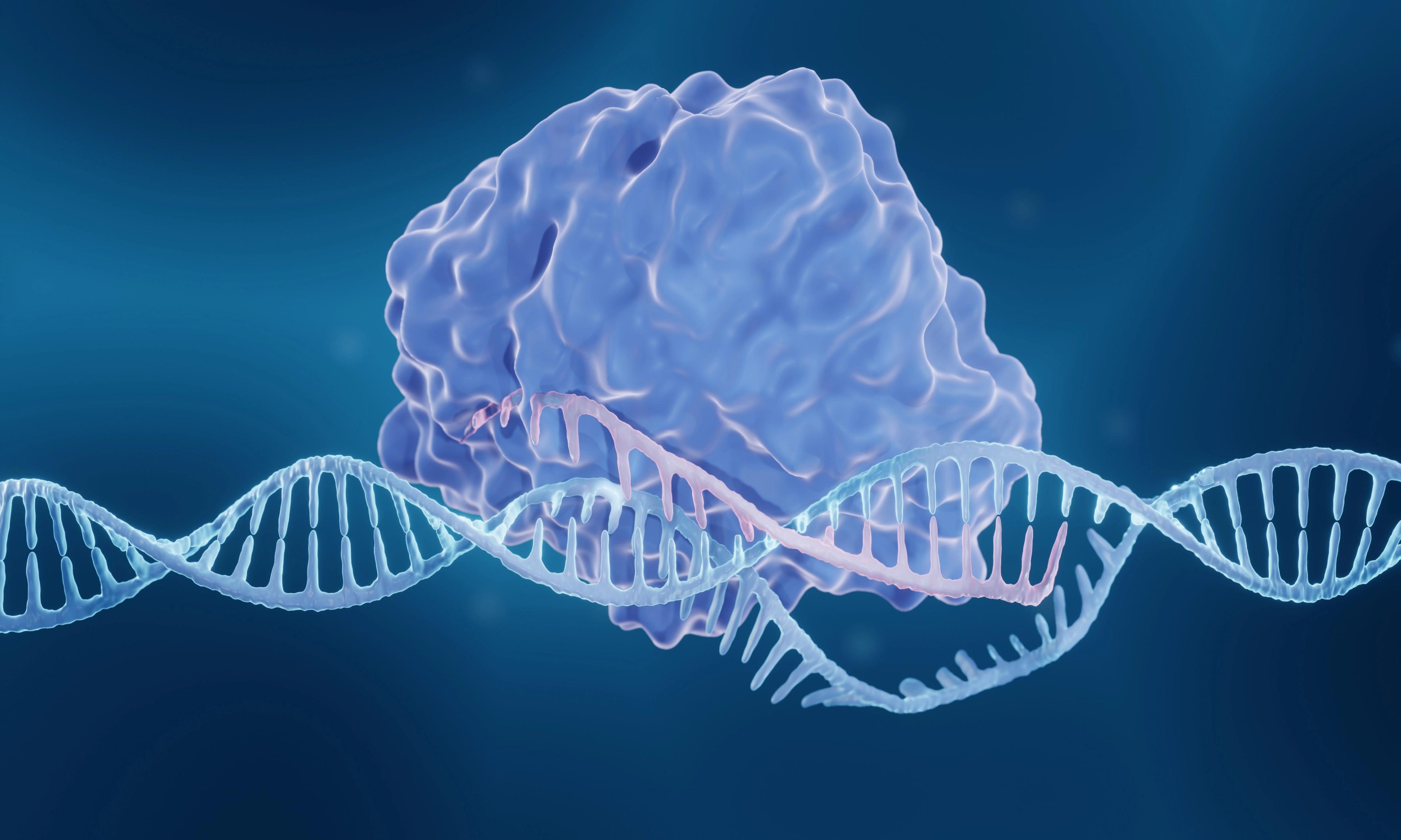
CyTRAK Orange
An orange probe staining both nucleus and cytoplasm.
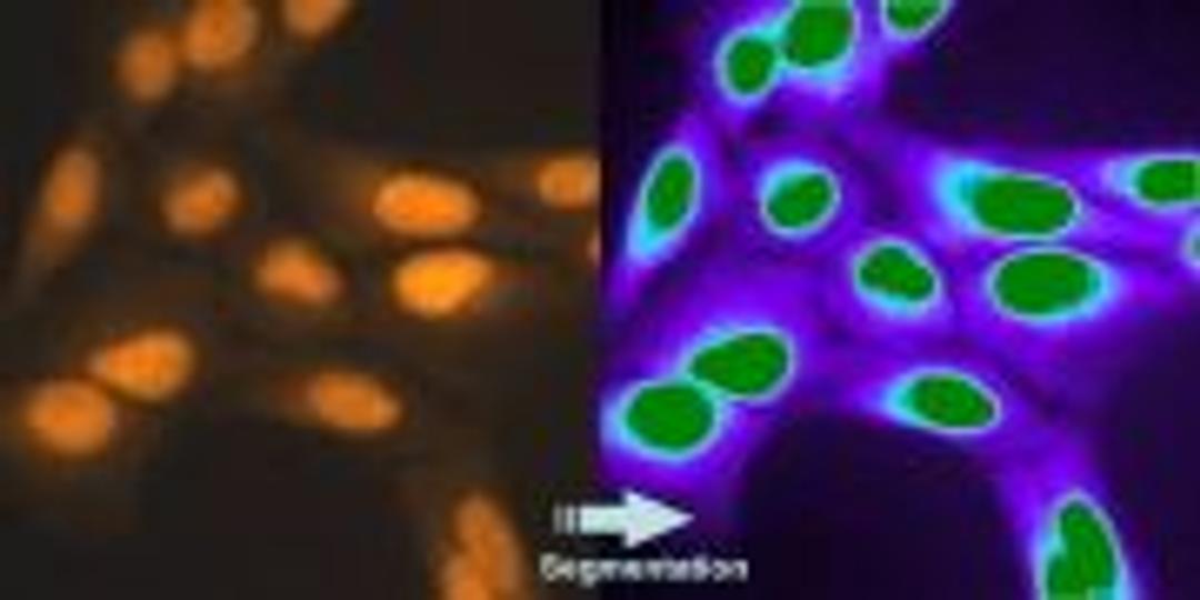
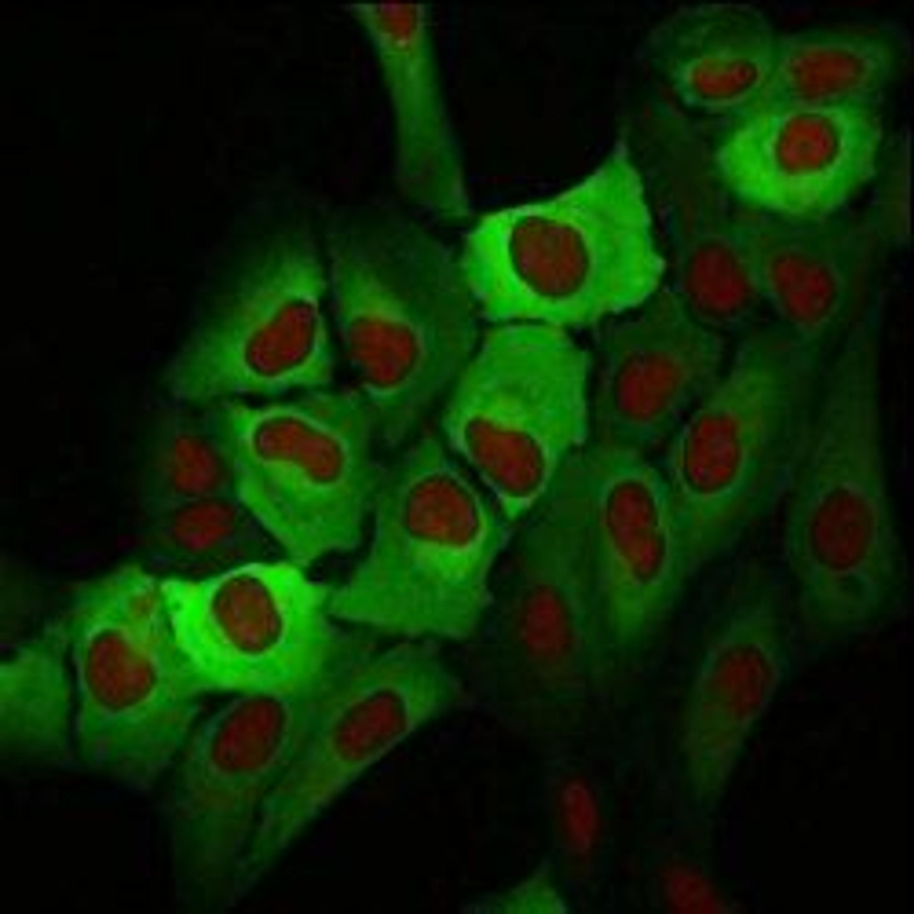
Live cell segmentation


The supplier does not provide quotations for this product through SelectScience. You can search for similar products in our Product Directory.
Very useful stain to interpret your microscopy data.
Visualize nuclei and cell cytoplasm by fluorescent confocal microscopy
Easy to use stain, relatively inexpensive, good stain to localize intracellular organelles and nuclei.
Review Date: 9 Sept 2021 | Biostatus Limited
Nice results.
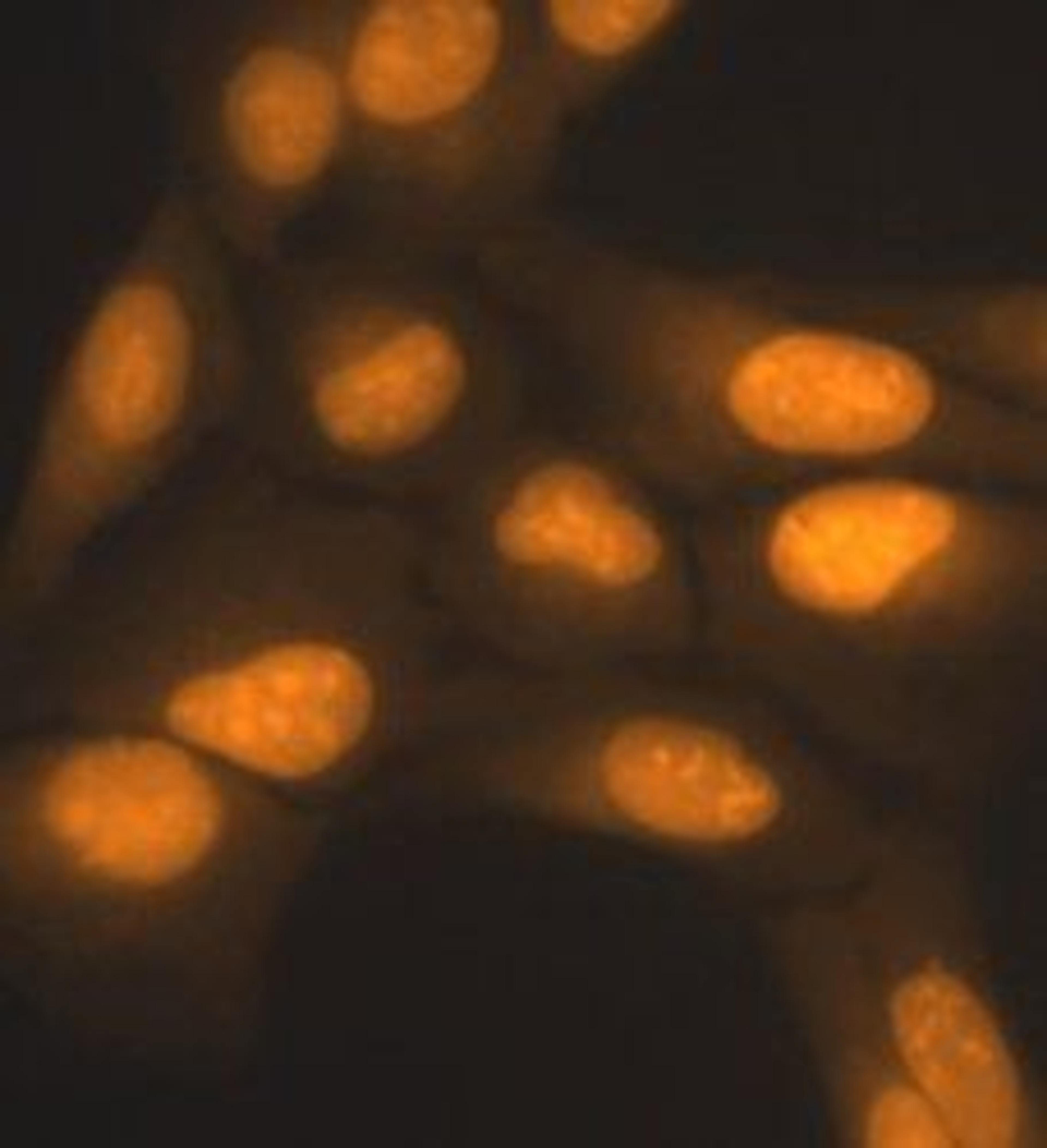
2-photon live-imaging
We used CyTRAK Orange to label nuclei of acute brain slices and performed 2-Photon live-imaging. The labeling is really easy and fast to process. This dye is very efficient to label nuclei in thick tissue (300 µm) and did not show any toxic effect.
Review Date: 18 May 2021 | Biostatus Limited
Manufacturer's Response
Dear Christelle, thank you for this review of CyTRAK Orange! We believe this is the first example of its use in 2-photon imaging and it was impressive to see detection through the full depth of 300 micron brain slices.
A fluorescent dye staining both nucleus and cytoplasm. It is water soluble and membrane permeant and can be used in LIVE cells in combination with other common fluors, especially GFP fusions, FITC-labelled antibodies and far-red dyes.
CyTRAK Orange™ can be used as a LIVE cell nuclear stain or as a cell location dye to define the perimeter of the cell in a variety of cell-based assays. It is highly compatible with existing protocols and can be used across a wide range of instrumentation platforms.
- Rapid staining of dsDNA/nuclei of LIVE or fixed cells
- Easy to use - no lyse, no wash, no RNase needed
- Ideal for use with GFP & FITC labels - fluoresces at Emλmax 615 nm
- Ideal for use as a cell location dye in HTS and HCS assays
- Photochemically and biologically very stable