Using Color Brightfield Microscopy and Image Stitching to Examine Fixed and Stained Tissues on Microscope Slides Using the Cytation™ 5 Cell Imaging Reader from BioTek®
3 Nov 2014The microscopic examination of samples and tissues is one of the most commonly used tools to investigate cellular structure of plant and animal tissues and is the hallmark of clinical diagnostic pathology. The tissues are typically stained with compounds to improve contrast resulting in colored specimens that can be optically examined. This application note describes the use of the Cytation™ 5 Cell Imaging Reader to perform color brightfield imaging on fixed and stained tissues on microscope slides.
Related Products
Request Quote for All Products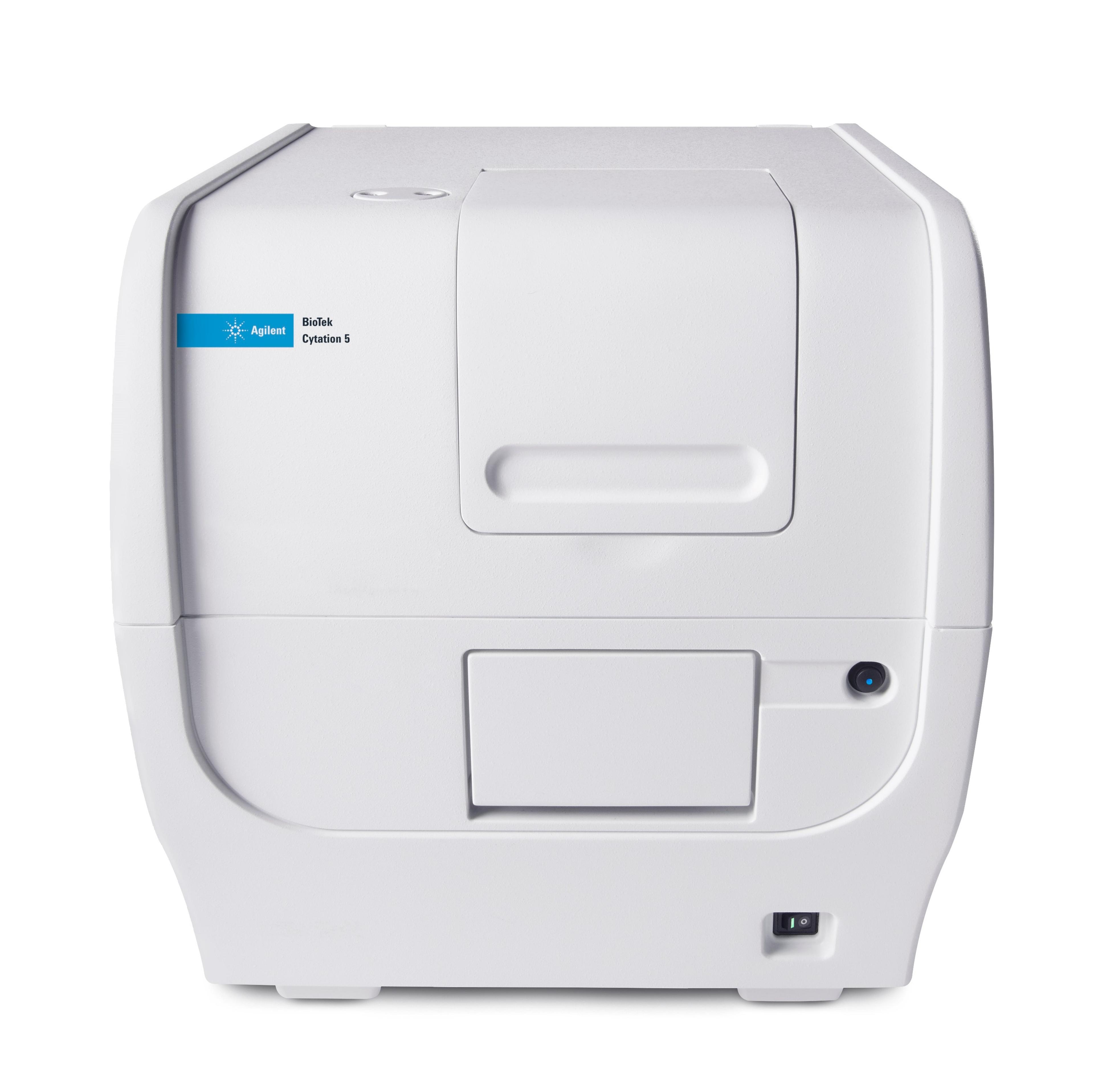
Agilent BioTek Cytation 5 Cell Imaging Multimode Reader
Agilent TechnologiesAgilent BioTek Cytation 5 is a uniquely integrated, configurable system that combines automated digital widefield microscopy with conventional multi-mode microplate detection to provide phenotypic cellular information and well-based quantitative data.